BIOLOGY Volume 1 - A Guide to General Biology - 2004
5. CELLS
5.9. Cell Membranes
5.9.8. Transport Across the Plasma Membrane
Although Plasma Membranes are typically only about 7 nm thick, they act as a barrier to ions and molecules, particularly polar (Water-soluble) molecules such as glucose or Amino Acids, because these substances are repelled by the non-polar (hydrophobic) Membrane Lipids. This barrier prevents The Cell's aqueous contents from escaping. Nevertheless, for various reasons, transport across membranes must occur to ensure:
1) the supply of nutrients;
2) the removal of metabolic end-products ("wastes");
3) the secretion of various useful substances;
4) the generation of ion gradients, which are essential for nervous and muscular activity;
5) the maintenance of an appropriate pH and proper ion concentrations within the cell, which are required for the efficient functioning of cellular Enzymes.
Here we discuss the Transport of substances across The Plasma Membrane, noting that Transport Across the membranes of cell Organelles is of a similar nature. There are four main mechanisms for substances to enter or leave the cell: diffusion, osmosis, Active Transport, and exocytosis or endocytosis. The first two processes are passive, meaning they require no Energy Expenditure; the latter two are active processes that involve the consumption of energy.
Diffusion and Facilitated Diffusion
Diffusion is defined as the net movement of substances from a region of higher concentration to a region of lower concentration down a diffusion gradient. It is a passive process that requires no energy expenditure and occurs spontaneously. If, for example, a bottle of perfume is left open in a closed room, the scent will gradually spread throughout the room until it is evenly distributed. This is caused by the random movement of molecules driven by their kinetic energy (energy of motion). Each type of molecule moves along its own diffusion gradient independently of other molecules. Oxygen, for example, diffuses from the Lungs into the Blood, while carbon dioxide diffuses in the opposite direction.
The rate of diffusion is primarily influenced by three factors.
1. The steepness of the diffusion gradient, i.e., the difference in concentration between point A and point B; the steeper the gradient, the higher the rate of diffusion. It is advantageous for a cell to maintain a steep diffusion gradient if rapid delivery of certain substances is required. In the lungs, for example, this is achieved by increasing the rate of Blood flow through them or by enhanced breathing.
2. The greater the surface area of the membrane through which the substance diffuses, the faster diffusion occurs. For Cells of roughly spherical shape, the surface-area-to-volume ratio decreases as the cell gets larger. This imposes constraints on cell size. A very large aerobic cell could not, for example, obtain oxygen quickly enough if it relied solely on diffusion. To increase the surface area available for uptake, some animal cells are equipped with microvilli.
3. The rate of diffusion decreases rapidly with increasing distance (it is inversely proportional to the square of the distance). Diffusion is therefore effective only over very short distances. This also imposes limitations on cell size. Since cells use diffusion for internal molecular transport, the diameter of most cells does not exceed 50 µm, and no part of the cell is further than 25 µm from its surface. An amino acid, for example, can travel a distance of a few micrometers in a matter of seconds, but it would take more than a day to travel a few centimeters. From this perspective, the extreme thinness of membranes is crucial, allowing molecules or ions to cross them rapidly.
The Factors affecting the rate of diffusion are summarized in Fick's law. It states that the rate of diffusion is proportional to the following expression:
Class="center">
So, which molecules can cross membranes by means of diffusion? Gases such as oxygen and carbon dioxide diffuse rapidly across membranes. Water molecules, although strongly polarized, are small enough to slip effortlessly between the hydrophobic phospholipid molecules. At the same time, ions and larger polar molecules are repelled by the hydrophobic Regions of the membrane and therefore diffuse across it extremely slowly. Other mechanisms are required for their entry into the cell.
Some ions and polar molecules enter the cell with the help of specialized transport Proteins, namely channel proteins and carrier proteins. The water-filled hydrophilic channels, or pores, of these proteins have a strictly defined shape that corresponds to a specific ion or molecule. Sometimes the channel is formed not within a single protein molecule, but between several adjacent molecules. Diffusion through channels can occur in both directions. This type of diffusion, facilitated by transport proteins, is called facilitated diffusion. Transport proteins that allow ions to pass are referred to as Ion Channels. Typically, ion channels are equipped with "gates," meaning they can open and close. Gated ion channels play a vital role in the conduction of nerve impulses.
In channel proteins, the shape is fixed (Fig. 5.16, B). It has been shown that the condition known as cystic fibrosis results from a defect in the protein that acts as a chloride ion channel. In carrier proteins, by contrast, the shape undergoes rapid changes, up to 100 cycles per second. They exist in two states, and their MECHANISM OF ACTION resembles a game of table tennis. Figure 5.17 illustrates how this mechanism Functions. The binding sites of the carrier protein face outward in one state ("ping") and inward toward the cell interior in the other ("pong"). The higher the concentration of dissolved molecules or ions, the greater the likelihood that they will become bound. If the concentration of the solute is higher outside than inside the cell, as in the example of glucose in Fig. 5.17, the net flux of this substance will be directed inward, and it will enter the cell. This is how glucose enters red Blood Cells. Transport of this kind exhibits all the Characteristic Features of diffusion, even though it is facilitated by the involvement of a protein. Another example of facilitated diffusion is the exchange of chloride and bicarbonate ions between red blood cells and Blood Plasma during the so-called chloride shift. This is one of the mechanisms that ensure the partial and selective permeability of membranes.

Fig. 5.17. Facilitated diffusion involving a carrier protein. The protein alternates between two states — "ping" and "pong". Since the concentration of glucose molecules (hexagons) is higher in the external environment, the net flux is directed inward in this case, moving down the diffusion gradient.
Osmosis
The Diffusion of Water through semipermeable membranes from a region of its high concentration to a region of low concentration is called osmosis. It is helpful to think of osmosis as a special type of diffusion in which only water molecules move. Let us consider the situation shown in Fig. 5.18. In this case, the solute molecules are too large to pass through the membrane pores, so equilibrium can only be achieved by the Movement of water molecules. Solution A has a higher concentration of water, so the net osmosis-driven flow of water is directed from A to B. Once equilibrium is reached, the net flow becomes zero. The tendency of water molecules to move from one place to another is measured by the water potential, denoted by the Greek letter ψ ("psi"). Water always moves from a region of high water potential to a region of low water potential. Solute molecules lower The water potential (in effect, they "dilute" the water!). The magnitude of this lowering is called the osmotic potential, ψo. Fig. 5.19 illustrates The Effect of various solutions on red blood cells. Osmosis in plant cells will be discussed in Chapter 13.
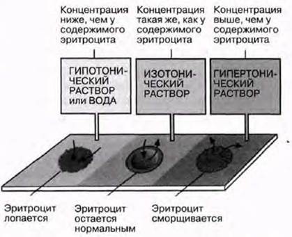
Fig. 5.18. Two solutions separated by a selectively permeable membrane.

Fig. 5.19. Behavior of red blood cells in solutions of different concentrations. A hypotonic solution has a higher water potential than the cell contents. As a result, water enters the cell via osmosis and bursts it, releasing the cell contents outward. A hypertonic solution has a lower water potential than the cell contents, causing water to leave the cell, which makes it shrink (crenate). In an isotonic solution, the water potentials of the solution and the cell contents are equal; therefore, no net movement of water occurs in either direction, and the cell volume remains normal and unchanged. Blood plasma must be isotonic with respect to red blood cells and other body cells.
5.4. Referring to Fig. 5.18, state which of the solutions has
a) a higher concentration of water molecules,
b) a higher concentration of solute molecules,
c) a higher water potential, and
d) a more negative osmotic potential?
e) Which of the two water potential values, -2000 kPa or -1000 kPa, is higher?
Active Transport
Active transport is the energy-driven transfer of molecules or ions across a membrane against a concentration gradient. Energy is required because the substance must move contrary to its natural tendency to diffuse in the opposite direction. This movement is typically unidirectional, whereas diffusion is reversible. The source of energy for active transport is ATP, a compound produced during Respiration that acts as an energy carrier within the cell. Consequently, active transport cannot take place in the absence of respiration.
Sodium ions (Na+), potassium ions (K+), and chloride ions (Cl-) predominate in extracellular and intracellular fluids. Fig. 5.20 shows that the concentrations of these ions inside human red blood cells and in blood plasma differ significantly. Inside red blood cells, as in most cells, the potassium concentration is much higher than outside. Another characteristic feature is that the intracellular potassium concentration exceeds the sodium concentration.

Fig. 5.20. Concentrations (in millimoles) of Na+, K+, and Cl- in red blood cells and their surrounding medium.
If respiration in red blood cells is inhibited by some specific Treatment, such as with cyanide, their ionic composition gradually changes and eventually matches that of the blood plasma. This demonstrates that these ions can passively diffuse across THE RED BLOOD cell plasma membrane, but under normal conditions, active transport—powered by energy supplied by respiration—maintains the concentrations shown in Fig. 5.20. In other words, sodium is actively pumped out of the cell, and potassium is actively pumped into it.
Active transport is carried out by carrier proteins located in the plasma membrane. Unlike the proteins involved in facilitated diffusion, these proteins require energy to undergo conformational changes. This energy is supplied by ATP generated during cellular respiration.
It was relatively recently discovered that the plasma membrane of most cells contains a sodium pump that actively extrudes sodium from the cell. In animal cells, the sodium pump is coupled with a potassium pump that actively takes up potassium ions from the external environment and transports them into the cell. This combined mechanism is called the sodium-potassium pump [the (Na+, K+)-pump]. Because this pump is present in almost all animal cells and performs A number of vital functions, it serves as a prime example of an active transport mechanism. Its physiological significance is evidenced by the fact that more than one-third of the ATP consumed by an animal cell at rest is used to pump sodium and potassium.
The pump is a specialized carrier protein spanning the entire thickness of the membrane (Fig. 5.21). Sodium and ATP bind to it from the inner side of the membrane, while potassium binds from the outer side. The transport of sodium and potassium across the membrane occurs as a result of Conformational Changes in this protein. Note that for every two potassium ions taken up, three sodium ions are pumped out of the cell. As a result, the cell interior becomes more negative relative to the external environment, establishing a potential difference across the Two Sides of the membrane. This restricts The entry of negatively charged ions (anions), such as chloride ions, into the cell. This exact mechanism explains why the concentration of chloride ions is lower in red blood cells than in blood plasma (Fig. 5.20), even though these ions can move in and out of cells via facilitated diffusion. Conversely, positively charged ions (cations) are attracted into the cell. Thus, both concentration and electrical charge are crucial in determining the direction in which ions move across the membrane.

Fig. 5.21. The sodium-potassium pump.
The sodium-potassium pump is essential for animal cells to maintain osmotic balance (osmoregulation). If it stops working, the cell will begin to swell and eventually burst. This happens because as sodium ions accumulate inside the cell, increasing amounts of water flow inward driven by osmotic forces. Obviously, Bacteria, Fungi, and plants, with their rigid cell walls, do not require such a pump. Animal cells also need it to maintain electrical activity in nerve and Muscle cells, and finally, for The active transport of certain substances, such as sugars and amino acids. High potassium concentrations are likewise required for Protein Synthesis, Glycolysis, Photosynthesis, and several other vital processes.
5.5. Try to explain the following observations.
a) If K+ ions are removed from the medium surrounding red blood cells, the influx of sodium into the cells and the efflux of potassium out of the cells increase dramatically.
b) If ATP is injected into cells, the outward flux of Na+ is enhanced.
Active transport is performed by all cells, but in certain cases, it plays a crucially important role. This is precisely the case in the epithelial cells lining the intestine and Kidney tubules, as the functions of these cells are intimately linked to secretion and absorption.
ACTIVE TRANSPORT IN THE INTESTINE. Upon being absorbed in the Small Intestine, Digestion products must cross the epithelial cells lining the intestinal wall. Subsequently, glucose, amino acids, and salts pass through the cells forming the walls of Blood Vessels, enter the bloodstream, and are carried by the blood to the Liver. Soon after a meal, the concentration of digestion products in the gut reaches a fairly high level, meaning that absorption is driven, to some extent, by diffusion. However, diffusion alone is far too slow here and must be supplemented by active transport. As shown in Fig. 5.22, this active transport is coupled with the operation of the (Na+, K+)-pump.

Fig. 5.22. Active transport of glucose across the plasma membrane of an intestinal or kidney cell. (Based on Fig. 36.12 from L. Stryer (1981) Biochemistry, 2nd ed., Freeman.)
Sodium pumped out of the cell by the sodium-potassium pump tends to diffuse back inward. The membrane contains a transport protein that requires both sodium and glucose to function. They are transported into the cell together via passive facilitated diffusion. Active Transport of Amino acids relies on a similar protein carrier—a "sodium-amino acid" symporter; the active component of this process is the continuous extrusion of sodium.
ACTIVE TRANSPORT IN NERVE AND MUSCLE CELLS. In nerve and muscle cells, the sodium-potassium pump establishes a potential difference across the plasma membrane, known as the Resting Potential (for Nerve Impulse propagation, see Ch. 17; for Muscle contraction, see Ch. 18).
A calcium pump operates in the membranes of the sarcoplasmic reticulum of muscle cells; here, calcium is actively pumped into the sarcoplasmic reticulum (a specialized form of The Endoplasmic reticulum) from the surrounding Cytoplasm. Muscle contraction is triggered by the rapid release of calcium in response to a nerve impulse.
ACTIVE TRANSPORT IN THE Kidneys. Active transport also takes place in the kidneys: sodium and glucose are actively transported out of the proximal convoluted tubules, while sodium is actively transported in the renal cortex. These processes are discussed in greater detail in Ch. 20.
ACTIVE TRANSPORT IN PLANTS. One example of active transport in plants is the uptake of sugars into the phloem, through which they are distributed to other PARTS OF THE plant (Ch. 13). This process plays an exceptionally vital role in leaves.
Endocytosis and exocytosis are two active processes by which various Materials are transported across the membrane either into cells (endocytosis) or out of cells (exocytosis) (Fig. 5.23).

Fig. 5.23. Endocytosis and exocytosis.
During endocytosis, the plasma membrane forms invaginations or outgrowths that subsequently pinch off to become vesicles or vacuoles1. There are two MAIN TYPES OF endocytosis.
1. Phagocytosis ("cell eating")—the engulfment of solid particles by cells. Specialized cells that carry out phagocytosis are called phagocytes; this function is performed, for example, by certain types of white blood cells that engulf bacteria. The membrane-bound sac enclosing the ingested particle is referred to as a phagocytic vacuole.
2. Pinocytosis ("cell drinking")—the uptake of liquid material by the cell. The resulting vesicles are often very small. In such cases, the process is termed micropinocytosis, and the vesicles are called micropinocytic vesicles. Human egg cells absorb nutrients from surrounding follicular cells precisely via this mechanism. In The Thyroid Gland, the hormone thyroxine is stored as thyroglobulin within specialized hollow structures (follicles). When the demand for thyroxine arises, follicular cells engulf thyroglobulin by pinocytosis, where it is converted into thyroxine and subsequently released into the blood. Pinocytosis is characteristic of a vast number of cells in both animals and plants (Fig. 5.10).
Exocytosis is the reverse of endocytosis. Through this mechanism, various materials are expelled from cells: indigestible solid residues are cleared from digestive vacuoles, and secretory cells release their products via "reverse pinocytosis." This is precisely how pancreatic enzymes, among others, are secreted (Fig. 5.29). In plant cells, exocytosis is used to export materials required for Cell wall construction (Fig. 5.30).
1 Vacuole—a fluid-filled membrane-bound sac. A vesicle is a small vacuole.
Last update: 06/08/2026
Editorial and Educational Adaptation: This material has been compiled based on the primary/original source text. The project team performed an editorial review, corrected technical inaccuracies, structured sections, and adapted the content for an educational format.
What was processed:
- elimination of formatting defects (OCR errors, structural breaks, corrupted characters);
- editorial organization of content;
- standardization of terminology in accordance with academic sources;
- verification of factual statements against the original source text.
All mentions of the author, publication year, and origin of the primary text have been preserved in accordance with the source.