Biochemistry: The Chemical Reactions of Living Cells, Volume 2 - D. Metzler 1980
On how electrons meet oxygen, how ATR is formed in the process, and some related phenomena
Electron transport chain and oxidative phosphorylation
Theories of oxidative phosphorylation and model experiments
Many schemes have been proposed for The formation of high-energy intermediates As a result of electron transfer. In this case, an analogy with substrate-level phosphorylation is natural, in which high-energy intermediates are formed during Electron transfer from substrate to substrate. As we have already seen (Ch. 8, Sec. 3,5), the aldehyde group of glyceraldehyde-3-phosphate is converted into an acyl phosphate, which, after transfer of the phosphate group to ADP, is released as a carboxylate group. In this process, the Free energy of aldehyde oxidation to a carboxyl group is coupled to ATP synthesis. The reaction differs from mitochondrial electron transfer in that the product, 3-phosphoglyceric acid, is not converted back to glyceraldehyde-3-phosphate. At the same time, the electron carriers of the Respiratory Chain must be regenerated in some cyclic process. This latter requirement necessitates the search for other mechanisms of Oxidative Phosphorylation.
a. Theories based on the specific Chemical properties of the electron-transferring coenzyme or prosthetic group
It is convenient to begin a review of the various theories of oxidative phosphorylation [82, 83] by referring again to equation (10-11)1). Lipmann [84] proposed a general scheme corresponding to this equation. The sequence of reactions begins with The addition of the Y'—OH group [group Y in equation (10-11)] across the appropriate double bond between carbon atoms in carrier BH2. Although isotope exchange reactions (Sec. D,5) rule out the possibility of either ADP or Pi functioning as Y, the assumption that a bound phosphate ion belonging, for example, to a phospholipid or coenzyme participates in this process remains attractive. The low-energy adduct Y—BH2 [equation (10-11)] is converted by oxidation into the compound Y~B, which is close in reactivity to an acyl phosphate or thioester.
It is important to consider that, regardless of The Nature of the compound Y~B, a fragment of the group Y remains bound after The transfer of Y to X. Thus, equation (10-11) will be more complete if Y is replaced by Y'OH. In this case, the resulting compound has the form X~Y', and the carrier remains in the form B—OH. To regenerate B, the hydroxyl group must be eliminated. In searching for a suitable chemical mechanism for these steps of oxidative phosphorylation, one problem is the requirement that such a hydroxyl group be easily eliminated.
Although in equation (10-11) compound Y adds to the reduced form of carrier B, followed by oxidation of B to the high-energy form of the oxidized carrier, it is also possible for Y to add to the oxidized form of carrier B. Then B—Y will be reduced to the form BH2~Y—the high-energy form of the reduced carrier. After reaction with group X [as in equation (10-11)], a modified reduced carrier B'H2 will remain, which upon subsequent elimination will revert to the form BH2, thereby completing the cycle. Thus, we should keep in mind the possibility of both oxidized and reduced high-energy forms of the carrier.
In one variant of Lipmann's general scheme, nucleophilic addition across the double bond of NADH is postulated [equation (8-48)]. The oxidative phosphorylation cycle would operate as shown in equation (10-14) [85, 86]. Note that the elimination of a Water molecule to regenerate NADH is the reverse of the reaction shown in equation (8-48); the equilibrium conditions in this case are unfavorable, which is a weak point of the scheme. One could also consider mechanisms based on the addition of Y—O- at the 6-position of NAD+2).
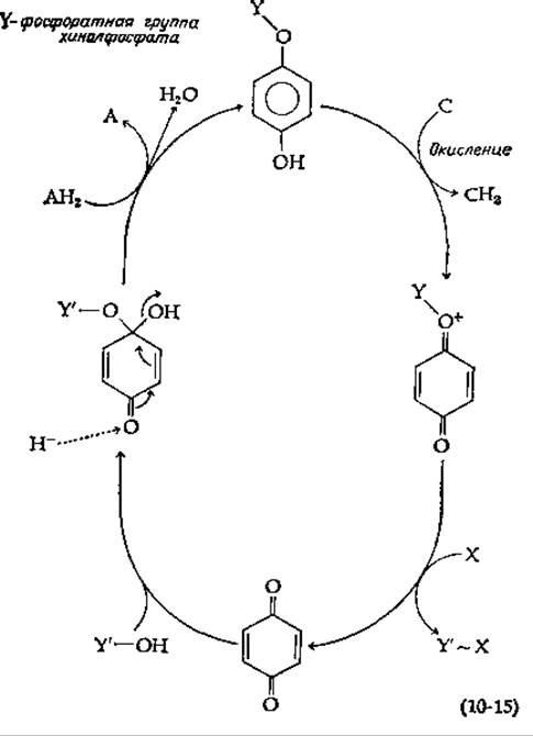
Many researchers have wondered whether the specific chemical properties of ubiquinone or other Quinones could be invoked to explain oxidative phosphorylation. Harrison [87] showed that oxidation of the phosphate ester of hydroquinone (quinol phosphate) in a suitable model system leads to the formation of a high-energy oxidized intermediate. In the presence of Pi [X in equation (10-15)], pyrophosphate (X~Y) is formed. (Alternatively, Y may be released in the form of a metaphosphate group capable of reacting with X.) An uncertain point in equation (10-15) remains The pathway of quinol phosphate formation. As shown in the equation, it can be formed by the addition of YOH across the carbonyl group of the oxidized quinone, followed by the loss of a hydroxyl ion during reduction by AH2.
1) Various mechanisms proposed for oxidative phosphorylation are reviewed by Lardy and Ferguson [82].
2) The reader may find it interesting to consider the following scheme: Y—O- adds at the 6-position of NAD+. The resulting adduct is oxidized (H- is removed from the 6-position). The new derivative transfers the Y group, forming X~Y and leaving 6-hydroxy-NAD+. The latter is reduced by carrier AH2 to 6-hydroxy-NADH, which, after tautomerization, loses OH- and is converted to NAD+.
Class="center">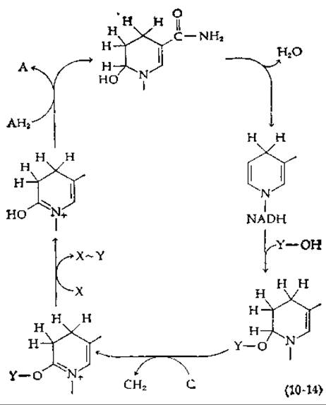
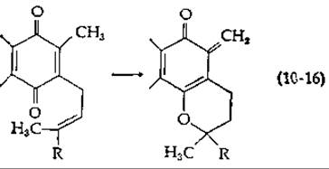
In a modified scheme [88], the loss of a proton from the methyl group of ubiquinone is postulated, with simultaneous ring closure to form a chromanol with an ortho-quinonoid Structure [quinone methide; equation (10-16)]. Phosphate addition and reduction can proceed in accordance with equation (10-15) (bottom center and left). Another theory postulates the functioning of the oxidized form of quinone as a reduced carrier [89].
Wang proposed a phosphorylation scheme based on the specific chemical properties of Cytochromes [equation (10-17)] [90, 91]. The reduced form of cytochrome with iron in the Fe(II) state [equation (10-17), top right] forms a coordinate bond with the imidazole group of the protein. Next, as a result of a two-electron oxidation by a carrier, one electron is removed from the iron and another from the imidazole group, forming a radical. The latter undergoes a coupling reaction with Y. Then, during a two-electron reduction by AH2, the intermediate radical and Fe(III) accept electrons, forming a high-energy derivative of the reduced form of the carrier. The latter transfers Y to X.

Another possibility is based on the results of The oxidation of thioesters by iodine [91, 92]. One can assume the participation of Methionine residues belonging to the electron carrier and undergoing cyclic transformation. There is every reason to believe that oxidative phosphorylation may be based on specific and as yet poorly understood chemical properties of mitochondrial iron-sulfur Proteins.
b. Theories based on Conformational Changes in proteins
The passage of electrons along the carrier chain can induce conformational changes in proteins, which can lead to the synthesis of high-energy intermediates. This view is supported by the small but clearly observable conformational changes in cytochrome c during Oxidation and reduction [12]. The close association of one protein with another, characteristic of The inner mitochondrial membrane, suggests that any conformational change induced at the electron transfer site can be transmitted through one or more proteins to a distant site (for example, to coupling factor F1), where ATP formation can occur.
With sufficiently large conformational Changes in the protein, a carboxyl group and an SH group may be brought into close proximity, leading to the spontaneous formation of a thioester bond. Then, when the protein returns to its relaxed conformation, the high-energy character of the thioester bond can be fully manifested, allowing it to enter into exchange reactions leading to ATP synthesis. Another possibility is the binding of ADP and inorganic phosphate by the protein at closely situated sites. Then, after appropriate conformational changes, these two components can be literally pressed together, with the spontaneous elimination of a hydroxyl ion and the formation of ATP and H2O.
A very interesting idea arising from a certain body of experimental data [93] is that ADP and Pi bind at adjacent sites, and within the hydrophobic environment of the F1 Active Site, spontaneous elimination of a water molecule occurs, forming tightly bound ATP. The driving force of the process may be the very tight binding of ATP to one of the protein conformers. Electron transfer can induce conformational changes that cause the release of the synthesized ATP by the F1 molecule.
Ideas regarding the conformational coupling of ATP synthesis and electron transfer become even more attractive when we recall that ATP is used in Muscles to perform mechanical work. In this case, ATP Hydrolysis is coupled with the relative movement of the protein Components of the Muscle (Box 10-E). Is it not reasonable to assume that ATP formation, in turn, occurs as a result of the movement of protein components induced in the mitochondrial membrane? The very sharp change in mitochondrial shape accompanying the transition between state 4 (ADP deficiency) and state 3 (active Respiration) has led some researchers to suggest that phosphorylation is inseparably linked to conformational Changes in membrane proteins [94]. Similar reasoning applies to phosphorylation in METABOLISM/14.html">Chloroplasts [95].
There is also a phonon theory, which is based not on conformational changes, but on specific lattice vibrations (for example, stretching vibrations of N—H bonds) excited in a membrane protein during electron passage [96]; these vibrations occupy the frequency range of 2700—5400 cm-1, i.e., 32—65 kJ∙mol-1 (Ch. 13, Sec. B, 3,a). As a result of such movement of protein molecules, appropriately positioned phosphates (possibly hydrogen-bonded to the amide groups of the main chain) can join together to form a pyrophosphate bond between them.
Box 10-E
Chemistry of Muscle contraction
While The properties of the Protein Assemblies found in muscle have been described in many interesting details (Ch. 4, Sec. E,1), the most important question remains open: how does the muscle machine use the free energy of ATP hydrolysis to perform mechanical worka,b? Based on Electron Cell/15.html">Microscopy and X-Ray Diffraction data, it was established that in the rigor state, all cross-bridges formed by Myosin heads are tightly attached to the thin Actin filaments. The addition of ATP, however, leads to the instantaneous detachment of the bridges from the thin filaments. In relaxed muscle, the thin filaments can move freely in the regions adjacent to the thick filaments, giving the muscle the property of a weakly stretched rubber band. However, muscle activation by a Nerve Impulse, accompanied by the release of Calcium Ions (Ch. 4, Sec. E,1), causes the thin filaments to slide between the thick ones, resulting in muscle shortening.
An activated muscle shortens only under relatively light loads; under higher loads, it maintains a constant length. Since the maximum muscle tension is proportional to the length of the region where the thin and thick filaments overlap, it is natural to attribute to the individual cross-bridges The Role of active centers that generate the force required for muscle contraction.

In Huxley's widely accepted "rowing hypothesis" model, cyclic changes in the state of cross-bridges are postulated. As shown in the accompanying figurec, in the initial state, the upper end of the myosin rod above the "hinge region", which projects 70—110 nm from the tail portion of the rod, moves freely in the space between the thick and thin filaments. Added ATP binds to the myosin heads, dissociating them from the thin filaments. The bound ATP is immediately hydrolyzed, but the resulting ADP and Pi remain bound in the Active Site of myosin. In this process, part of the free energy of ATP hydrolysis is stored, possibly in the form of corresponding conformational changes in the myosin HEAD. In the absence of calcium ions, the release of ADP and Pi and their replacement by new ATP occurs only very slowly. Thus, myosin itself possesses rather weak ATPase activity. It is assumed that in an activated muscle, the head containing the ATP Cleavage products binds to the actin subunit via cross-bridges positioned at right angles to the thin filaments. Then, the energy stored in the myosin head (or in actin) is expended to drive conformational changes, as a result of which the angle of attachment of the myosin head to the thin filament changes from 90° to approximately 45°. These changes can be observed directly by electron microscopy. In the absence of ATP (rigor), the heads form a serrated structure, binding to the actin filaments at an angle of about 45°. This change in the binding angle is considered sufficient to displace the actin filament by ~ 10 nm (the length of two actin subunits). Subsequently, Pi and ADP are released from the head-binding site; they are replaced by ATP, which causes dissociation of the Actomyosin complex. Meanwhile, other cross-bridges attach to the thin filaments, preventing them from returning to their original position.
Huxley's model commands considerable respect and is reasonably well supported by experimental evidence. However, it does not show precisely how the energy of ATP hydrolysis is coupled to conformational changes. It is possible that the elementary cycle of muscle contraction is based on entirely different processes. The idea has also been proposed that the conformational changes occur neither in the myosin head nor in actin, but in the "hinge" region of myosin. In this region, There is a segment of 200—300 residues containing A large number of positively charged side chains, and the α-Helix formed by these residues appears to be rather unstable. Possibly, interaction with ATP induces a cooperative conformational change in the protein, as a result of which the α-helix in the mentioned segment is converted into a "random coil", in which the two peptide chains of the myosin rod adopt a more extended conformationd,e. Apparently, the influx of calcium ions is sufficient to destabilize the structure and trigger conformational changes. The subsequent reaction with actin must induce reverse conformational changes, causing the chain segment to shorten again and pull the thin filament. Perhaps this kind of cycle, consisting of reversible stretching and shortening of the chain, may seem unconvincing to the reader; however, The Use of precisely such a "helix–coil" transition in Collagen made it possible to construct an interesting mechanism shown in the accompanying figuref. The collagen "pulley" first passes through a concentrated solution of lithium bromide, where the collagen undergoes contraction, and then through water, in which the concentrated salt that has penetrated the fiber is diluted, causing the helical collagen chains to regain the random coil conformation. This machine actually works, using the free energy of dilution of lithium bromide as an energy source.

The interest of several researchers is focused on determining whether the most important details of the muscle contraction mechanism reside in acting. For example, it has been suggested that ATP hydrolysis causes a shortening of several percent in 15—20 actin molecules simultaneously, which is sufficient for the total displacement of 10 nm required for contraction. According to another point of view, the cross-bridges are not part of the contractile mechanism but serve merely as a kind of "latch". It is known that a muscle contracts with almost no change in volume, and therefore anything that causes thickening of the sarcomere will lead to its shortening. It has been suggested that after ATP hydrolysis, negatively charged phosphate groups bind to Actin filaments and that the resulting electrostatic repulsion causes lateral Swelling of the sarcomereh. Several studies have re-emphasized the possibility that the energy of ATP cleavage is transformed (via Resonance transfer) into vibrational energy of amide bonds in the α-helical regions of myosini,k. This vibrational energy can be transmitted over long distances along the hydrogen-bond networks present in proteins and is somehow utilized in the contractile process. Although this idea may seem somewhat artificial, it reminds us that myosin rods, as well as thin filaments, should not be thought of as inert material. We do not know at present where the contractile springs are located in the system; it is possible that one of these speculative schemes will prove correct.
The reader may ponder for themselves what kind of mechanics is required to cleave ATP and produce contraction. In doing so, it is useful to look at The structure of ATP itself. First of all, note that the triphosphate group contains many negative charges that mutually repel each other. Imagine further what must happen when an ATP molecule displaces ADP and Pi from the actin-bound myosin head. This may disrupt the protein–protein interaction; most likely, electrostatic repulsion is induced at some specific point on their contact surface. Think about the formation of ATP during OXIDATIVE PHOSPHORYLATION AND the potential role of protons in ATP synthesis (Sec. D, 9,c). Could protons exert some effect on the protein surrounding the ATP molecule in the reverse process? Think about the action of Mg2+ complexed with the polyphosphate group of ATP, and also what might happen if a Ca2+ ion binds to an adjacent protein group. Consider the evidence for possible phosphorylation of protein side chains during intermediate Stages of the process. What would happen if a Histidine side chain, hydrogen-bonded to the peptide backbone at the terminal region of a helix, were phosphorylated? The author of this book has been unable to integrate all these considerations into a coherent Mechanism of muscle action, but perhaps one of the readers will succeed?
Although ATP serves as the immediate energy source for working muscle, its concentration is only about 5 mM. However, muscle also contains phosphagen, an N-phosphate derivative of a guanidino compound. In mammalian muscles, the phosphagen is creatine phosphate. In invertebrates, this role is played by Arginine phosphate or related compounds.

The group transfer potential for creatine phosphate is —43.1 kJ∙mol-1. Consequently, the transfer to form ATP occurs spontaneously, with a ∆G' value of —8.6 kJ∙mol-1. Creatine phosphate is present in muscle at a concentration of 20 mM, serving as a reservoir of high-energy phosphoryl groups and maintaining a high "energy charge" in the adenylate System of the muscle (creatine Biosynthesis is described in Ch. 14, Sec. B,3).
a Tonomura Y., Oosawa F., Annu. Rev. Biophysics Bioeng., 1, 159—190
b Taylor E. W., Annu. Rev. Biochem., 41, 577—616 (1972).
c Figure adapted from Mannherz H. G., Leigh I. B., Holmes K. C., Rosenbaum G., Nature (London), New Biol., 241, 226—229 (1973).
d Harrington W. F., PNAS, 68, 685—689 (1971).
e Burke M., Himmelfarb S., Harrington W. F., Biochemistry, 12, 701—710 (1973).
f Steinberg I. Z., Opłatka A., Katchalsky A., Nature (London), 210, 568— 571 (1966).
g Laid K., J. Theor. Biol., 44, 117—130 (1974).
h Ashley R., J. Theor. Biol., 36, 339—354 (1972).
i McClare C. W. F., J. Theor. Biol., 35, 569—595 (1972).
k Davydov A. S., J. Theor. Biol., 38, 559—569 (1973).
c. Theories Based on the Proton Gradient
Taking into account the negative outcome of all attempts to find high-energy intermediates, as well as the obvious requirement for an intact membrane, Mitchell proposed the chemiosmotic theory of oxidative phosphorylation in 1961 [97, 98]. This theory also accounts for the existence of energy-dependent processes, such as the accumulation of cations by Mitochondria. The fundamental principles of Mitchell's theory are illustrated in Fig. 10-12. It is assumed that there is a proton pump in the inner mitochondrial membrane driven by electron flow: this pump ejects protons from the matrix across the membrane. The idea of pumping protons via Electron transport is not new in itself; it had previously been suggested that this mechanism underlies the accumulation of Hydrochloric acid in The Stomach. As indicated in Fig. 10-12, the oxidized carrier B acquires two protons upon reduction to the BH2 form. These protons do not necessarily have to come from the reduced carrier AH2, and Mitchell suggested that they are taken up from the solution on the inner side of the membrane, i.e., from the matrix side. Then, when BH2 is reoxidized by carrier C, the protons are released, but on the outer side of the membrane. Mitchell presented Evidence indicating the required Stoichiometry of the process: for every two protons passing through the membrane, one ATP molecule is synthesized. It follows that three different proton pumps, corresponding to the three phosphorylation sites, must be integrated into the Electron Transport Chain.
The postulated proton pumps must lead either to the accumulation of a significant excess of protons in the intermembrane space, with a consequent decrease in pH, or to the accumulation of protons on the membrane itself. The second scenario would correspond to the case where counterions X- do not cross the membrane along with the protons. As a result, a Membrane Potential is generated—a phenomenon well established for nerve cell membranes (Ch. 5, Sec. B,3).
According to the central postulate of the chemiosmotic theory, the membrane contains an oriented ATPase or ATP synthase that utilizes the free energy of the proton gradient to synthesize ATP (Fig. 10-12). Given that ∆G' (pH 7) for ATP synthesis is + 34.5 kJ∙mol-1, and assuming that the passage of two protons through the ATPase is required to form one ATP molecule, it can be calculated that the necessary pH gradient [Equation (3-25)] would be 34.5/(2∙5.708) = 3.0 pH units at 25 °C. However, at a phosphorylation state of 104 M-1, the pH gradient would have to be ~5 units. It has been shown experimentally that electron transport indeed induces a pH difference and that an artificially induced pH gradient (across the mitochondrial membrane) leads to ATP synthesis. However, pH gradients of the required magnitude were not observed. At the same time, if the membrane is charged as shown in Fig. 10-12, without the accumulation of excess protons in the medium, a potential arises across the membrane, which can be utilized for ATP Synthesis in the same way as a proton gradient. A pH gradient of 3.0 units at 25 °C would be equivalent to a membrane potential ∆ψ ~0.177 V [Equation (3-63)]. The change in free energy upon the passage of a single ion across the membrane is expressed by the equation

The combined effect of the pH gradient and the membrane potential (both expressed in volts) is called the proton-motive force, which is described by the following equation:

where RT/F = 0.0592 at 25 °C. Since the membrane potential of both Mitochondria and chloroplasts is extremely difficult to measure directly, this postulate has not undergone the necessary verification1). Nevertheless, Mitchell's hypothesis has stimulated numerous experiments and is generally regarded as one of the most important current concepts in membrane biology.
How, then, can we explain the accumulation of ions by mitochondria? As shown in the lower part of Fig. 10-12, electroneutrality can be maintained in two ways. The protons translocated to the outside can be balanced by a parallel flow of counterions X-. On the other hand, if for every two protons moving outward, a cation such as Ca2+ enters, neutrality will also be maintained, but this must result in the accumulation of Ca2+ ions within the mitochondria. It has been shown experimentally that electron transport is indeed accompanied by cation accumulation. Not only calcium ions accumulate; in the presence of an appropriate ionophore (Ch. 5, Sec. B, 2,c), "energy-dependent" accumulation of potassium ions can be observed [99].
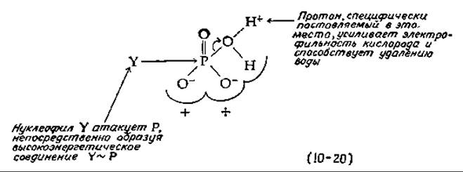
Is it possible, at least conceptually, to envision the design of a proton pump and an oriented ATPase driven by electron flow? We will consider only one purely hypothetical model. For a nucleophile Y to form a high-energy bond Y~P by direct attack on the phosphorus atom of Pi, an OH- ion must be removed. At pH 7, the probability of such a reaction is very low, but it can become significant at lower pH. Thus, we can imagine that the function of the oriented ATPase is to capture a proton and specifically hold it near the oxygen atom that is to be eliminated [Equation (10-20)]. But how can a proton be directed to the exact Location needed? It could probably pass through a channel in the membrane that delivers it to the required site. Perhaps it is even easier to imagine that the proton is specifically generated at the desired site directly during The electron transport process.
1) The membrane potential is estimated indirectly, for example, from the distribution of K+ ions on opposite sides of mitochondrial membranes in the presence of valinomycin [98a] or using Dyes that change fluorescence depending on the membrane potential [98b]. Membrane vesicles of E. coli Cells have also been studied [98c], but the interpretation of the results remains controversial [98d].

FIG. 30-32 Principal Features of the chemiosmotic theory of oxidative phosphorylation.
Now let us consider what the oxidation of the iron atom in cytochrome from Fe2+ to Fe3+ will lead to. It is quite natural to imagine that the removal of an electron will trigger a chain of electron movements in the coordinated histidine, in the adjacent peptide, and in other polar groups [Equation (10-21)], resulting in the appearance of an extra proton near the phosphate.
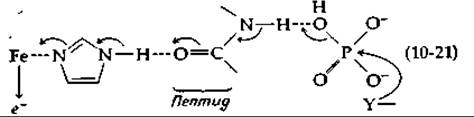
One can imagine that this chain of rearrangements will reach the neighboring protein and extend to the active site of the Ft protein. Thus, we have come full circle and are once again searching for something like a "high-energy intermediate." Many biochemists have a feeling that when Oxidative phosphorylation is fully explained, it will turn out that there was some truth in all of today's competing views.
Last update: 06/08/2026
Editorial and Educational Adaptation: This material has been compiled based on the primary/original source text. The project team performed an editorial review, corrected technical inaccuracies, structured sections, and adapted the content for an educational format.
What was processed:
- elimination of formatting defects (OCR errors, structural breaks, corrupted characters);
- editorial organization of content;
- standardization of terminology in accordance with academic sources;
- verification of factual statements against the original source text.
All mentions of the author, publication year, and origin of the primary text have been preserved in accordance with the source.