Human Biochemistry Volume 1 - Murray R. 1993
Bioenergetics and Metabolism of Carbohydrates and Lipids
Oxidation and Biosynthesis of Fatty Acids
Biosynthesis of Saturated Fatty Acids
Previously, Cleavage processes were thought to be the exact reverse of synthesis pathways (e.g., Glycogenolysis versus Glycogenesis), and fatty acid synthesis was viewed simply as the reverse of their oxidation.
It is now established that the mitochondrial system for FATTY ACID Biosynthesis—which involves a somewhat modified ß-Oxidation sequence—only elongates medium-chain Fatty acids already present in the Organism. In contrast, the complete de novo biosynthesis of palmitic acid from acetyl-CoA occurs actively outside the Cell/35.html">Mitochondria via an entirely different pathway. An active system responsible for elongating fatty acid chains is located in The Endoplasmic reticulum.
Extra-mitochondrial system for de novo fatty acid biosynthesis (Lipogenesis)
This enzyme system is found in the soluble (cytosolic) fraction of Cells from many Organs, particularly the Liver, Kidneys, Brain, Lungs, Mammary Glands, and adipose tissue. Fatty acid biosynthesis requires NADPH, ATP, Mn2+, and HCO-3 (as a source of CO2); acetyl-CoA serves as the substrate, and palmitic acid is the end product. The cofactor requirements for biosynthesis and ß-oxidation differ significantly.
Formation of malonyl-CoA
The first step in fatty acid biosynthesis, catalyzed by acetyl-CoA carboxylase and driven by ATP energy, is the carboxylation of acetyl-CoA, with bicarbonate serving as the source of CO2. The enzyme requires the vitamin biotin for its function (Fig. 23.5). This multi-enzyme complex consists of a variable number of identical subunits, each containing biotin, biotin carboxylase, a carboxybiotin-carrying protein, transcarboxylase, and an allosteric regulatory center. The reaction proceeds in two stages: (1) ATP-dependent carboxylation of biotin (Fig. 20.4), and (2) transfer of the carboxyl group to acetyl-CoA, yielding malonyl-CoA. Acetyl-CoA carboxylase is activated by citrate and inhibited by long-chain acyl-CoA derivatives. The active form of the enzyme readily polymerizes to form filaments composed of 10–20 protomers.
The synthase complex catalyzing fatty acid formation
Class="center">
Fig. 23.5. Biosynthesis of malonyl-CoA. E — acetyl-CoA carboxylase.
There are two types of synthase complexes catalyzing fatty acid biosynthesis, both located in the soluble fraction of The Cell. In Bacteria, plants, and lower animals such as Euglena, all individual Enzymes of The synthase system exist as autonomous Polypeptides, with acyl radicals linked to one of them, known as the acyl carrier protein (ACP). In Yeast, mammals, and birds, the synthase system is a multienzyme complex that cannot be dissociated into its components without losing activity, and the ACP is an integral part of this complex. Both bacterial ACP and the ACP of the multienzyme complex contain the vitamin pantothenic acid in the form of 4'-phosphopantetheine (see Fig. 17.6). Within the synthase system, ACP acts in place of CoA. The fatty acid synthase complex is a dimer (Fig. 23.6). In animals, the monomers are identical and consist of a single polypeptide chain encompassing 6 fatty-acid-synthesizing enzymes and an ACP with a reactive SH-group belonging to 4'-phosphopantetheine. Located immediately adjacent to this group is another sulfhydryl group belonging to a Cysteine residue within 3-ketoacyl synthase (the condensing enzyme), which is part of the other monomer (Fig. 23.6). Because both sulfhydryl groups are required for synthase activity, the complex is active only as a dimer.

Fig. 23.6. Multienzyme complex catalyzing fatty acid synthesis. The complex is a dimer composed of two identical polypeptide monomers, 1 and 2. Each monomer comprises 6 individual enzymes and an acyl carrier protein (ACP). Cys—SH — cysteine thiol group. The sulfhydryl group of 4'-phosphopantetheine on one monomer lies in close proximity to the analogous cysteine sulfhydryl group of ketoacyl synthase on the other monomer, indicating a HEAD-to-tail arrangement of the monomers. The exact sequence of enzymes within the monomers has not been definitively established and is shown here according to Tsukamoto. Each monomer contains all the enzymes required for fatty acid biosynthesis; however, it is not a functional unit on its own (the functional unit comprises fragments of both monomers, with half of one monomer interacting with the "complementary" half of the other). The synthase complex simultaneously synthesizes two fatty acid molecules.

Fig. 23.7. Biosynthesis of long-chain fatty acids. This diagram illustrates how The addition of a single malonyl residue elongates the acyl chain by 2 carbon atoms. Cys — cysteine residue; Pp — 4'-phosphopantetheine. The Structure of fatty acid synthase is shown in Fig. 23.6. ① and ② — individual monomers of fatty acid synthase. Two acyl chains are synthesized simultaneously on a single dimer, utilizing two pairs of —SH groups; in each pair, one group belongs to Pp and the other to Cys.
In the initial step of the process, a priming acetyl-CoA molecule interacts with the cysteine —SH group via transacylase (Fig. 23.7). Under the Influence of the same transacylase, malonyl-CoA reacts with the neighboring —SH group belonging to 4'-phosphopantetheine located on the ACP of the opposite monomer. This reaction yields an acetyl (acyl) malonyl enzyme. 3-Ketoacyl synthase catalyzes the reaction between the enzyme-bound acetyl group and the methylene group of malonyl with the release of CO2, producing a 3-ketoacyl enzyme (acetoacetyl enzyme); this frees up the cysteine sulfhydryl group previously occupied by the acetyl group. Decarboxylation drives the reaction to completion and serves as the driving force of biosynthesis. The 3-ketoacyl group is successively reduced, dehydrated, and reduced again to form the corresponding saturated acyl-S-enzyme. These reactions are analogous to those of ß-oxidation; notable differences include the fact that biosynthesis yields the D(—)-isomer of the 3-hydroxy acid rather than the L(+)-isomer, and NADPH rather than NADH serves as the hydrogen donor in the reduction steps. Next, a new malonyl-CoA molecule reacts with the phosphopantetheine —SH group, shifting the saturated acyl residue onto the now-free cysteine —SH group. This reaction cycle repeats 6 more times, with each incoming malonate residue extending the carbon chain until a saturated 16-carbon acyl radical (palmitoyl) is formed. The palmitoyl moiety is then released from the multienzyme complex by the sixth enzyme of the complex, thioesterase (deacylase). Free palmitic acid must be converted into its active acyl-CoA derivative before entering any other metabolic pathway. This activated palmitate is then typically esterified to formacylglycerols (Fig. 23.8).
The mammary gland contains a specialized thioesterase specific for C8, C10, or C12 acyl residues destined for milk Lipids. In ruminant mammary glands, this enzyme is an integral part of the fatty acid synthase complex.
Apparently, a single dimeric synthase complex contains 2 active sites that function independently, resulting in the simultaneous production of 2 molecules of palmitic acid.
Organizing all the enzymes of this metabolic pathway into a single multienzyme complex ensures high catalytic efficiency and prevents competition from other processes, thereby achieving cellular compartmentalization of the pathway without The Need for additional permeability barriers.
The overall net reaction for The biosynthesis of palmitic acid from acetyl-CoA and malonyl-CoA is given below:
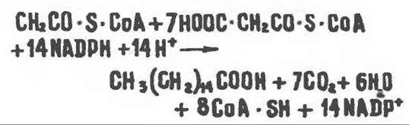
The primer molecule of acetyl-CoA provides carbons 15 and 16 of the palmitic acid chain. All subsequent two-carbon units are added via malonyl-CoA. In The Liver and mammary glands of mammals, butyryl-CoA can also serve as a primer. If propionyl-CoA acts as the primer, odd-numbered long-chain Fatty acids are synthesized. Such fatty acids are particularly characteristic of ruminants, in which propionic acid is produced in the rumen by microbial Fermentation.

Fig. 23.8. The metabolic fate of palmitate.
Sources of reducing equivalents and acetyl-CoA. NADPH is utilized as a coenzyme in the reduction of both 3-ketoacyl and 2,3-unsaturated acyl derivatives. The hydrogen required for reductive fatty acid biosynthesis is generated during the oxidative Reactions of the Pentose Phosphate Pathway. Notably, Tissues with an active pentose phosphate pathway are exceptionally proficient at lipogenesis (e.g., the liver, adipose tissue, and Cytology/practical/135.html">Lactating mammary gland). Furthermore, both metabolic pathways operate outside the mitochondria within the cell, meaning The transfer of NADPH/NADP between them is unimpeded by membranes or other barriers. Additional sources of NADPH include the malate-to-Pyruvate conversion catalyzed by the "malic enzyme" (NADP-malate dehydrogenase) (Fig. 23.9), as well as an extra-mitochondrial reaction catalyzed by isocitrate dehydrogenase (though its physiological contribution is likely minor).
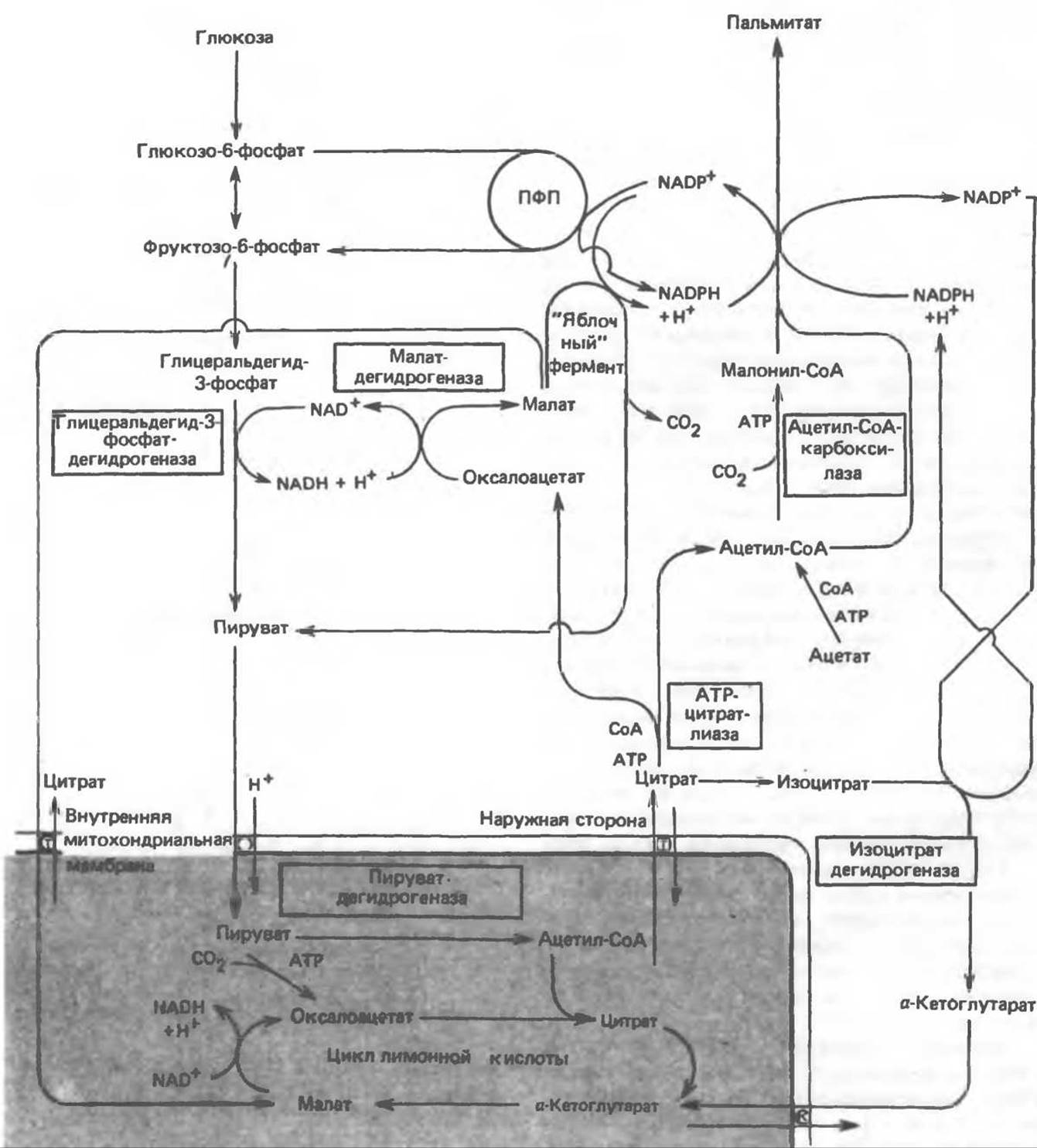
Fig. 23.9. Sources of acetyl-CoA and NADPH for lipogenesis. PPP — pentose phosphate pathway; T — tricarboxylate carrier system; K — $\alpha$-ketoglutarate carrier system
Acetyl-CoA, which serves as the building block for fatty acid synthesis, is generated in the mitochondria from CARBOHYDRATES via pyruvate oxidation. However, acetyl-CoA cannot freely cross the extramitochondrial compartment membrane, which is the primary site of fatty acid biosynthesis. Under conditions of good Nutrition, the activities of extramitochondrial ATP-citrate lyase and the "malic" enzyme increase in parallel with the activities of enzymes involved in fatty acid biosynthesis. It is currently believed that the pathway utilizing pyruvate in lipogenesis proceeds through a citrate intermediate. This metabolic pathway involves Glycolysis, followed by The oxidative decarboxylation of pyruvate to acetyl-CoA within the mitochondria, and a subsequent Condensation reaction with oxaloacetate to yield citrate, which is an intermediate of The Citric Acid Cycle. Citrate is then transported into the extramitochondrial compartment, where ATP-citrate lyase, in the presence of CoA and ATP, catalyzes its cleavage into acetyl-CoA and oxaloacetate. Acetyl-CoA is converted into malonyl-CoA (Fig. 23.5) and enters the biosynthesis of palmitic acid (Fig. 23.9). Oxaloacetate can be reduced to malate by NADH-dependent malate dehydrogenase; subsequently, a reaction catalyzed by the "malic" enzyme produces NADPH, which supplies the hydrogen required for lipogenesis. This metabolic process ensures the transfer of reducing equivalents from extramitochondrial NADH to NADP. Alternatively, malate can be transported back into the mitochondria, where it is converted into oxaloacetate. It should be emphasized that the operation of the mitochondrial citrate (tricarboxylate) transport system requires malate, which is exchanged for citrate (see Fig. 13.16).
In ruminants, the levels of ATP-citrate lyase and the "malic" enzyme in lipogenic tissues are quite low. This is presumably because the primary source of acetyl-CoA in these animals is acetate produced in the rumen. Since acetate is activated to acetyl-CoA extramitochondrially, it does not need to enter the mitochondria and be converted into citrate prior to incorporation into the long-chain fatty acid biosynthetic pathway. Due to the low activity of the "malic" enzyme in ruminants, NADPH generation catalyzed by extramitochondrial isocitrate dehydrogenase assumes particular importance.

Fig. 23.10. Microsomal fatty acid chain elongation system (elongase system).
The Microsomal Fatty Acid Chain Elongation System (Elongase)
Microsomes appear to be the primary site for the elongation of long-chain fatty acids. Fatty acid acyl-CoA derivatives are converted into products containing two additional carbon atoms, with malonyl-CoA acting as the acetyl group donor and NADPH as the reducing agent. The intermediates of this pathway are CoA thioesters. Saturated ($C_{10}$ and higher) and Unsaturated fatty acids can serve as primer molecules. Starvation inhibits The process of fatty acid chain elongation. During The formation of nerve cell myelin sheaths in the brain, the elongation of stearyl-CoA is sharply accelerated, resulting in The production of $C_{22}$ and $C_{24}$ fatty acids, which are constituents of Sphingolipids (Fig. 23.10).
Boyer Р. D. (ed.). The Enzymes, 3rd ed., Vol. 16 of Lipid Enzymology, Academic Press, 1983.
Debeer L. J., Mannaerts G. P. The mitochondrial and peroxisomal pathways of Fatty acid oxidation in rat liver, Diabete Metab. (Paris), 1983, 9, 134.
Goodridge A.G. Fatty acid synthesis in eukaryotes, Page 143. In: BIOCHEMISTRY OF LIPIDS and Membranes, Vance D. E., Vance J. E. (eds.), Benjamin/Cummings, 1985.
Gurr M.I., James A. I. Lipid Biochemistry: An Introduction, 3rd ed., Wiley, 1980.
Pandе S. V., Parvin R. Page 143. In: Carnitine Biosynthesis, METABOLISM, and Functions, Frenkel R. A., McGarry J. D. (eds.), Academic Press, 1980.
Schulz H. Oxidation of Fatty acids, Page 116. In: Biochemistry of Lipids and Membranes, Vance D. E., Vance J. E. (eds.), Benjamin/Cummings, 1985.
Singh N.. Wakil S.J., Stoops J.K. On the question of halfor fullsite reactivity of animal fatty acid synthetase, J. Biol. Chem., 1984, 259, 3605.
Tsukamoto Y. et al. The architecture of the animal fatty acid synthetase complex, J. Biol. Chem., 1983, 258, 15312.
Various authors. Disorders characterized by evidence of abnormal Lipid Metabolism. In: The Metabolic Basis of Inherited Disease, 5th ed., Stanbury J. B. et al. (eds.), McGraw-Hill, 1983.
Last update: 06/08/2026
Editorial and Educational Adaptation: This material has been compiled based on the primary/original source text. The project team performed an editorial review, corrected technical inaccuracies, structured sections, and adapted the content for an educational format.
What was processed:
- elimination of formatting defects (OCR errors, structural breaks, corrupted characters);
- editorial organization of content;
- standardization of terminology in accordance with academic sources;
- verification of factual statements against the original source text.
All mentions of the author, publication year, and origin of the primary text have been preserved in accordance with the source.