Human Biochemistry, Volume 1 - Murray R. 1993
Bioenergetics, Carbohydrate and Lipid Metabolism
Oxidation and Biosynthesis of Fatty Acids
Fatty Acid Oxidation
Free Fatty acids
“Free fatty acids” (FFAs) refer to fatty acids in their unesterified form; they are sometimes called non-esterified fatty acids (NEFAs). In Blood Plasma, long-chain FFAs form a complex with albumin, whereas within The Cell they associate with the fatty acid-binding protein, known as Z-protein; in reality, they are virtually never truly free. Short-chain Fatty acids are more Water-soluble and exist either as un-ionized acids or as fatty acid anions.
Activation of fatty acids
Much like in glucose METABOLISM, a fatty acid must first be converted into an active derivative through an ATP-dependent reaction before it can interact with Enzymes catalyzing further transformations. This is the sole stage in Fatty acid oxidation that requires energy in the form of ATP. In the presence of ATP and coenzyme A, the enzyme acyl-CoA synthetase (thiokinase) catalyzes The conversion of a free fatty acid into an “active fatty acid,” or acyl-CoA, driven by the Cleavage of a high-energy phosphate bond.
Class="center">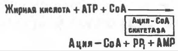
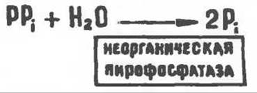
The presence of inorganic pyrophosphatase, which hydrolyzes the high-energy phosphate bond in pyrophosphate, drives the activation process to completion. Consequently, the Activation of a single fatty acid molecule ultimately consumes two high-energy phosphate bonds.
Acyl-CoA synthetases are located in The Endoplasmic reticulum, as well as within Mitochondria and on their outer membrane. Several acyl-CoA synthetases have been described in the literature; they exhibit Specificity for fatty acids of particular chain lengths.
The Role of carnitine in fatty acid oxidation
Carnitine (γ-trimethylamino-β-hydroxybutyrate), (CH3)3N+—CH2—CH(OH)—CH2—COO-, is a widely distributed compound,
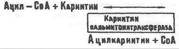
particularly abundant in Muscle tissue. It is synthesized from Lysine and Methionine in The Liver and Kidneys. Although the activation and oxidation of lower fatty acids can occur in mitochondria independently of carnitine, long-chain acyl-CoA derivatives (or FFAs) cannot penetrate the mitochondria and undergo oxidation unless they are first converted into acylcarnitine derivatives. The outer face of The inner mitochondrial membrane harbors the enzyme carnitine palmitoyltransferase I, which transfers long-chain acyl groups to carnitine, yielding acylcarnitine. The latter is capable of crossing into the mitochondrial matrix, where the enzymes catalyzing β-oxidation reside.
A proposed mechanism explaining the involvement of carnitine in mitochondrial fatty acid oxidation is illustrated in Fig. 23.1. Additionally, mitochondria contain another enzyme, carnitine acetyltransferase, which catalyzes The transfer of short-chain acyl groups between CoA and carnitine. The physiological function of this enzyme remains unclear; it may facilitate The transport of acetyl groups across the mitochondrial membrane.
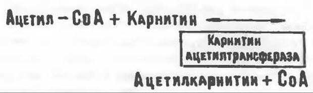
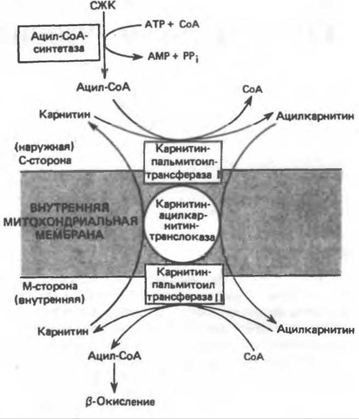
Fig. 23.1. The role of carnitine in transporting long-chain fatty acids across the inner mitochondrial membrane. Long-chain acyl-CoA cannot traverse the inner mitochondrial membrane directly, whereas acylcarnitine—formed via the action of carnitine palmitoyltransferase I—readily crosses it. Carnitine-acylcarnitine translocase acts as a transport system that translocates acylcarnitine molecules across the inner mitochondrial membrane in exchange for free carnitine molecules. Subsequently, upon the action of carnitine palmitoyltransferase II, localized on the matrix side of the inner mitochondrial membrane, acylcarnitine interacts with CoA. This regenerates acyl-CoA in the mitochondrial matrix and releases free carnitine.
β-Oxidation of Fatty acids
An Overview is provided in Fig. 23.2. During fatty acid β-oxidation, two carbon atoms are sequentially cleaved from the carboxyl end of the acyl-CoA molecule. The carbon chain is broken between the α(2) and β(3) carbon atoms, which gives rise to the term β-oxidation. The resulting two-carbon fragments are molecules of acetyl-CoA. For instance, the Complete oxidation of palmitoyl-CoA yields 8 molecules of acetyl-CoA.
Sequence of reactions
A group of enzymes collectively known as “fatty acid oxidases” resides in the mitochondrial matrix in close proximity to the Respiratory Chain located in the inner mitochondrial membrane. This system catalyzes The oxidation of acyl-CoA to acetyl-CoA, which is coupled to the phosphorylation of ADP to ATP (Fig. 23.3).
Following the translocation of the acyl moiety across the mitochondrial membrane via the carnitine transport system and the transfer of the acyl group from carnitine back to CoA, two hydrogen atoms are removed from the 2(α) and 3(β) carbon atoms in a reaction catalyzed by acyl-CoA dehydrogenase. The product of this reaction is a Δ2-trans-enoyl-CoA. This enzyme is a flavoprotein containing FAD as its prosthetic group. Its oxidation within the mitochondrial respiratory chain proceeds via another flavoprotein known as electron-transferring flavoprotein (see p. 123). Next, Hydration of the double bond takes place, producing 3-hydroxyacyl-CoA, a reaction catalyzed by Δ2-enoyl-CoA hydratase. Subsequently, 3-hydroxyacyl-CoA is dehydrogenated at the 3rd carbon atom to form 3-ketoacyl-CoA, catalyzed by 3-hydroxyacyl-CoA dehydrogenase with NAD acting as a coenzyme. 3-Ketoacyl-CoA is then cleaved between the second and third carbon atoms by 3-ketothiolase (acyl-CoA acetyltransferase), yielding acetyl-CoA and an acyl-CoA derivative shortened by two carbon atoms compared to the original molecule. This thiolytic cleavage requires an additional molecule of CoA. The resulting shortened acyl-CoA re-enters the β-oxidation cycle at reaction step 2 (Fig. 23.3). Through this pathway, long-chain fatty acids are completely degraded to acetyl-CoA (C2 fragments), which are subsequently oxidized to CO2 and H2O in The Citric Acid Cycle operating within the mitochondria.

Fig. 23.2. Schematic overview of fatty acid β-oxidation.
Oxidation of odd-chain fatty acids
The β-oxidation of fatty acids with an odd number of carbon atoms culminates in The formation of a three-carbon intermediate, propionyl-CoA, which is subsequently converted into succinyl-CoA, an intermediate of The Citric Acid cycle (see also Fig. 20.2).
Energetics of Fatty Acid Oxidation
As a result of Electron transport along the respiratory chain from reduced flavoprotein and NAD, 5 energy-rich phosphate bonds are synthesized (see Ch. 13) for every 7 (out of 8) acetyl-CoA molecules produced during the ß-Oxidation of palmitic acid (7 x 5 = 35). A total of 8 acetyl-CoA molecules are formed, and each of them, upon entering the citric acid cycle, drives the synthesis of 12 energy-rich bonds. Overall, per palmitate molecule, this pathway generates 8 x 12 = 96 energy-rich phosphate bonds. Accounting for the two bonds required for fatty acid activation, this yields a total of 129 energy-rich bonds per mole, or 129 x 30.5 = 3935 kJ. Given that the Free energy of combustion of palmitic acid is 9791 kJ/mol, approximately 40% of the energy is conserved as phosphate bonds during fatty acid oxidation.
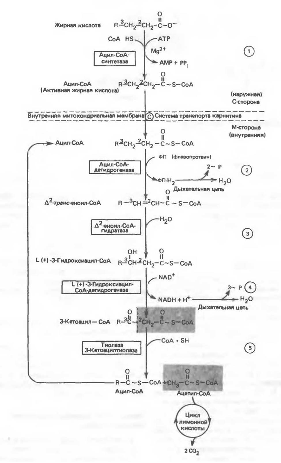
Fig. 23.3. ß-Oxidation of fatty acids. Long-chain acyl-CoA undergoes stepwise shortening through cycle-after-cycle enzymatic reactions 2–5; each cycle releases an acetyl-CoA molecule, catalyzed by thiolase (reaction 5). When a four-carbon acyl radical remains, it yields two molecules of acetyl-CoA via reaction 5.
Peroxisomal Fatty Acid Oxidation
In Peroxisomes, fatty acid ß-oxidation proceeds via a modified pathway. The products of oxidation in this case are acetyl-CoA and H2O2, with the latter being formed at the step catalyzed by a flavoprotein-linked dehydrogenase. This oxidation pathway is not directly coupled with phosphorylation and ATP formation, but it facilitates The breakdown of very long-chain fatty acids (e.g., C20, C22); it is induced by high-fat diets or the administration of hypolipidemic drugs such as clofibrate. Peroxisomal enzymes do not act on short-chain fatty acids, and the ß-oxidation process stops upon the formation of octanoyl-CoA. Octanoyl and acetyl groups are subsequently exported from peroxisomes as octanoylcarnitine and acetylcarnitine and are oxidized in mitochondria.
α- and ω-Oxidation of Fatty Acids
ß-Oxidation is the principal pathway of fatty acid Catabolism. However, it has recently been discovered that α-oxidation of fatty acids—namely, the successive removal of single-carbon units from the carboxyl end of the molecule—takes place in Brain Tissues. This process involves CoA-containing intermediates and is not accompanied by the generation of energy-rich phosphate bonds.
ω-Oxidation of fatty acids is normally very minor. This type of oxidation, catalyzed by hydroxylases in the presence of cytochrome P-450 (see p. 123), takes place in the endoplasmic reticulum. The —CH3 group is converted into a —CH2OH group, which is then oxidized to —COOH, resulting in the formation of a dicarboxylic acid. The latter is broken down via ß-oxidation, typically down to adipic (C6) and suberic (C8) acids, which are subsequently excreted in the urine.
Clinical Aspects
Ketosis develops at a high rate of fatty acid oxidation in the liver, particularly when it occurs against a Background of carbohydrate deficiency (see p. 292). Such a condition arises during high-fat diets, starvation, Diabetes Mellitus, ketosis in lactating cows, and Pregnancy toxemia (ketosis) in sheep. The causes underlying impaired fatty acid oxidation are outlined below.
Carnitine deficiency occurs in newborns, most frequently premature infants; it is caused either by impaired carnitine Biosynthesis or by renal carnitine "leaking". Carnitine losses can occur during hemodialysis; patients suffering from organic aciduria lose significant amounts of carnitine, which is excreted from the body as conjugates with organic acids. To compensate for the loss of this compound, some patients require a special diet containing carnitine-rich foods. Signs and symptoms of carnitine deficiency include episodes of hypoglycemia resulting from impaired Gluconeogenesis secondary to defective fatty acid oxidation, diminished ketone body production accompanied by elevated plasma free fatty acid (FFA) levels, muscle weakness (myasthenia), and lipid accumulation. Treatment involves oral administration of carnitine. The symptoms of carnitine deficiency closely resemble those of Reye's syndrome; however, carnitine levels are normal in the latter condition. The cause of Reye's syndrome remains unknown.
A decrease in hepatic carnitine palmitoyltransferase activity leads to hypoglycemia and lowered plasma ketone body levels, whereas a reduction in muscle carnitine palmitoyltransferase activity impairs fatty acid oxidation, resulting in periodic muscle weakness and myoglobinuria.
Jamaican vomiting sickness occurs in humans after ingestion of unripe ackee fruit (Blighia sapida), which contains the toxin hypoglycin, an inactivator of acyl-CoA dehydrogenase, thereby inhibiting ß-oxidation.
Dicarboxylic aciduria is characterized by The excretion of C6–C10 dicarboxylic acids and The Development of hypoglycemia without an increase in ketone body levels. The cause of this disorder is a deficiency of medium-chain acyl-CoA dehydrogenase in the mitochondria. This impairs ß-oxidation and enhances the ω-oxidation of long-chain fatty acids, which are shortened to medium-chain dicarboxylic acids and eliminated from the body.
Refsum's disease is a rare neurological disorder caused by the tissue accumulation of phytanic acid, derived from phytol, a constituent of dietary plant chlorophyll. Phytanic acid contains a methyl group at the third carbon atom, which blocks its ß-oxidation. Normally, this methyl group is removed via α-oxidation, but patients with Refsum's disease have an inherited defect in the α-oxidation system, leading to the tissue accumulation of phytanic acid.

Fig. 23.4. Reaction sequence for the Oxidation of Unsaturated fatty acids, exemplified by linoleic acid. ∆4-cis-Fatty acids or fatty acids yielding ∆4-cis-enoyl-CoA enter this pathway at the stage indicated in the scheme.
Zellweger syndrome (cerebrohepatorenal syndrome) is a rare inherited disorder characterized by the complete absence of peroxisomes in all tissues. Patients with Zellweger syndrome accumulate C26–C38 polyenoic acids in their brains because peroxisomal deficiency prevents the oxidation of very long-chain fatty acids.
Oxidation of Unsaturated fatty acids
Unsaturated fatty acid CoA derivatives undergo ß-oxidation steps until, depending on THE POSITION OF the double bonds, either a ∆3-cis-acyl-CoA or ∆4-cis-acyl-CoA derivative is formed (Fig. 23.4); subsequently, ∆3-cis-acyl-CoA is isomerized by ∆3-cis → ∆2-trans-enoyl-CoA isomerase to yield ∆2-trans-enoyl-CoA. The latter then enters the standard ß-oxidation pathway. Any ∆4-cis-acyl-CoA derivative—either formed during the oxidation of linoleic acid (Fig. 23.4) or entering the cycle at this stage—is converted by acyl-CoA dehydrogenase into ∆2-trans-∆4-cis-dienoyl-CoA; this intermediate is then converted into ∆3-trans-enoyl-CoA by the NADP-dependent enzyme ∆2-trans-∆4-cis-dienoyl-CoA reductase. Next, ∆3-cis → ∆2-trans-enoyl-CoA isomerase acts on the ∆3-trans double bond, producing ∆2-trans-enoyl-CoA, which serves as a standard intermediate in ß-oxidation.
Microsomal Peroxidation of Polyunsaturated Fatty Acids
NADPH-dependent peroxidation of unsaturated fatty acids is catalyzed by enzymes localized in microsomes (see p. 124). Antioxidants, such as BHT (butylated hydroxytoluene) and α-tocopherol (vitamin E), inhibit microsomal Lipid Peroxidation.
Last update: 06/08/2026
Editorial and Educational Adaptation: This material has been compiled based on the primary/original source text. The project team performed an editorial review, corrected technical inaccuracies, structured sections, and adapted the content for an educational format.
What was processed:
- elimination of formatting defects (OCR errors, structural breaks, corrupted characters);
- editorial organization of content;
- standardization of terminology in accordance with academic sources;
- verification of factual statements against the original source text.
All mentions of the author, publication year, and origin of the primary text have been preserved in accordance with the source.