Principles of Biochemistry Volume 1 - A. Lehninger 1985
Biomolecules
Cells
Viruses as supramolecular parasites
Our Structure/133.html">Discussion of Cells as the fundamental units of living matter would be incomplete without considering Viruses. Although not strictly alive, viruses are supramolecular complexes formed via biological pathways that are capable of self-Replication within appropriate host cells. A virus consists of a nucleic acid molecule surrounded by a protective protein coat, or capsid. Viruses exist in two distinct states. Outside of their host cells, they are essentially inanimate particles known as virions, possessing a defined shape, precise dimensions, and a specific chemical composition. Some viruses can even be crystallized, behaving in this state much like extremely large molecules. However, once a viral particle (or its nucleic acid) enters a specific host Cell, its mode of existence changes dramatically: it becomes an intracellular parasite. The viral nucleic acid carries the Genetic information that dictates the entire STRUCTURE OF THE intact virion. It commandeers the host cell's biochemical machinery, disrupting normal cellular operations and redirecting The Cell's Enzymes and Ribosomes away from synthesizing normal cellular components toward producing vast numbers of new progeny Viral Particles. Consequently, the infection of a host cell by a single virion can result in the generation of dozens or even hundreds of new viral particles (Fig. 2-24). In some host-virus systems, these newly formed virions are released as the host cell eventually dies and undergoes lysis. In other systems, the newly synthesized viral nucleic acid remains within the host cell, sometimes exerting only a minor effect on cell viability; frequently, however, this leads to profound alterations in both the Morphology and function of the host cell. Certain viruses contain DNA, whereas others contain RNA.
Class="center">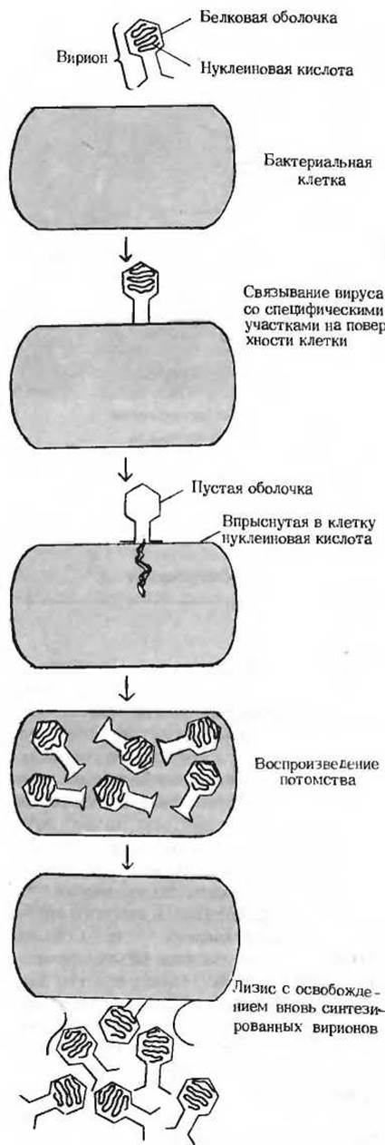
Fig. 2-24. Replication of a bacteriophage within a host cell.
Hundreds of different viruses are known, each specific to particular types of host cells. Hosts may include animal, plant, or bacterial cells (Table 2-3). Viruses that specifically infect Bacteria are called Bacteriophages, or simply phages (derived from the Greek word meaning "to eat" or "to devour"). The capsid of some viruses may be constructed from a single type of protein molecule, as seen in the tobacco mosaic virus—one of the simplest viruses and the first to be crystallized (Fig. 2-25). Other viruses may contain dozens or even hundreds of Different types of Proteins. Viral dimensions vary over a wide range. For instance, one of the smallest viruses, bacteriophage ΦX174, has a diameter of 18 nm, whereas one of the largest, the vaccinia virus, approaches the size of the smallest bacteria. Viruses also differ widely in shape and structural complexity. Among the most complex is bacteriophage T4 (Fig. 2-25), which infects E. coli. Phage T4 features a HEAD, a tail, and an intricate set of tail fibers; when injecting viral DNA into the host cell, these structures work in concert like a stinger or a hypodermic syringe. Figure 2-25 and Table 2-3 provide data on the size, shape, and mass of various viral particles, as well as the types and molecular weights of their constituent Nucleic Acids. Some viruses are exceptionally pathogenic to humans. These include the causative agents of smallpox, poliomyelitis, Influenza, the common cold, infectious mononucleosis, and shingles. Furthermore, latent viruses are widely believed to be responsible for certain cancers in animals. Viruses play an increasingly vital role in biochemical research, as they provide extraordinarily valuable insights into Chromosome structure, the mechanisms of enzymatic nucleic acid synthesis, and The regulation of genetic information transfer.
Table 2-3. Properties of Selected Viruses
|
Virus |
Nucleic Acid |
Particle Mass, 106 Daltons |
Length, nm |
Shape |
|
E. coli bacteriophages — ΦX174 |
DNA |
6 |
18 |
Polyhedral |
|
T4 |
DNA |
220 |
200 |
Tadpole-shaped |
|
λ (lambda) |
DNA |
50 |
120 |
Tadpole-shaped |
|
MS2 |
RNA |
3.6 |
20 |
Polyhedral |
|
Plant viruses Tobacco mosaic virus |
RNA |
40 |
300 |
Rod-shaped |
|
Tomato bushy stunt virus |
RNA |
10.6 |
28 |
Polyhedral |
|
Animal viruses Poliovirus |
RNA |
6.7 |
30 |
Polyhedral |
|
Simian virus 40 (SV40; induces tumors in newborn animals) |
DNA |
28 |
45 |
Spherical |
|
Adenovirus (causes common cold symptoms) |
DNA |
200 |
70 |
Polyhedral |
|
Smallpox virus |
DNA |
4000 |
250 |
Spherical |
All cells are bounded by a Plasma Membrane and contain Cytoplasm, ribosomes, and a nuclear region or nucleus. The size and shape of cells are constrained by the rates of physical diffusion of nutrient molecules and oxygen, as well as by the cell's surface-area-to-volume ratio. There are two Major Classes of cells: prokaryotic and eukaryotic. Prokaryotes, which include bacteria and blue-green Algae, are small, structurally simple cells characterized by the absence of a membrane-bounded nucleus enclosing their genetic material. They possess a Cell wall and a plasma membrane, and some are equipped with flagella for locomotion. The cytoplasm of Prokaryotic Cells lacks membrane-enclosed Organelles, but contains ribosomes and nutrient granules. Prokaryotic cells grow and divide very rapidly. The bacterium Escherichia coli, the most thoroughly studied prokaryote, has proven exceptionally useful for biochemical and genetic investigations.
Eukaryotic cells are much larger than prokaryotic cells, having volumes 1,000 to 10,000 times greater. In addition to a distinct, membrane-bounded nucleus containing multiple Chromosomes, eukaryotic cells harbor numerous membrane-enclosed organelles. These include Mitochondria, whose function is to oxidize cellular "fuel" and generate ATP, as well as METABOLISM/14.html">Chloroplasts (in photosynthetic cells), which capture light energy and utilize it to convert CO2 into glucose. It is widely theorized that Mitochondria and chloroplasts evolved from bacteria. Eukaryotic organelles also include The Endoplasmic reticulum, which channels and transports secretory products to the Golgi apparatus, where they are packaged and exported from the cell. Lysosomes contain degradative enzymes, whereas Peroxisomes compartmentalize enzymes involved in the formation and breakdown of peroxides, isolating them from the rest of the cellular contents. The eukaryotic cytoplasm contains at least three distinct types of microfilaments, along with microtubules. Microfilaments, microtubules, and the microtrabecular lattice collectively form a flexible internal framework known as the Cytoskeleton. Many animal cells bear Cilia and flagella, whose whip-like or rotational movements are driven by pairs of internal microtubules. Ribosomes are also present in eukaryotic cells, existing either freely in the cytoplasm or bound to the outer surface of the rough endoplasmic reticulum. Furthermore, the external surface of animal cells features specialized recognition sites that bind specific cells and Hormones.
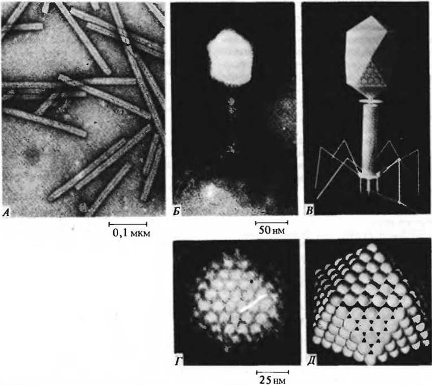
Fig. 2-25. (A) Rod-shaped tobacco mosaic virus. (B) Electron micrograph and (C) molecular model of bacteriophage T4, a complex virus resembling a tadpole in shape. Following the attachment of the phage's terminal fibers to specific sites on the E. coli cell wall, DNA from the phage head is injected through the tail structure into the cell. (D) Electron micrograph and (E) tennis-ball model of an adenovirus, whose capsid consists of 252 protein subunits forming an icosahedron (a polyhedron with 20 faces).
Viruses are inanimate supramolecular structures. Each viral particle consists of a single nucleic acid molecule enclosed within a protein coat. Viruses are capable of infecting specific host cells, subverting their machinery to replicate viral particles According to the genetic instructions encoded within the viral nucleic acid. The Study of viruses has yielded invaluable insights into the biochemical mechanisms governing The transfer of genetic information.
Introduction/47.html">Further Reading
Textbooks
Curtis, H. Biology, 3rd ed. New York: Worth Publishers, 1979. A beautifully written and illustrated general biology textbook.
Dyson, R. D. Cell Biology: A Molecular Approach, 2nd ed. Boston: Allyn and Bacon, 1978. A textbook with a strong biochemical orientation.
Fawcett, D. W. The Cell, 2nd ed. Philadelphia: W. B. Saunders, 1981. Features comprehensive Cytology/cytology/93.html">ELECTRON MICROGRAPHS OF various Cells and Tissues.
Karp, G. Cell Biology. New York: McGraw-Hill, 1979. Places special emphasis on the molecular aspects of genetic information transfer.
Ledbetter, M. C., and K. R. Porter. Introduction to the Fine Structure of Plant Cells. New York: Springer-Verlag, 1970.
Loewy, A. G., and P. Siekevitz. Cell Structure and function, 3rd ed. New York: Holt, Rinehart and Winston, 1979.
Selected Short Books and Monographs
Luria, S. Life: The Unfinished Experiment. New York: Charles Scribner's Sons, 1973. A Nobel laureate in molecular biology provides an Overview and critique of contemporary biology.
Margulis, L. Origin of Eukaryotic Cells. New Haven, Conn.: Yale University Press, 1970. An engaging elaboration of a prominent evolutionary theory.
Thomas, L. The Lives of a Cell: Notes of a Biology Watcher. New York: Viking Press, 1974. Captivating essays on cellular life written by a distinguished biomedical researcher.
Selected Articles on Special Topics in Cell Biology
Berg Н. How Bacteria Survive, Sсi. Am., 233, 36-44, August (1975).
Capaldi R. A. A Dynamic Model of Cell Membranes, Sei. Am., 230, 26-33, March (1974).
Everhart T. E., Hayes T. L. The Scanning Electron microscope, Sсi. Am., 226, 54-69, January (1972).
Mazia D. The Cell Cycle, Sсi. Am, 230, 54-64, January (1974).
Porter K.R., Tucker J. B. The Ground Substance of the Living Cell, Sсi. Am, 244, 56-67, March (1981).
Satir P. How Cilia Move, Sсi. Am, 231, 44-63, October (1974).
Sloboda R. D. The Role of Microtubules in Cell Structure and Cell Division, Amer. Sсi, 68, 290-298 (1980).
Wessells N. K. How Living Cells Change Shape, Sсi. Am, 225, 76-85, October (1971).
To understand the molecular logic of cells, we must learn to evaluate the properties and interactions of Biomolecules both qualitatively and quantitatively. We must also be able to analyze complex phenomena occurring within the living cell by reducing them to their simplest underlying components and processes. To this end, a series of problems is provided at the end of each chapter to illustrate key biochemical principles. Some problems have concrete numerical solutions characterizing the dimensions of molecules and cells or the rates of biochemical processes. Other problems require analyzing a given biochemical structure or process, demanding the application of fundamental biochemical principles and some careful thought. Certain problems are relatively straightforward with unambiguous solutions, whereas others call for a more advanced approach. Solving problems is the best way to gain a solid, working grasp of the fundamentals of biochemistry.
The following books are recommended to help readers familiarize themselves with biochemical problems and develop problem-solving skills.
Montgomery R., Swenson С. A. Quantitative Problems in Biochemical Sciences, 2d ed. Freeman, San Francisco, 1976. Segel L. Biochemical Calculations, 2d ed., Wiley, New York, 1976. Enzyme kinetics is covered with exceptional clarity. Wood W. В., Wilson J. Н.. Benbow R. M., Hood L. E. Biochemistry: A Problems Approach, 2d ed., Benjamin, Menlo Park, Calif., 1981. This unique book covers a wide range of problems, examining major topics in biochemistry and providing A large number of diverse Selection/41.html">Review Questions and numerically solved problems. The core topics follow roughly the same sequence as in this textbook.
Below are several problems relating to the material in Chapter 2. Solving them will help clarify the geometric and numerical relationships that characterize cellular structure and function. For ease of reference and discussion, each problem has its own title.
1. Small size of cells and their components. Using the data in Table 2-2, roughly calculate the number of (a) Liver cells, (b) mitochondria, and (c) Myoglobin molecules that could fit in a single layer on the tip of a pin (0.5 mm in diameter). Assume all structures are spherical. The area of a circle is пr2, where п = 3.14.
2. Number of solute molecules contained in the smallest known cells. The smallest known cells are Mycoplasmas—spherical cells approximately 0.33 µm in diameter. Their tiny size allows mycoplasmas to easily pass through filters that retain larger bacteria. One mycoplasma species, Mycoplasma pneumoniae, can cause primary atypical Pneumonia.
(a) The primary energy source for mycoplasmas is D-glucose, at an intracellular concentration of about 1.0 mM. Calculate the number of glucose molecules contained in a single cell. Avogadro's number (the number of molecules in 1 mole of an unionized substance) is 6.02∙1023. The volume of a sphere is 4/3пr3.
(b) The intracellular fluid of mycoplasmas contains 10 g of hexokinase (molecular weight 100,000) per liter. Calculate the molar concentration of hexokinase, the first enzyme in the energy-yielding Glycolysis pathway.
3. Components of E. coli. E. coli cells are cylindrical, with a height of 2 µm and a diameter of 0.8 µm. The volume of a cylinder is calculated using the formula r, where h is the height of the cylinder.
(a) What is the weight of a single E. coli cell if its density (due primarily to Water) averages 1.1 g/cm3?
(b) The protective cell wall of E. coli is 10 nm thick. What percentage of the bacterium's total volume does The cell wall comprise?
(c) E. coli grows and multiplies rapidly because its cells contain about 15,000 spherical protein-synthesizing particles, or ribosomes (18 nm in diameter). What fraction of the total cell volume do the ribosomes account for?
4. Genetic information in E. coli DNA. The genetic information contained in DNA is encoded by a linear sequence of key words called codons. Each codon consists of a specific sequence of three NUCLEOTIDES (three Base Pairs in double-stranded DNA) and corresponds to a single amino acid residue in a protein. E. coli DNA has a very high molecular weight—approximately 2.5∙109. The average Molecular Weight of a base pair is 660, with each base pair contributing 0.34 nm to the overall length of the DNA molecule.
(a) Using these data, calculate the length of the E. coli DNA molecule. Compare this length to the dimensions of the cell. How does it manage to fit inside the cell?
(b) Calculate the maximum number of proteins that could be encoded by the E. coli DNA molecule, assuming an average E. coli protein consists of 400 Amino Acids.
5. High Metabolic Rate in bacteria. Bacteria exhibit a significantly higher metabolic rate compared to animal cells. Under optimal conditions, a bacterial cell typically doubles in size and divides every 20 minutes, whereas animal cells require about 24 hours to do so. Because of this high metabolic rate, bacteria need a large surface-area-to-volume ratio.
a) Why should the maximum metabolic rate depend on The ratio of cell surface area to volume?
b) Calculate the surface area-to-volume ratio for a spherical bacterium, Neisseria gonorrhoeae (0.5 µm in diameter), which causes Gonorrhea. Compare this value with the surface area-to-volume ratio of a spherical amoeba, a large Eukaryotic Cell with a diameter of 150 µm.
c) Estimate the body surface area-to-volume ratio for an average human weighing 70 kg (model The Human Body as a sphere and a set of cylinders). Compare this value with the surface area-to-volume ratio of bacteria.
6. Strategies for increasing cell surface area. Certain cells whose primary function is to absorb nutrients from their environment (such as the cells lining the lumen of the Small Intestine or plant ROOT Hair cells) are exceptionally well-suited for this role because their nutrient-absorbing surface area is greatly expanded by microvilli. Suppose an epithelial cell lining the intestinal lumen is spherical (20 µm in diameter). Because only a portion of the cell faces the intestinal lumen, assume that microvilli cover an area equal to 25% of the total cell surface area. Furthermore, assume that the microvilli are cylindrical, 1.0 µm high and 0.1 µm in diameter, and arranged in a regular lattice with a center-to-center spacing of 0.2 µm between adjacent microvilli. The surface area of a sphere is
Problem 6
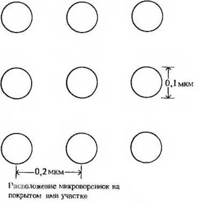
4пr2. Based on these data, calculate: a) the number of microvilli in the covered region, b) the surface area of this region without microvilli, c) the total surface area including the microvilli, and d) the percentage increase in the absorption capacity (determined by the cell's surface area-to-volume ratio) provided by the presence of microvilli.
Last update: 06/08/2026
Editorial and Educational Adaptation: This material has been compiled based on the primary/original source text. The project team performed an editorial review, corrected technical inaccuracies, structured sections, and adapted the content for an educational format.
What was processed:
- elimination of formatting defects (OCR errors, structural breaks, corrupted characters);
- editorial organization of content;
- standardization of terminology in accordance with academic sources;
- verification of factual statements against the original source text.
All mentions of the author, publication year, and origin of the primary text have been preserved in accordance with the source.