Principles of Biochemistry, Volume 1 - A. Lehninger 1985
Biomolecules
Globular Proteins: Structure and Function of Hemoglobin
Chapter Summary
Globular Proteins, judging by the results of studies on their shape and size, have compactly folded polypeptide chains. X-ray crystallography of Myoglobin and other small single-chain proteins, such as cytochrome c, Lysozyme, and Ribonuclease, shows that each of these proteins is characterized by a definite tertiary Structure, i.e., a specific way the polypeptide chain folds in space. In all globular proteins, The polypeptide chains are very tightly folded, so that little, if any, space remains inside the protein molecule for Water molecules. Almost all hydrophobic R-groups are hidden inside the molecule and shielded from interaction with water, while most ionic R-groups are located On the surface in a hydrated state, facing the aqueous environment. The Tertiary Structure of the folded polypeptide chain is stabilized by a variety of non-covalent interactions (especially hydrophobic interactions between non-polar R-groups), electrostatic attraction between oppositely charged R-groups, and Hydrogen Bonds. All these interactions, being weak by nature, are collectively very strong. In some globular proteins, disulfide cross-links play a role in forming and stabilizing the tertiary structure. The information determining the tertiary structure of proteins is encoded in the Amino Acid Sequence of their polypeptide chains, as evidenced by the fact that Homologous proteins from different species share not only many invariant amino acid residues but also the same conformation. Proof of this assertion is that many denatured globular proteins that have lost their characteristic biological activity are capable of spontaneously renaturing with full restoration of their biological activity.
Oligomeric globular proteins, containing two or more polypeptide chains, are larger molecules with more complex structures compared to single-chain proteins, and are often endowed with regulatory properties. The way individual polypeptide chains (subunits) are packed in an oligomeric protein molecule is called its quaternary structure. X-ray crystallography of Hemoglobin and other Oligomeric Proteins has shown that they also have a very compact structure, with most hydrophobic R-groups located inside the globule and most hydrophilic R-groups on the outside. In the hemoglobin molecule, which consists of two α-chains and two β-chains, there are only a few contacts between identical chains, but many contacts that bind the α- and β-subunits together to form α1β1 and α2β2 pairs. The oxygen saturation curve of hemoglobin is sigmoidal, making hemoglobin well-suited for binding oxygen in the Lungs and releasing it in peripheral Tissues. Myoglobin, unlike hemoglobin, has a much higher affinity for oxygen and is characterized by a hyperbolic oxygen saturation curve, which enables it to store oxygen in Muscles. Oxygen binds more easily to hemoglobin at higher pH values and low CO2 concentrations; conversely, the release of oxygen from hemoglobin is favored by lower pH values and high CO2 concentrations. These relationships, as well as the regulatory effect of 2,3-diphosphoglycerate (DPG) binding to hemoglobin on its oxygen affinity, are due to the presence of four specific binding sites on hemoglobin for O2, CO2, H+ ions, and DPG, as well as changes in The quaternary structure of hemoglobin during the oxygenation-deoxygenation cycle. Thus, hemoglobin subunits, like the subunits of other oligomeric proteins, are capable of transmitting signals about regulatory interactions through Conformational Changes in the protein molecule. Changes in The amino acid sequence of globular proteins caused by Gene Mutations, such as the replacement of two amino acid residues in the hemoglobin molecule in Sickle Cell anemia, can cause significant changes in protein conformation and, consequently, affect its biological Functions.
Books
Cantor C. R., Schimmel P. R. Biophysical Chemistry, Part I, The Conformation of Biological Macromolecules, Freeman, San Francisco, 1980.
Dickerson R.E., Geis I. Hemoglobin: Structure, Function, Evolution, and Pathology, Benjamin/Cummings, Menlo Park, Calif., 1982.
Fermi G., Perutz M. Atlas of Molecular Structures in Biology, vol. 2, Hemoglobin and Myoglobin, Oxford University Press, New York, 1981.
Glusker J.P., Trueblood K.N. Crystal Structure Analysis: A Primer, Oxford University Press, New York, 1972.
Haschemeyer R., Haschemeyer A. H. Proteins: A Guide to Study by Physical and Chemical Methods, Wiley, New York, 1973.
Schulz G. E., Schirmer R. H. Principles of Protein Structure, Springer-Verlag, New York, 1979.
Articles
Anfinsen C.B. Principles That Govern the Folding of Polypeptide Chains, Science, 181, 223-230 (1973).
Cerami A., Peterson C.M. Cyanate and Sickle Cell Disease, Sci. Am., 232, 44, April (1975) (offprint 1319).
Dickerson R. E. Cytochrome C and the Evolution of Energy METABOLISM, Sci. Am., 242, 236, March (1980).
Ingram V.M. Gene Mutation in Hb: The Chemical Difference Between Normal and Sickle Cell Hemoglobin, Nature, 180, 326–328 (1957). Discovery of the amino acid substitution in hemoglobin S.
Kendrew J.C. The Three-Dimensional Structure of a Protein Molecule, Sci. Am., 205, 96-111, December (1961) (offprint 121).
Koshland D. E., Jr. Protein Shape and Biological Control, Sci. Am., 229, 52, October (1973).
Pauling L., Itano H., Singer S.J., Wells I. C. Sickle Cell Anemia: A Molecular Disease, Science, 110, 543–548 (1949). Classic description of electrophoretic differences between Hemoglobins A and S.
Perutz M.F., Hemoglobin Structure and Respiratory Transport, Sci. Am., 239, 92, December (1978).
Perutz M. E., Lehmann H. Molecular Pathology of Human Hemoglobin, Nature, 219, 902-909 (1968).
1. Formation of bends and intrachain cross-links in polypeptide chains.
Indicate where bends or turns in the chain are possible in the polypeptide shown below. Where can intrachain disulfide cross-links form?
Class="center">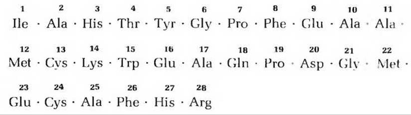
2. Location of specific Amino Acids in globular proteins. Based on X-ray crystallography data of myoglobin and other small single-chain globular proteins, A number of generalizations have been made regarding the folding of polypeptide chains in soluble proteins. Based on these generalizations, indicate the most likely location (inside or on The surface of the native globular protein molecule) of the amino acid residues of aspartic acid, leucine, Serine, valine, glutamine, and Lysine. Explain your answer.
3. Formation of functioning proteins from linear polymers. A protein possesses biological activity only if it has the correct three-dimensional structure. Protein Synthesis is based on information contained in a linear, i.e., one-dimensional, coding sequence of DNA. In accordance with this information, Ribosomes assemble a linear, one-dimensional sequence of amino acids. Given these facts, explain how biologically active proteins with a specific three-dimensional structure can form in Cells. Provide some experimental data supporting your explanations.
4. Disulfide cross-links and protein polypeptide chain folding. The hypothesis that the folding pattern of a protein polypeptide chain (i.e., its secondary and tertiary structures) is determined by its linear amino acid sequence can be tested by allowing unfolded protein molecules to spontaneously refold. By determining the biological activity of the protein before unfolding its chains and after their folding (renaturing Treatment with 8 M urea). If urea is then removed by dialysis and suitable conditions are created for The formation of new disulfide cross-links, ribonuclease activity is restored to 95–100%. The scheme of this experiment is shown in Fig. 8-8. Below are the results of similar Experiments on the renaturation of other proteins:

1) Based on the assumption of random disulfide bond formation.
a) If the formation of four disulfide cross-links during ribonuclease chain folding occurred As a result of completely random interactions between Cysteine residues, one would expect the enzyme activity after renaturation to be only 1% of the original activity. Why would it be so low?
b) The activity of ribonuclease, lysozyme, and alkaline phosphatase recovered after renaturation of these proteins is much higher than would be expected based on the assumption of random disulfide cross-link formation. Explain this observation.
c) One of the proteins listed in the table, namely Insulin, clearly stands out from the others. The observed activity of insulin after its renaturation is very low and practically coincides with the activity predicted based on the assumption of random disulfide bond formation. What can be concluded about the native structure of insulin based on this observation? Try to imagine how the native structure of insulin is formed.
5. Number of polypeptide chains in an oligomeric protein. A certain amount (660 mg) of an oligomeric protein with a Molecular Weight of 132,000 was treated with an excess of 2,4-dinitrofluorobenzene in a weakly alkaline medium until the chemical reaction was complete. Then, the peptide bonds of the protein were subjected to complete Hydrolysis by heating the protein in the presence of concentrated HCl. The hydrolysate contained 5.5 mg of the following compound:
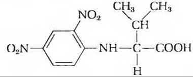
No other 2,4-dinitrophenyl derivatives formed in the reaction with α-amino groups of amino acids were detected.
a) Explain why these data can be used to determine the number of polypeptide chains in an oligomeric protein.
b) Calculate the number of polypeptide chains in this protein.
6. Molecular weight of hemoglobin. The first indication that proteins far exceed the Organic compounds known at that time in molecular weight was obtained more than 100 years ago. For example, even then it was known that hemoglobin contains 0.34 wt % of iron.
a) Based on this information, determine the minimum molecular weight of hemoglobin.
b) Subsequent experiments showed that the true molecular weight of hemoglobin is 64,500. What information can be derived from this regarding the number of iron atoms in hemoglobin?
7. Hemoglobin packing in human erythrocytes. Human Blood contains 160 g of hemoglobin per 1 L of blood. There are about 5.0∙109 erythrocytes per 1 mL of blood. Although each erythrocyte is biconcave disc-shaped, for simplicity of calculation we will consider them simply as cylinders of the following dimensions:
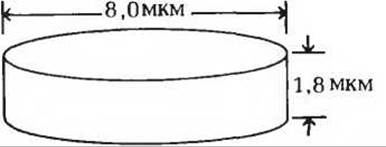
a) Calculate The amount of hemoglobin (by weight) contained in a single erythrocyte.
b) How many hemoglobin molecules are contained in a single erythrocyte?
c) Calculate the volume of a single erythrocyte.
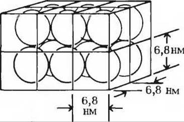
d) Hemoglobin is a globular protein whose molecule has a diameter of 68 nm. What fraction of the total erythrocyte volume is occupied by hemoglobin?
e) The ratio of the total volume of hemoglobin to the total volume of an erythrocyte (see item d above) does not give a clear picture of how tightly hemoglobin molecules are packed in The Cell. It should be remembered that when packing spherical molecules, the empty space between the spheres always constitutes a significant fraction of the total volume. Assuming that hemoglobin in the erythrocyte is packed in a cubic lattice, as shown in the figure, calculate the total volume of the lattice occupied by hemoglobin molecules in a single erythrocyte, and compare it with the volume of the erythrocyte. How tightly packed are the hemoglobin molecules in the erythrocyte?
f) In light of your answer to the previous question (item e), evaluate whether hemoglobin molecules in erythrocytes are located close enough to each other to interact. If so, could the interaction between hemoglobin S molecules in sickle cells affect their shape?
8. The Role of myoglobin in oxygen storage by tissues.
a) Animal tissues contain about 70% water by weight. The normal concentration of oxygen in tissue water is 3.5∙10-5 M. Calculate the amount of oxygen that can be stored in 1 kg of tissue as a gas dissolved in water.
b) Most mammalian tissues contain myoglobin, which serves to store oxygen. In humans, the highest concentration of myoglobin is found in Heart tissue, where it accounts for 0.7% of the total tissue weight. Calculate the amount of oxygen (in grams) that can be stored in 1 kg of human Cytology/cytology/34.html">Cardiac Muscle tissue. Compare your result with the answer to the question in part a.
c) The skeletal muscles of marine mammals capable of remaining underwater for extended periods contain significantly more myoglobin than those of all other vertebrates, with its concentration being proportional to the duration of the animal's submersion. Fresh seal meat left exposed to air for some time was found to contain 0.15 g of oxygen per 1 kg of wet weight. Calculate the percentage of myoglobin in the seal's muscles.
9. Comparison of The properties of hemoglobins in maternal and fetal erythrocytes. Studies of Oxygen transport in pregnant females have shown that the oxygen saturation curves for maternal and fetal blood, obtained under identical conditions, differ significantly. This phenomenon is due to the presence in fetal erythrocytes of hemoglobin (hemoglobin F, a2у2), which differs in structure from normal hemoglobin A (a2ß2) found in maternal erythrocytes.
a) Under physiological conditions, which hemoglobin exhibits a higher affinity for oxygen—hemoglobin A or hemoglobin F? Explain your answer.
b) What is the physiological Significance of the fact that the two hemoglobins have different affinities for oxygen? Explain your answer.

c) If 2,3-bisphosphoglycerate (BPG) is thoroughly removed from preparations of hemoglobin A and F, their oxygen saturation curves shift to the left (i.e., the oxygen affinity of the hemoglobins increases). However, the oxygen affinity of hemoglobin A then becomes higher than that of hemoglobin F. If BPG is added back to the hemoglobin preparations, the oxygen saturation curves return to their original appearance as shown in the figure. What effect does BPG have on the oxygen affinity of hemoglobin? How can the difference in oxygen affinity between maternal and fetal hemoglobins be explained based on the information given above?
10. Identification of mutant hemoglobin. A preparation of mutant hemoglobin was subjected to tryptic hydrolysis, followed by peptide mapping. It was revealed that the mutant hemoglobin differs from normal hemoglobin A by containing a lysine residue instead of an asparagine residue in one of the Peptides.
a) Why is hemoglobin hydrolyzed with Trypsin?
b) Which of the mutant hemoglobins listed in Table 8-4 could the studied hemoglobin be?
c) How could this mutant hemoglobin be identified more quickly and simply?
11. How to distinguish hemoglobin C from hemoglobin S? When two blood samples—one containing hemoglobin C and the other hemoglobin S—were stored in a refrigerator, their respective labels were lost. How can one determine which sample contains hemoglobin C and which contains hemoglobin S (see Table 8-4)?
Last update: 06/08/2026
Editorial and Educational Adaptation: This material has been compiled based on the primary/original source text. The project team performed an editorial review, corrected technical inaccuracies, structured sections, and adapted the content for an educational format.
What was processed:
- elimination of formatting defects (OCR errors, structural breaks, corrupted characters);
- editorial organization of content;
- standardization of terminology in accordance with academic sources;
- verification of factual statements against the original source text.
All mentions of the author, publication year, and origin of the primary text have been preserved in accordance with the source.