ZOOLOGY OF INVERTEBRATES IN THREE VOLUMES - BOOK 2 - G.I. Shcherbak - 1996
PHYLUM ARTHROPODA
Arthropods represent the most species-rich phylum in the animal kingdom. While over 1.5 million arthropod species have been described to date, scientists estimate that the actual number of extant species may reach 3 to 5 million.
Arthropods have successfully colonized every habitat within the biosphere and, alongside certain vertebrates, have evolved the capacity for active flight (as seen in the majority of insects). Their life forms, feeding strategies, and environmental adaptations are exceptionally diverse. It is difficult to find any habitat devoid of arthropods. They inhabit all seas and oceans—from the Water Column to the benthic zone, and at various depths within marine sediments; they thrive in freshwater bodies of all types; and numerous groups have adapted to terrestrial existence. The ROOT-permeated soil layer, the soil surface, and plant Tissues are likewise densely populated by arthropods. Many species are animal parasites. Nutritionally, they encompass herbivores, predators, saprophages, and parasites. There is virtually no natural organic substance on Earth that is not consumed by some arthropod.
Arthropods share many features with segmented worms, particularly polychaetes. G. Cuvier, a prominent scientist of the past century, united these animals into a single phylum, Articulata. However, through a series of key aromorphoses, arthropods attained a higher degree of Tissue and organ differentiation, most notably in their Nervous System and Sensory Organs. Certain groups exhibit complex behaviors, such as brood care, sophisticated construction activities, social structures, and even communication systems (as observed in advanced Hymenoptera).
Like Annelids, arthropods are metameric animals whose bodies consist of a series of segments; however, arthropod metamerism is distinctly heteronomous. Groups of similar segments are consolidated into body regions known as tagmata. Most commonly, three tagmata are distinguished: the HEAD, Thorax, and abdomen. In arachnids, the head and thorax are fused into a single tagma, the cephalothorax; trilobites and myriapods possess only two tagmata—the head and trunk. Of particular significance is The process of cephalization—The formation of a head tagma where sensory organs (antennae, eyes) and structures for the preliminary Processing of food (mouthparts) are concentrated.
The head comprises the head lobe, or acron, and several anterior segments. The majority of researchers consider the acron to be homologous to the prostomium of annelids. As for the number of head segments, however, a consensus is lacking. According to various sources, the arthropod head comprises anywhere from 4 to 8 or even more segments, with recent studies suggesting 6. The number of segments making up the thorax and the abdomen—which terminates in the anal lobe or telson (the homolog of the pygidium)—varies considerably, yet a general evolutionary trend toward stabilization and reduction in segment number is observed across the phylum.
Tagmatization also drives the Integration of the internal contents of segments; certain organs expand at the expense of equivalent ones. Specifically, a concentration of nervous system ganglia into a centralized Brain is observed.
A hallmark feature of arthropods, from which the phylum derives its name, is The Structure of their appendages. These consist of individual articles linked flexibly by joints, forming multi-articulated levers capable of complex and precise movements. In accordance with the tagmatization of the body, arthropod appendages have undergone functional specialization. The head bears appendages dedicated to sensory Functions—antennae (one or two pairs)—while several other pairs have been modified into mouthparts involved in capturing, holding, and grinding food (upper and lower jaws, or chelicerae and partially pedipalps). Appendages of the thoracic tagma primarily perform locomotor functions (swimming, crawling, walking, running, jumping), though these are sometimes supplemented by others (respiratory, sensory). Abdominal appendages are reduced in many groups; only in trilobites and certain crustaceans (Class Malacostraca) are they adapted for swimming, while one or two pairs are frequently specialized as genital appendages for Fertilization and reproduction.
In its most complete form, an arthropod appendage consists of a basal portion (protopodite) from which two branches diverge: an outer branch (exopodite) and an inner branch (endopodite); in addition, aquatic forms bear a gill-like outgrowth (epipodite). In primitively aquatic arthropods (such as crustaceans), certain appendages retain a biramous structure, whereas in most others, one branch undergoes reduction, rendering the appendages uniramous (as seen in the walking legs of tracheates).
An important characteristic distinguishing arthropods from annelids and shaping the fundamental traits of their Organization is the presence of a rigid cuticle. This layer serves not only a protective function but also acts as an external Skeleton (exoskeleton) to which Muscles attach.
The cuticle is composed of Proteins, Amino Acids, Lipids, Glycoproteins, phenols, pigments, and water (up to 40%). However, its most characteristic component is Chitin. This high-molecular-weight polysaccharide is composed of glucose monomers. While resembling Glycogen or Cellulose, chitin differs from these compounds in that each monomer molecule is linked via an NH group to an acetyl group ($O=C-CH_3$). Its chemical name is poly-N-acetyl-D-glucosamine. Chitin is a tough and chemically stable substance. True chitin is found exclusively in arthropods, whereas other animals (certain polychaetes, Mollusks, hydroids, etc.) and Fungi contain chitinoids—substances structurally and chemically similar, yet distinct from chitin. In arthropods, chitin does not occur in a pure state but is bound to the protein arthropodin (actually a mixture of proteins), forming an insoluble complex with chitin. A second characteristic cuticular protein is resilin, which resembles rubber, is highly elastic, and is concentrated in areas where skeletal flexibility is required. Chitin molecules possess a fibrous structure that imparts elasticity, flexibility, and strength to the cuticle. Depending on the arthropod group, chitin accounts for 1% to 90% of the dry weight of the cuticle. The hardness of the cuticle is determined by the hardening of its proteins, which transform into particularly durable sclerotins. The process of sclerotization closely parallels the tanning of leather under METABOLISM/18.html">The Influence of specialized tanning agents.
The arthropod cuticle features regions covered by thick, rigid, non-stretchable cuticle (sclerites) alternating with soft, elastic, stretchable areas (membranes). The alternation of these regions enables the mobility of the body and its appendages. Most segments (excluding head segments) typically feature four sclerites: a dorsal tergite, a ventral sternite, and a pair of lateral pleurites, flexibly joined by elastic membranes. In the appendages, the individual segments are encased in rigid cuticle, while the articulations consist of membranes, permitting relative movement. An internal skeleton—consisting of inward projections of the sclerites known as apodemes—is also present, serving as attachment sites for muscles. The cuticle not only envelops the entire arthropod body but also lines the foregut, hindgut, and the tracheal system in tracheates.
Morphologically, the cuticle comprises several layers that differ in fine Structure and Chemical composition. Although these layers bear different names across various arthropod classes, the most
commonly distinguished are the outer layer, or epicuticle, and the inner layer, or procuticle, which is further subdivided into an exocuticle and an endocuticle. The hypodermal Cells lie beneath the endocuticle. An epicuticle is primarily present in terrestrial arthropods, rendering their integument impermeable to water. Each cuticular layer has a heterogeneous structure and is itself composed of multiple sublayers. The entire thickness of the endo- and exocuticle is penetrated by numerous pore canals containing extensions of hypodermal cells.
The musculature of arthropods does not form a continuous muscular body wall; instead, it is represented by discrete bundles connecting the movable Regions of the cuticular skeleton—the sclerites or appendage segments. These Muscle bundles attach to internal cuticular ingrowths, which, as noted, function as an exoskeleton. Arthropods thus exhibit a novel type of locomotion anchored to an external skeleton rather than a Skin-muscle tube, as seen in worms. Almost all arthropod musculature, with the exception of certain visceral muscles, is striated. This confers a distinct advantage, as striated muscles contract more rapidly than smooth muscles. Thanks to this locomotor architecture, arthropods are capable of exceptionally rapid (relative to their size) and varied movements, with many groups (the majority of insects) also achieving flight.
The body cavity of arthropods is a mixocoel. During embryonic development, paired metameric coelomic sacs are initially formed, much like in annelids. Subsequently, the walls of these coelomic sacs break down into individual cells, and the coelomic cavities merge with remnants of the primary body cavity to form a mixed body cavity, or mixocoel, which lacks a true cellular lining. This forms a system of lacunar or slit-like spaces situated between the Internal Organs. Mesodermal cells derived from the walls of the coelomic sacs later give rise to muscles, Blood Cells, the fat body, and other mesodermal structures. Adult arthropods lack a true coelom, with only vestigial remnants persisting in some forms—such as the antennal and maxillary glands of crustaceans, the coxal glands of arachnids, and certain other structures. A fluid known as hemolymph circulates within the mixocoel, functioning simultaneously as coelomic fluid and blood.
The Digestive System of arthropods consists of three regions: an ectodermal foregut, an entodermal midgut, and an ectodermal hindgut. Each of these regions is further differentiated in accordance with the animal's feeding habits. A defining feature of the arthropod digestive tract that distinguishes
them from other animal phyla is the modification of anterior body appendages into mouthparts specialized for holding and mechanically processing food. In many cases, supplementary to the mouthparts, the foregut features a specialized region for mechanical food processing (the gastric mill in Malacostraca or the gizzard in insects). The foregut frequently serves for temporary food storage as well, such as the crop in butterflies and bees. In most terrestrial arthropods, Salivary Glands empty into the foregut. The midgut gives rise to diverse outgrowths that increase its surface area (hepatic caeca in crustaceans and arachnids, pyloric appendages in insects).
The Primary processes of Digestion AND ABSORPTION take place within the midgut and its caeca. The hindgut, particularly in terrestrial arthropods, is likewise differentiated into regions performing distinct functions, chief among them being the reabsorption of water from feces and its return to the hemolymph—a critical adaptation for water conservation. The intestine of terrestrial arthropods also plays a role in osmoregulation and the Elimination of Metabolic waste products, a function served by specialized tubular outgrowths arising from the posterior region of the midgut (in arachnids) or the anterior portion of the hindgut (in insects and myriapods)—namely, the Malpighian tubules.
The excretory system of primitively aquatic forms (crustaceans, horseshoe crabs) is represented by modified paired coelomoducts, which bear different names depending on their Location (antennal, maxillary, or coxal glands) and function similarly to the nephridia of annelids. In terrestrial arthropods, this function is assumed by Malpighian tubules operating in conjunction with the hindgut into which they empty. Malpighian tubules absorb metabolic wastes dissolved in water, converting them into insoluble compounds, while the hindgut facilitates the reabsorption of water and essential solutes, returning them to the body cavity fluid.
Unlike that of annelids, the Circulatory system OF arthropods is open and partially reduced. It retains only the principal vessels—the dorsal vessel, occasionally a ventral vessel, and a few lateral vessels—while entirely lacking capillaries and small vessels. Instead, a central pulsing organ, The Heart, is developed. All major vessels are Arteries that empty directly into the body cavity. The same fluid, hemolymph, circulates through both the vessels and the body cavity, bathing the internal organs directly.
Respiratory organs in arthropods are highly diverse. Very small arthropods with thin integuments inhabiting water or extremely humid environments can respire across the entire body surface. Larger arthropods with thick cuticles possess specialized respiratory structures whose surfaces are covered by a thin cuticle through which gas exchange occurs. In aquatic arthropods, Respiration is carried out by gills, which are typically modified appendages or parts thereof (epipodites). In terrestrial arthropods, the respiratory organs include book Lungs, which are also considered modified appendages (in arachnids), and tracheae (found in some arachnids, myriapods, and insects). Book lungs are deep, pouch-like invaginations with folded walls that open externally via narrow slits. Tracheae are fine, typically branching tubes opening to the exterior through small spiracles and ramifying throughout the body to supply oxygen directly to individual cells.
The Nervous System of arthropods is constructed on the same plan as that of annelids, consisting of a supraesophageal ganglion (or brain), circumesophageal connectives, and a ventral nerve cord. The architecture of the supraesophageal ganglion is highly complex, comprising three regions: the anterior protocerebrum, the middle deutocerebrum, and the posterior tritocerebrum. The protocerebrum possesses the most intricate structure. The brain processes sensory information received from Sense Organs and coordinates the animal's behavior. The ventral nerve cord frequently exhibits a concentration of ganglia, resulting in the formation of more complex compound ganglia. In some instances, all ganglia fuse into a single centralized ganglionic mass, as seen in crabs or houseflies.
Most arthropods possess well-developed sense organs (tactile organs, chemoreceptors, equilibrium receptors, and eyes). Arthropod eyes fall into two categories: simple eyes, possessing a single lens, and compound, or faceted, eyes, which are composed of numerous—sometimes several thousand—closely apposed visual units known as ommatidia. Each ommatidium perceives only a single point of the visual field before it, with the result that the compound eye produces a mosaic image composed of countless discrete points. Faceted eyes are characteristic of horseshoe crabs, the majority of crustaceans and insects, and extinct trilobites.
The rigid cuticle of arthropods is insensitive to mechanical stimuli, meaning that the sense of Touch and chemical Senses are localized to specific areas of the integument where the cuticle is thin or perforated. In arthropods with a thin cuticle, such as caterpillars or soft-bodied regions in other species, touch is perceived by sensitive Nerve Cells with free endings located beneath the cuticle, which detect any contact or deformation of the integument. On rigid PARTS OF THE body, touch is mediated by tactile sensilla (Fig. 49). Such a sensillum consists of a hollow cuticular Hair (seta) movably articulated with the cuticle by a thin membrane. A channel runs beneath the hair, piercing the thickness of the cuticle. A sensory nerve Cell process passes through this channel and attaches to the Base of the hair. At the slightest touch, the hair deflects, deforming the articulating membrane and stimulating the sensory nerve ending. In addition to the sensory neuron, each sensillum includes two other cells involved in its formation. Tactile sensilla are scattered across the entire body of the arthropod, but are most abundant on the antennae, legs, segment boundaries, and leg joints. Sensilla responsible for chemical senses (olfactory and gustatory receptors) are built on a similar principle, though their cuticular structures can vary widely. A mandatory feature of chemoreceptors is the presence of microscopic Pores in the cuticular part of the sensillum, allowing chemical molecules to pass through and interact with the endings of sensory Neurons.
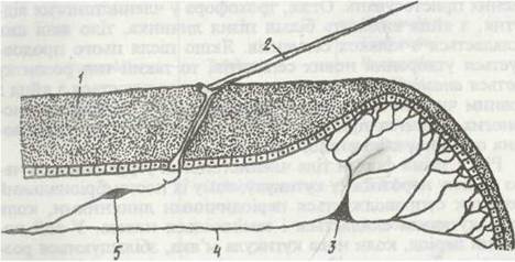
Fig. 49. Tactile endings in arthropods:
1 - sclerite cuticle; 2 - seta (hair); 3 - sensory cell with numerous endings on a thin membrane; 4 - its central process; 5 - sensory cell at the base of the hair
Arthropods reproduce exclusively sexually. Most of them are dioecious, though hermaphrodites are also known. Pronounced Sexual Dimorphism—external differences between females and males—is frequently present. The STRUCTURE OF THE Reproductive System varies among different groups and will be discussed in the respective sections. Fertilization is either internal or spermatophoric, involving encapsulated packets of sperm (spermatophores) that the male introduces into the female's genital openings or attaches to them.
Arthropods are characterized by a specialized type of development. Their eggs are very rich in yolk, which surrounds the egg Cell Nucleus; consequently, Cleavage is superficial and incomplete. It is restricted to a specific region of the egg surface known as the germ band. Arthropod development exhibits embryonization, with early stages protected within the egg envelope. This phenomenon serves a protective function, as early developmental stages are the most vulnerable and lack defensive adaptations. Thus, a trochophore stage is absent in arthropods; instead, a more advanced larva hatches from the egg, its body already consisting of several segments. If the formation of new segments continues post-hatching, this type of development is called anamorphosis; if the animal hatches with the full number of segments, it is termed epimorphosis. In some arthropods, development is direct, meaning a fully formed individual hatches from the egg, albeit of a smaller size.
Growth and changes in body shape in arthropods are constrained by the rigid, inelastic cuticle, which is why their postembryonic development is accompanied by periodic molts, during which the old cuticle is shed and replaced by a new one. Body growth occurs during the brief period when the new cuticle is still soft. Molting is regulated by the neuroendocrine system.
As can be seen, in the structure of their major Organ Systems, arthropods are closely related to annelids; however, they differ primarily in The Development of a chitinous cuticle that functions as an exoskeleton. This evolutionary shift drove major restructurings of the ancestral body plan inherited from polychaete-like precursors. Their mode of locomotion changed, with the coelom losing its skeletal support role while retaining its transport function. The coelomic sacs broke down, giving rise to a hemocoel (a mixed body cavity). Concurrently, the circulatory system became open, communicating directly with the hemocoel. The skin-muscle tube, which provides peristaltic locomotion in annelids, disintegrated. In its place, a new Locomotor System evolved, consisting of striated muscle bundles and hinged, jointed appendages. The functional specialization and differentiation of these appendages led to the grouping of segments into tagmata. Finally, the development of a thick cuticle impermeable to gas exchange necessitated the evolution of specialized respiratory organs.
The phylum Arthropoda is divided into four subphyla: Branchiata (or Crustacea), Tracheata, Trilobitomorpha, and Chelicerata.
Last update: 13/08/2026
Editorial and Educational Adaptation: This material has been compiled based on the primary/original source text. The project team performed an editorial review, corrected technical inaccuracies, structured sections, and adapted the content for an educational format.
What was processed:
- elimination of formatting defects (OCR errors, structural breaks, corrupted characters);
- editorial organization of content;
- standardization of terminology in accordance with academic sources;
- verification of factual statements against the original source text.
All mentions of the author, publication year, and origin of the primary text have been preserved in accordance with the source.