INVERTEBRATE ZOOLOGY IN THREE VOLUMES - BOOK 2 - H.Y. Shcherbak - 1996
PHYLUM SIPUNCULIDA
CLASS SIPUNCULIDA
Sipunculans range in size from 15 mm to 60 cm. In most species, the proboscis does not exceed the trunk in length, yet it is significantly narrower, capable of retracting rapidly into the trunk and everting outwards. At the anterior end of the proboscis lies the Mouth opening, surrounded by short tentacles covered with ciliated epithelium (Fig. 45).
Class="center">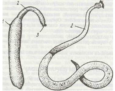
Fig. 45. Sipunculans:
a - Phascolosoma margaritaurea; b - Golfingia vulgare; c - Dendrostomum pyroides; 1 - anus; 2 - proboscis; 3 - circumoral tentacles
The body surface (except for the tentacles) is covered by a thin yet rather dense cuticle layer, beneath which lies a single-layered epithelium rich in cutaneous glands. Hook-like or awl-like outgrowths are often scattered randomly or arranged in regular rings on the proboscis surface. The trunk surface is covered with numerous small Skin papillae associated with Nerve Cells (sensory structures), or with the ducts of cutaneous glands opening onto them. Sipunculans are typically colored in yellowish or brownish tones.
The muscular sac is continuous, formed by three Muscle layers: an outer circular, a middle diagonal, and an inner longitudinal layer.
Proboscis retractor Muscles, located within the body cavity, separate from the longitudinal muscle layer. Their number varies among different species; for instance, Phascolosoma margaritaceum has four. The retractors attach to the body wall near the Base of the proboscis and extend to its anterior end, where they anchor to a specialized muscle ring at the base of the tentacles.
The body cavity is an unsegmented coelom, occupying the entire trunk and most of the proboscis. At the anterior end of the trunk, a circular canal separates from the coelom and connects with the coelomic canals of the tentacles. Researchers view this Structure not as an independent segment, but as a demarcated section of the general coelom functionally associated with the hydraulic extension of the circumoral tentacles. Peritoneal epithelium lines the inner walls of the integumentary-muscular sac, surrounds the intestine forming a mesentery, and also lines the ring and coelomic canals of the tentacles.
The coelomic fluid participates in locomotion. It is pumped by the musculature from one end of the body to the other, causing peristaltic-like contraction waves to pass along the body surface, thereby driving movement. Contraction of the proboscis retractors causes its rapid withdrawal into the trunk, whereas eversion is driven by the pressure of the coelomic fluid generated by the contraction of the trunk circular muscles.
The coelomic fluid has a pinkish color and contains various cellular elements. It acts as a hydroskeleton, maintains the constancy of the internal environment, transports substances, and provides respiratory and protective Functions, thus playing The Role of Blood.
The MAIN TYPES OF cells in the coelomic fluid (Fig. 46) include: amoebocytes with pseudopodia, which phagocytize various microorganisms and accumulate solid Metabolic waste products in the form of yellow granules supplied from Organs to the body cavity, among other functions; and hemocytes—flattened red cells containing hemerythrin, an iron-containing pigment structurally similar to Hemoglobin, whose function is Oxygen transport.
They consist of a spherical stationary Cell and one or more flat ciliated cells. Originating from the peritoneal epithelium, they are initially attached to it by a stalk, but later detach and swim actively in the coelomic fluid using cilia. The ciliated cells secrete a sticky substance that binds together solid metabolic products, inclusion-bearing amoebocytes, and the urns themselves into clusters, which are subsequently eliminated via nephromixia. The discs consist of numerous (2–64) stationary cells arranged in a single layer to form a non-motile plate (disc). When the intestinal wall is damaged by coarse food (sharp sand particles, etc.), the discs are transported by amoebocytes to the affected site, where they form a peculiar "patch" (protective function).

Fig. 46. Formed elements of the coelomic fluid of Sipunculus
The Digestive System (Fig. 47) appears as a long, highly convoluted tube significantly exceeding the body in length. It begins with the mouth opening at the tip of the proboscis and ends with the anus located in the anterior third of the body on the dorsal side. The system comprises ectodermal foregut (Esophagus) and hindgut, and an entodermal midgut. The esophagus is suspended from the body wall by a specialized muscular strand. The midgut is extremely long; it first extends posteriorly, then loops anteriorly forming numerous coils, and wraps around a specialized supporting muscle attached to the body wall at one end near the anus and at the other near the posterior pole. The hindgut is short and is also anchored to the body walls by several muscles. Consequently, in sipunculans, the mouth and anus are closely positioned, and the intestine forms a loop. Digestive Enzymes are secreted by unicellular glands in the midgut walls; large digestive glands are absent.
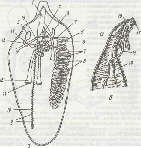
Fig. 47. Internal anatomy of Phascolosoma margaritaceum (dorsal dissection)
Burrowing sipunculans pass mud or sand through their intestine, digesting organic matter contained within these substrates, along with various Protozoans, small crustaceans, and other microinvertebrates. Sipunculans hiding in various shelters use their tentacles to capture infusorians, turbellarians, worm larvae, and the like from their surroundings. Some species become commensals of other marine inhabitants, particularly holothurians and polychaetes.
The excretory system consists of one to three nephromixia, which discharge both metabolic waste and reproductive products to the exterior. Each nephromixia appears as an elongated sac connected to the body cavity by a ciliated funnel-shaped opening and opens externally via a specialized pore located anterior to the anus. Chloragogen cells covering The surface of the midgut participate in the removal of metabolic wastes.
The Circulatory system in sipunculans is absent; the coelomic fluid performs the FUNCTIONS OF BLOOD.
Respiration occurs across the entire body surface, and the respiratory pigment is contained, as previously noted, within the coelomic fluid cells—the hemocytes.
The Nervous system consists of a small paired supraesophageal ganglion (Brain) lying above the esophagus, from which numerous nerves branch out to the tentacles, the circumoral body region, and the intestine. Connectives extend from the brain, encircle the esophagus, and join beneath it; from this junction originates the ventral nerve cord, which lies within the body cavity on the inner wall of the integumentary-muscular sac and extends to the posterior end of the body. It lacks ganglia, with nerve cells distributed along the entire length of the cord.
Sense Organs are poorly developed. Sensitive sensilla are scattered across the entire body surface, with the highest concentration on the tentacles. At the anterior end of the proboscis, There is a pair of so-called nuchal organs, which are specialized papillae covered with sensitive ciliated epithelium. These are presumably chemoreceptors.
Sipunculans are dioecious animals. Paired Gonads, appearing as folded ridges, originate from the peritoneal epithelium and are located near the bases of the proboscis retractor muscles on the ventral side of the mid-trunk. Already at early Selection/3.html">Stages of development, Germ Cells enter the body cavity, where they mature and are released to the outside via nephridiostomes (nephromyxia), where Fertilization takes place. Cleavage of the egg is spiral and determinate.
The trochophore (Fig. 48) features an apical plate with long cilia, a pair of eyespots near it, two ciliary bands—the prototroch and the metatroch—and between them, the mouth opening leading to the foregut rudiment. The midgut initially appears as a solid mass without a lumen. The anus and hindgut develop later on the DORSAL SIDE OF the body, below the metatroch. The pair of mesodermal bands is unsegmented.
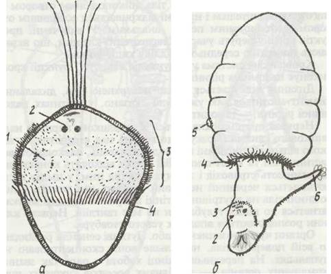
Fig. 48. Larval stages of Sipunculida:
a - swimming trochophore of Phascolosoma vulgare; b - crawling pelagosphaera of Sipunculus polyrnyotus;
1 - mouth; 2 - eyes; 3 - prototroch; 4 - metatroch; 5 - anus; 6 - sole
Trochophore larvae lead a planktonic lifestyle, which facilitates the dispersal of the animals by ocean currents. During this period, the larvae do not feed, surviving instead on yolk reserves.
Metamorphosis is accompanied by the disproportionate elongation of the trochophore. Its posterior region grows extensively, causing the anus to shift near the anterior end, while the gut grows and forms a loop. The mesodermal bands do not undergo segmentation but instead split into two layers: one envelops the gut to form the mesentery, while the other lines the integumentary-muscular sac. This process gives rise to an unsegmented coelom. The septum separating the trunk and tentacular coelomic compartments forms later, as the larva transforms into an adult. At the end of metamorphosis, the larval organs (ciliated bands, eyespots, etc.) degenerate, and the larva settles to the bottom to begin a crawling existence on the substrate.
In some species, such as *Sipunculus polymyotus*, the larvae possess a specialized ciliated lobe at the anterior end—the sole (Fig. 48, b). Such larvae are capable of both swimming and crawling. This structure subsequently disappears.
In Indonesia, locals consume dried sipunculans of the species *Sipunculus edulis* as a delicacy.
Last update: 13/08/2026
Editorial and Educational Adaptation: This material has been compiled based on the primary/original source text. The project team performed an editorial review, corrected technical inaccuracies, structured sections, and adapted the content for an educational format.
What was processed:
- elimination of formatting defects (OCR errors, structural breaks, corrupted characters);
- editorial organization of content;
- standardization of terminology in accordance with academic sources;
- verification of factual statements against the original source text.
All mentions of the author, publication year, and origin of the primary text have been preserved in accordance with the source.