Review of Medical Physiology - William F. Ganong 2002
Circulation
Body Circulating Fluids
Blood - Hemostasis
Hemostasis is The process of clot formation in damaged Blood Vessels, aimed at preventing blood loss while maintaining blood in a fluid state within the vascular lumen. The balance between coagulation and anticoagulation is restored through complex, interrelated systemic mechanisms. In addition, this balance is influenced by local factors in various Organs.
Response to Injury
Injury to small blood vessels triggers a series of events (Fig. 27-24) that lead to clot formation (hemostasis). As a result, the damaged vessel is sealed, which helps prevent further blood loss. The initial event is vasoconstriction and The formation of a temporary platelet hemostatic plug (platelet plug) due to platelet binding to Collagen and their subsequent aggregation. This is followed by The conversion of the platelet plug into a definitive thrombus.
Constriction of injured arterioles or small Arteries can be so pronounced that it leads to their obliteration. Vasoconstriction occurs as a result of the release of serotonin and other vasoconstrictor substances from platelets adhering to the walls of damaged vessels. It has been noted that the transection of arteries even of the caliber of the radial arteries results in a constriction that may arrest Hemorrhage. However, this does not mean that the ligation of an injured vessel can be delayed. Furthermore, in cases of longitudinal or ragged arterial injuries, vasoconstriction fails to achieve occlusion, and bleeding continues.
Class="center">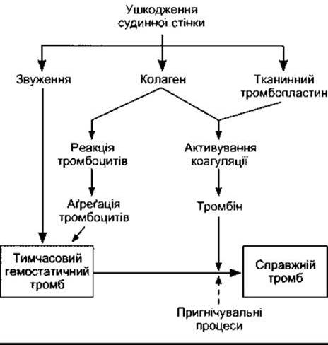
Fig. 27-24. Reactions involved in hemostasis. The dashed arrow indicates inhibition (modified and reproduced with permission from Deykin D: Thrombogenesis, N Engl J Med 1967;267 622).
Table 27-10. Some Proteins synthesized by the Liver: physiological Functions and properties1
Name |
Main function |
Binding features |
Serum or plasma concentration |
Albumin |
Binding and Transport of substances; osmotic regulation |
4500-5000 mg/dL |
|
Not established; possibly involved in inflammation |
Trace; elevated in inflammation |
||
a1-Antiprotease |
Inhibitor of Trypsin and other proteases |
Serum and tissue fluid proteases |
1.3-1.4 mg/dL |
a-Fetoprotein |
Osmotic regulation; binding and transport protein2 |
Hormones, amino acids |
Normally present in fetal blood |
a2-Macroglobulin |
Inhibitor of serum endoproteases |
Proteases |
150-420 mg/dL |
Antithrombin III |
Inhibitor of intrinsic coagulation system proteases |
1:1 protease binding |
17-30 mg/dL |
Ceruloplasmin |
Copper transport |
Six copper atoms/mol |
15-60 mg/dL |
C-reactive protein |
Not established; involved in inflammation |
Complement C1q |
<1 mg/dL; elevated in inflammation |
Fibrinogen |
Precursor of fibrin in hemostasis |
200-450 mg/dL |
|
Haptoglobin |
Binding and transport of free Hemoglobin |
1:1 hemoglobin binding |
40-180 mg/dL |
Hemopexin |
Binds Porphyrins, particularly heme, for recycling |
1:1 with heme |
50-100 mg/dL |
Transferrin |
Iron transport |
Two iron atoms/mol |
3.0-6.5 mg/dL |
Apolipoprotein B |
Assembly of lipoprotein particles |
Lipid transporter |
|
Angiotensinogen |
Precursor of the pressor peptide angiotensin II |
||
Proteins, clotting factors II, VII, IX, X |
20 mg/dL |
||
Antithrombin C, protein C |
Inhibition of blood coagulation |
||
Insulin-like growth factor I |
Transmitter of Growth Hormone anabolic effects |
IGF-I receptor |
|
Steroid hormone-binding globulin |
Steroid carrier protein in blood |
3.3 mg/dL |
|
Thyroxine-binding globulin |
Thyroid hormone carrier protein in blood |
1.5 mg/dL |
|
Transthyretin (thyroxine-binding prealbumin) |
Thyroid hormone carrier protein in blood |
Thyroid hormones |
25 mg/dL |
1 Reproduced with permission from McPhee SJ et al: Pathophysiology of Disease, 3rd ed. McGraw-Hill, 2000.
2 The function of a-fetoprotein is not fully understood, but it is attributed to these functions due to its structural similarity to albumin.
The Coagulation Mechanism
The temporary hemostatic plug is converted into a definitive thrombus through the action of fibrin. The coagulation mechanism responsible for fibrin formation involves a cascade of reactions in which Enzymes are activated, which in turn activate Other Enzymes. In the past, this system was complicated by inconsistencies in nomenclature; however, the adoption of a numerical nomenclature for most clotting factors (see Table 27-9) has helped avoid this.
The fundamental reaction in blood coagulation is the Conversion of the soluble plasma protein fibrinogen into insoluble fibrin (Fig. 27-25). This reaction involves the Cleavage of two pairs of Polypeptides from each fibrinogen molecule. This leaves a moiety—the fibrin monomer—which then polymerizes with other monomer molecules to form fibrin. Initially, fibrin exists as a meshwork of interlacing fibers. Subsequently, through the formation of covalent cross-links, it is transformed into a dense aggregate (stabilization). This reaction is catalyzed by activated factor XIII and requires Ca2+. The conversion of fibrinogen to fibrin is catalyzed by Thrombin. Thrombin is a Serine protease generated from a circulating precursor, prothrombin, under METABOLISM/18.html">The Influence of activated factor X. It also exerts other effects, including the activation of platelets, endothelial Cells, and leukocytes, by acting on at least one G protein-coupled receptor.
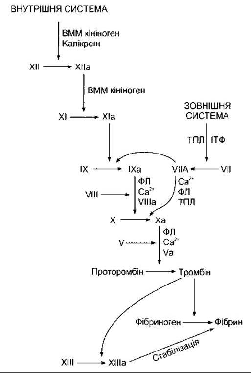
Fig. 27-25. Mechanism of blood coagulation; a - active form of a clotting factor; HMW - high molecular weight; PL - platelet phospholipid; TPL - tissue thromboplastin; TFPI - tissue factor pathway inhibitor.
Activation of factor X can occur through the reactions of two systems: the intrinsic and extrinsic pathways (see Fig. 27-25). The initial reaction of the intrinsic (blood) system involves the conversion of inactive factor XII to active factor XII (XIIa). This activation is catalyzed by high-molecular-weight kininogen and kallikrein (see Chapter 31) and is initiated in vitro by the contact of blood with negatively charged surfaces such as Glass and collagen fibers. Activation in vivo occurs via contact with collagen fibers underlying the vascular endothelium. Active factor XII then triggers the activation of factor XI, and active factor XI activates factor IX. Active factor IX forms a complex with active factor VIII, which is activated upon dissociation from von Willebrand factor. The complex of factors IXa and VIIIa activates factor X. Phospholipids from aggregated platelets (PL) and Ca2+ are required for the full activation of factor X. The extrinsic (tissue) system is initiated by the release of tissue thromboplastin, a protein-phospholipid mixture that activates factor VII. Tissue thromboplastin and factor VII lead to the activation of factors IX and X. In the presence of PL, Ca2+, and factor V, activated factor X catalyzes the conversion of prothrombin to thrombin. The extrinsic pathway is inhibited by tissue factor pathway inhibitor, which forms a quaternary Structure with TPL, factor VIIa, and factor Xa.
Anticoagulant Mechanisms
The blood's thrombogenic tendency is counteracted in vivo by reactions designed to prevent thrombus formation within the vascular lumen and to dissolve established thrombi. These reactions involve an interplay between thromboxane A2, which promotes platelet aggregation, and prostacyclin, which inhibits it. This interplay promotes clot formation upon vascular injury while preserving the integrity of the endothelial layer (see Chapter 31).
Antithrombin III is a circulating protease inhibitor that binds to serine proteases of the coagulation system and blocks their clotting activity. This binding is facilitated by heparin, a natural anticoagulant consisting of a mixture of sulfated Polysaccharides with a Molecular Weight of 15,000-18,000. Clotting factors whose activity is attenuated include the active forms of factors IX, X, XI, and XII. The endothelium also plays a crucial role in preventing the propagation of thrombi within blood vessels. All endothelial cells, except those of the cerebral microvasculature, produce thrombomodulin—a thrombin-binding protein—and express it on their surface. In circulating blood, thrombin acts as a procoagulant, activating factors V and VIII. However, upon binding to thrombomodulin, it becomes an anticoagulant because the thrombomodulin-thrombin complex activates protein C (Fig. 27-26). Activated protein C (APC), together with its cofactor protein S, inactivates factors V and VIII, and also suppresses tissue plasminogen activator inhibitor activity, thereby enhancing plasmin formation.
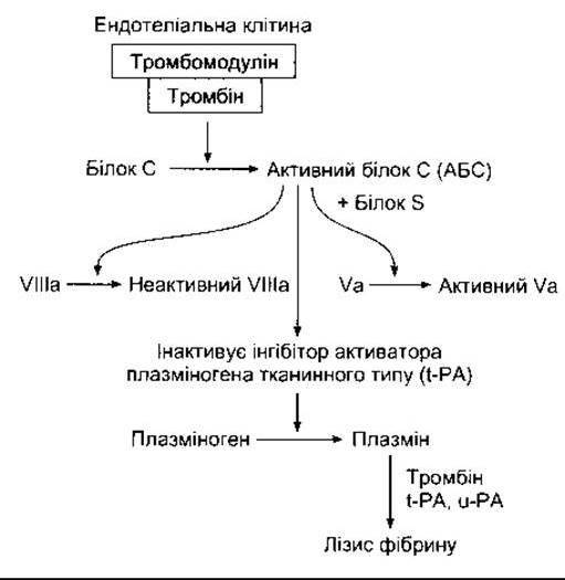
Fig. 27-26. The fibrinolytic System and Its regulation by protein C.
Plasmin (fibrinolysin) is the active component of the plasminogen (fibrinolytic) system (see Fig. 27-26). This enzyme causes the lysis of fibrin and fibrinogen, yielding fibrinogen degradation products (FDPs), which inhibit thrombin. Plasmin is formed from an inactive precursor (plasminogen) under the action of thrombin and tissue-type plasminogen activator (t-PA). It is also activated by urokinase-type plasminogen activator (u-PA). Knockout of either the t-PA or u-PA Gene in mice results in minor fibrin deposition and delayed clot lysis. Knockout of both genes leads to massive fibrin deposition and impaired wound healing (see Chapter 33). Characteristic growth and fertility defects are also observed, as the plasminogen system not only dissolves blood clots but also influences Cell migration and ovulation.
Human plasminogen consists of a 560-amino-acid heavy chain and a 241-amino-acid light chain. The amino-terminal glutamate heavy chain forms five loops, each held together by three Disulfide Bonds (Fig. 27-27). These loops are called kringles because they resemble the Danish pastry of the same name. Kringles serve as Lysine-binding sites that attach the molecule to fibrin and other clotting factors, and they are also characteristic of prothrombin. The conversion of plasminogen to plasmin involves the t-PA-catalyzed Hydrolysis of the bond between Arg 560 and Val 561.
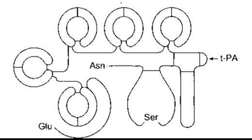
Fig. 27-27. Structure of human plasminogen. Note the N-terminal Glu and C-terminal Asn, as well as the five loop structures (kringles). Under the influence of t-PA, hydrolysis occurs at the site indicated by the arrow. As a result, the C-terminal light chain separates from the N-terminal heavy chain, while the disulfide bonds remain intact. All of this leads to the activation of the molecule (modified and reproduced with permission from Bachman F, in: Thrombosis and Hemostasis. Verstraete M et al [editors]. Leuven University Press, 1987).
Plasminogen receptors are localized On the surface of various cells and are particularly abundant on endothelial cells. The binding of plasminogen to specific receptors triggers its activation. Consequently, the intact blood vessel wall features a mechanism that actively counteracts thrombus formation.
Today, human t-PA is produced via Recombinant DNA technology and widely used in clinical practice (as the therapeutic agent alteplase). When administered within the first few hours to patients with myocardial infarction, the drug induces thrombus lysis in the coronary arteries. Fibrinolytic agents used in the Cytology/cytology/16.html">Early stages of myocardial infarction Treatment also include the bacterial enzyme streptokinase (see Chapter 32).
There is a group of homologous proteins known as annexins, which influence coagulation and Fibrinolysis. About 20 annexins have been described, with 10 of them identified in mammals. One of these, annexin II, forms a "platform" on endothelial cells where the Components of the fibrinolytic system interact and fibrinolysis takes place. Annexin V forms a protective shell around phospholipids involved in coagulation, thereby exerting an antithrombotic effect. However, the precise physiological roles of the various annexins are not yet fully understood.
Anticoagulants
As noted above, heparin is a natural anticoagulant that potentiates the effects of antithrombin III. It also acts as a cofactor for lipoprotein lipase (see Chapter 17). Protamine, a protein with strongly basic properties, binds irreversibly to heparin and is used clinically to neutralize it. Depolymerization of unfractionated heparin yields low-molecular-weight heparin fragments (with an average molecular weight of 5,000). Today, low-molecular-weight heparins are increasingly used in clinical practice because they offer a longer half-life and more predictable effects.
Under in vivo conditions, dropping the Ca2+ concentration low enough to significantly impair blood coagulation would be fatal. In vitro, however, blood clotting can be prevented by adding oxalates, which form insoluble salts with Ca2+, or chelating agents that bind Ca2+. Coumarin derivatives, such as dicumarol and warfarin, are also effective anticoagulants as they block the effects of vitamin K. Vitamin K serves as a cofactor for the enzyme that catalyzes the conversion of glutamic acid residues into y-Carboxyglutamic acid residues. Six of the proteins involved in blood coagulation undergo this conversion of glutamic acid residues to y-carboxyglutamic acid residues before entering the bloodstream, making them vitamin K-dependent. These include factors II (prothrombin), VII, IX, and X, along with proteins C and S (see above).
Hemostatic Disorders
Disorders of thrombus formation can be caused by platelet abnormalities (see above), but hemorrhagic disorders generally result from a selective deficiency in most clotting factors (Table 27-11). Hemophilia A, caused by a factor VIII deficiency, is relatively common. In this condition, treatment with plasma-derived factor VIII concentrates yields positive results; however, historically, the administration of such products often led to HIV transmission. Today, highly purified preparations are available, including factor VIII produced via recombinant DNA technology. In addition to promoting platelet adhesion (see above), von Willebrand factor also forms a complex with factor VIII and regulates its plasma levels. Congenital deficiency of von Willebrand factor leads to bleeding disorders (von Willebrand disease).
Table 27-11. Examples of disorders caused by blood clotting factor deficiencies
Factor deficiency |
Clinical syndrome |
Cause |
I |
Afibrinogenemia |
Associated with placental abruption during Pregnancy; occasionally congenital |
II |
Hypoprothrombinemia (tendency toward hemorrhage in liver disease) |
Impaired hepatic synthesis, mostly due to vitamin K deficiency |
V |
Parahemophilia |
Congenital |
VII |
Hypoconvertinemia |
Congenital |
VIII |
Hemophilia A (classic hemophilia) |
Congenital defect resulting from various Mutations in the X-chromosome gene encoding factor VIII; an X-linked hereditary disease |
IX |
Hemophilia B (Christmas disease) |
Congenital |
X |
Stuart-Prower factor deficiency |
Congenital |
XI |
PTA deficiency |
Congenital |
XII |
Hageman trait |
Congenital |
The absorption of vitamin K, like that of other Fat-soluble vitamins, is impaired in obstructive jaundice due to the absence of Bile in the intestine and the subsequent malabsorption of fats (see Chapter 26). This results in a deficiency of clotting factors, which can lead to hemorrhages. The formation of blood clots within the lumen of blood vessels is called thrombosis (as opposed to physiological extravascular clot formation). Thrombosis represents a serious medical problem and occurs most frequently when blood flow is sluggish, such as in the Veins OF THE lower extremities following surgery or childbirth. This happens because slowed blood flow fails to wash away activated clotting factors, causing them to accumulate. Thrombosis also develops in the coronary and cerebral arteries at sites of intimal atherosclerotic plaques and endocardial lesions. Thrombi frequently obstruct arterial Blood supply to the affected organs. Furthermore, fragments of thrombi (emboli) can break loose, travel through the bloodstream to distant sites, and cause tissue damage. For example, thrombi originating in lower extremity veins can occlude the pulmonary artery or its branches (Pulmonary Embolism), while thrombi from the left ventricle in myocardial infarction (mural thrombosis) can cause cerebral or peripheral arterial embolism.
Table 27-12. Approximate protein content in human Lymph1
Source of lymph |
Protein content, g/dL |
Choroid plexus |
0 |
Ciliary body |
0 |
2 |
|
2 |
|
4 |
|
Gastrointestinal tract |
4.1 |
4.4 |
|
Liver |
6.2 |
1 Data from JN Diana
Congenital absence of protein C leads to uncontrolled intravascular blood clotting and typically results in death during infancy. If diagnosed in a timely manner and treated with blood products rich in protein C, these clotting disorders can be reversed. Another quite common cause of thrombosis is APC resistance, which arises from a point mutation in the factor V gene that prevents the inactivation of factor V by APC. Mutations involving protein S and antithrombin III have also been described and lead to thrombosis, though they occur less frequently.
Disseminated intravascular coagulation is a severe complication of septicemia, massive tissue injury, and other conditions characterized by fibrin deposition within blood vessels and widespread thrombosis in small and medium-sized vessels. The consumption of platelets and clotting factors simultaneously triggers severe bleeding. This syndrome is widely believed to be primarily driven by enhanced thrombin generation resulting from elevated tissue factor activity without a commensurate increase in tissue factor pathway inhibitor activity.
Last update: 10/08/2026
Editorial and Educational Adaptation: This material has been compiled based on the primary/original source text. The project team performed an editorial review, corrected technical inaccuracies, structured sections, and adapted the content for an educational format.
What was processed:
- elimination of formatting defects (OCR errors, structural breaks, corrupted characters);
- editorial organization of content;
- standardization of terminology in accordance with academic sources;
- verification of factual statements against the original source text.
All mentions of the author, publication year, and origin of the primary text have been preserved in accordance with the source.