Orthopedics - Oleksa A.P. 2006
Congenital and Acquired Deformities of the Lower Extremity
Congenital and Acquired Hip Deformities
Hip Dysplasia and Congenital Hip Dislocation
The term displasia coxae congenita refers to the intrauterine underdevelopment of all elements of the hip joint, including the acetabulum, femoral HEAD, Joint Capsule, ligaments, and Muscles. This condition occurs in 16 per 1,000 newborns, with only 3–4 of these developing femoral dislocation.
The hip joint develops from a block of mesenchymal Cell condensates that form the joint components separately around the 11th week of fetal development.
Among the elements of the hip joint, the acetabulum is of primary importance. Its development is driven by the functional stimulation of the femoral head. In a 3- to 4-month-old fetus, the acetabulum is oval in shape and angled both anteriorly and posteriorly. The acetabular roof is inclined at a 30° angle, and the limbus (labrum acetabuli) complements its margin in a rim-like fashion.
Following birth, the normally developing acetabulum gradually assumes a round shape, while the cartilaginous limbus remains pliable due to its slow ossification. Acetabular formation is completed by the age of ten, after which its shape remains unchanged. The triradiate (Y-shaped) acetabular Cartilage fuses around the age of 14–15 in girls and 15–16 in boys.
Initially, the femoral head is ellipsoid in shape, and the femoral neck is short and wide. It acquires its normal shape after 5–6 years. At first, the fetal neck and head exhibit slight retroversion (4–10°), which subsequently rotates forward during development due to the physiological Torsion of the proximal Femur. At birth, the Head and Neck show significant anteversion (25° or more), which progressively decreases during growth and torsion, stabilizing at 8–11° in adulthood without further changes. Additionally, the neck-shaft angle of the femur changes during child growth, reaching 127° in maturity.
Simultaneously with bone development, the soft-tissue elements of the hip joint and muscles develop. However, the fetal hip joint does not always develop normally, resulting in underdevelopment, or Dysplasia.
Hip dysplasia can be bilateral, but it is more frequently unilateral and predominantly affects females (5:1), particularly those whose mothers had this condition.
Genetically inherited dysplasia has been confirmed by observations in numerous families. If dysplasia is detected in one child, the risk of its occurrence in subsequent children is 10 times higher than in healthy families. Furthermore, The Development of dysplasia can be triggered by hormonal imbalances that cause laxity in the ligamentous-capsular apparatus of the hip joint. Dunn (1976), and Andren and Borglin (1961) pointed to elevated levels of estrone and 17β-estradiol in urine, while Wilkinson (1993) experimentally induced dysplasia and hip dislocation through estrogen administration.
Cases of fetal hip dysplasia also occur in primiparas, associated with uterine rigidity and firm Abdominal muscles (Green E.N., Griffin P., 1982). Undoubtedly, mechanical factors contribute to dysplasia and even congenital hip dislocation, which commonly occurs in breech presentations and hydrodynamic Amniotic Fluid disorders.
For a long time, there was no clinical Classification of hip dysplasia, although numerous studies focused on the etiopathogenesis of mechanical-developmental dysplasia. The term "congenital defect" has been largely replaced by "developmental defect" (Burger B.J. et al., 1990; Greenhill B. et al., 1990; Atar D. et al., 1993).
Koszla refers to physiological dysplasia, meaning that all elements of the hip joint—especially the acetabulum—must undergo a growth-related transition from an imperfect state to full joint formation. However, not all fetuses achieve complete normal hip development prior to birth.
Stanislavlijevic (1982) outlines three critical periods during which the risk of dysplasia arises. The first period occurs at the 12th week of intrauterine development, when under METABOLISM/18.html">The Influence of various conditions, the femur is adducted, placing significant pressure on the acetabular limbus. Any disruption in the Development of the acetabular roof creates conditions for the Displacement of the femoral head.
The second period begins when fetal hip movements commence (at the 18th week of development). This causes overstretching of the joint capsule and ligaments, while hypertonicity of the rectus femoris Muscle serves as a favorable factor for femoral head displacement.
The third factor occurs in cases of breech presentation or hydrodynamic and hormonal disorders that provoke dysplasia.
During examinations of newborns, Wynne-Davies (1970), and Carr, Jefferson, and Benson (1993) established that most children with dysplasia exhibited a degree of hip instability compared to a control group. However, this cannot be considered a definitive sign of dysplasia due to individual variations in ligamentous-capsular laxity. Therefore, the primary diagnostic sign of dysplasia is considered to be an underdeveloped acetabular roof. Thus, newborns fall into two groups: one with joint instability resulting from ligamentous-capsular laxity, and another with acetabular underdevelopment manifested by an inclined roof. In both cases, the femoral head is susceptible to displacement.
Dega (1932) and Mau et al. (1971) previously noted that one type of dysplasia is caused by intrauterine malposition, while another is driven by tissue laxity known as the "contracture syndrome" (Fajt, Poul). In the syndrome caused by malposition, limited hip mobility—particularly abduction or leg spreading—predominates, leading to secondary underdevelopment of the acetabular roof if left untreated. This is especially common in children with hereditary dysplasias, where genetic factors combine with contractures and laxity of the ligament-capsule apparatus, which is responsible for stabilizing the femoral head. Nevertheless, within the contracture group, only a small number of children sonographically display signs of acetabular roof dysplasia.
Harmful factors such as obstetric assistance provided to a mother with a narrow pelvis are short-term and of minor significance. Long-acting pathological factors are far more damaging. Breech deliveries occur in 3–4% of cases, representing longitudinal fetal positioning where the baby is born buttocks-first, with the legs flexed at the hips or all joints. In this scenario, the femur is strongly flexed, adducted, and laterally rotated, causing the lesser trochanter to abut against the anterior rim of the acetabulum. This creates a lever mechanism that forces the femoral head out of the socket, resulting in overstretching of the joint capsule-ligament apparatus and subsequent joint instability.
Among diagnosed cases of hip dysplasia, 12 to 50% of infants are born via breech delivery (Dunn P.M., 1976; Garvey M., Donoghue V.B., Gorman W. et al., 1992; Walter R.S. et al., 1992). Breech presentation creates unfavorable conditions for fetal development, thus increasing the incidence of developmental dysplasia of the hip (DDH) (Dunn W., 1976; Harcke H., Grissom L., 1994).
Consequently, breech presentation is of major clinical significance and represents a key risk factor that provokes hip dysplasia and potential femoral head displacement.
Therefore, in hip dysplasia, the femoral head can become displaced both intrauterinely and during the post-neonatal period. This can be diagnosed via Ultrasonography, which is the most cost-effective screening method.
Kubacki et al. (1998) performed ultrasonography of the hip joints in post-term infants with high birth weight and breech presentation, as well as in premature infants with low birth weight. Evaluations were conducted using Graf's ultrasound classification criteria, as presented in Table 4.
These authors established that post-term newborns with high birth weight were more prone to dysplasia. Low-birth-weight infants showed no pathological dysplasia. In both groups, maternal parity (whether the mother was primiparous or multiparous) had no significant impact.
Female predominance was also observed in both physiological and pathological hip dysplasias.
During examinations of infants in their first week of life who sonographically exhibited signs of physiological dysplasia, 90% showed gradual spontaneous stabilization. In the remaining 10%, the joints progressed to pathological dysplasia. Only a very small percentage of newborns examined between days 2 and 7 displayed signs of pathological dysplasia that rapidly acquired classical features of joint underdevelopment.
Based on these findings, Kubacki and co-authors suggest that post-term infants (gestational age over 42 weeks) and those weighing over 4 kg should be classified as high-risk, although some authors (Gardiner M.H., Clarke P.M.N., 1990) consider this assertion debatable.
However, it should be noted that sonography is a sufficient tool for determining fetal position, identifying risk groups, and detecting hip dysplasia and dislocation.
Sonography in newborns During the first week of life, with follow-up monitoring up to three months, reduces the number of children requiring Treatment. To clarify the Diagnosis of developmental hip dysplasia, it is strictly forbidden to perform X-ray examinations in three-month-old infants, or even older ones. Furthermore, computed tomography should not be used, as the radiation dose reaches 3–4 rads.
Magnetic Resonance imaging provides a three-dimensional representation of the hip joint, which can reveal THE POSITION OF the cartilaginous labrum of the acetabulum, displacement of the ligamentum teres, joint capsule adhesion, and all bone and soft-tissue periarticular disorders; however, it should not be used unless absolutely necessary.
Early Clinical symptoms of developmental hip dysplasia:
1. Asymmetry of the Skin folds on the thighs (adductor suprapatellar, gluteal, popliteal) or an unequal number of folds in unilateral dysplasia. This sign is unreliable, as it also occurs in healthy infants, but it serves as a ground to suspect dysplasia.
2. Shortening of the lower extremity, determined by aligning the legs straightened or flexed at 90° at the Hip and knee joints.
3. Lateral Rotation of the extremity, visible when the infant is in a free, relaxed position. This is particularly pronounced in unilateral dysplasia.
4. Limited abduction of the legs (thigh abduction, Fig. 130) flexed at 90° at the hip and knee joints. This symptom is the most reliable, and the degree of limited thigh abduction depends on the level of the femoral head within the acetabulum. Figure 131 shows the angles of possible normal leg abduction. If there is hip subluxation, abduction is sharply limited, whereas in dysplasia it is less limited.
Class="center">Table 4. Criteria for ultrasonographic evaluation of the hip joint according to Graf
|
Type |
Bony socket shape |
Bony acetabular roof |
Cartilaginous acetabular roof |
Angle α |
Angle β |
|
I A |
normal |
sharp |
narrow and long |
>60 |
<55 |
|
I B |
normal |
blunt |
short with a broad base |
>60 |
>55 |
|
II A <3 |
Relatively normal |
rounded |
wide and covers the femoral head |
50-59 |
>55 |
|
II B >3 |
sloped with defects |
rounded |
wide, but does not yet cover the head |
50-59 |
>55 |
|
II C |
defective |
rounded, transitioning to sloped-flat |
wide, but does not yet cover the head |
43-49 |
>55 |
|
D |
with very pronounced defect |
rounded, transitioning to sloped-flat |
displaced upward with joint decentralization |
43-49 |
70-77 |
|
III A |
poor |
flatly sloped |
displaced upward with joint decentralization |
<43 |
>77 |
|
III B |
poor |
sloped |
displaced upward with joint decentralization |
<43 |
>77 |
|
IV |
poor |
very sloped-flat |
pushed upward by the femoral head |
<43 |
>77 |
5. The sign of reduction and subluxation of the hip (Putti-Marx sign). The infant's flexed thighs are grasped with the hands so that the thumbs lie on the Medial surface of the thighs, while the other fingers rest in the region of the greater trochanter. During gradual leg abduction, one can feel the femoral head reduce into the acetabulum, and subluxate upon thigh adduction. After a few such movements, a "click" can be detected (Ortolani-Marx sign). These signs can be observed in both physiological and pathological dysplasia during the first days and weeks of the infant's life, since with the increase in muscle tone by three months of age, these symptoms of physiological dysplasia disappear.

Fig. 130. Limitation of thigh abduction.
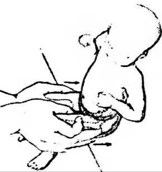
Fig. 131. When pressure is applied to adducted and flexed thighs, posterior displacement of the hip can be felt on the side of joint dysplasia.
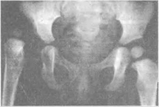
Fig. 132. Radiograph of both hip joints with Congenital Dislocation of the right femur.
In pathological dysplasia and congenital hip dislocation, skin fold asymmetry, shortening and lateral rotation of the thigh, and especially the limitation of its abduction, are so pronounced that clinical diagnosis presents no difficulty.
In addition, in congenital hip dislocation, Palpation may reveal prominence of the greater trochanter, displacement of the femoral head, and a vacant fossa in the femoral triangle (Scarpa's triangle), which causes the sign of a persistent pulse when pressure is applied to the femoral artery below Poupart's ligament.
Once developmental hip dysplasia is suspected based on clinical symptoms, the diagnosis is refined sonographically. However, in the vast majority of cases, general practitioners confirm the diagnosis of dysplasia or congenital hip dislocation radiographically, which serves as tangible documentation. Radiography of both hip joints is performed on a single film with the lower extremities adducted and straightened, using gonadal shielding.
There are various radiological diagnostic schemes (Reinberg's, Putti's, Ombredanne's, etc.), which can be summarized by the following signs:
1. The degree of inclination (slope) of the acetabular roof (acetabular index), which is normally about 30° (Fig. 132). On the radiograph, a horizontal Hilgenreiner line (line of Köhler) is drawn with a pencil, passing through the triradiate cartilage in the center of the acetabula. Thereafter, from a point on this line located at the center of the acetabulum, a second tangent line is drawn to the upper edge of the socket. If this angle is greater than 30°, it indicates dysplasia. The difference in angle measurements is particularly clear in unilateral acetabular dysplasia and head subluxation.
2. Lateral displacement of the femur, i.e., lateral shifting of the femur. To determine this displacement, three vertical lines are drawn perpendicular to Hilgenreiner's line: one middle line divides the pelvis (through the middle of the symphysis and sacrum) into two equal parts, while two lateral parallel lines are drawn tangent to the femurs. Normally, the distance between the middle line and the lateral lines should be equal. If the femur is displaced laterally, the distance on that half of the pelvis is greater.
3. Upward displacement of the femur ("height h"), determined by measuring the distance from the horizontal Hilgenreiner line to the apices of the femoral radiographic shadows. On the side of hip subluxation, this distance is smaller than on the healthy side, whereas in the case of dislocation, the apex of the femoral shadow may be level with or even above Hilgenreiner's line.
4. Disruption of Shenton's line, which is normally traced along the inferior margin of the pubic bone shadow (from the side of the obturator foramen) and then smoothly arches along the inferomedial margin of the femoral neck. Connecting the ends of the arch with a similar smooth line forms an oval. When the femur is displaced upward, the smooth line is broken and fails to match the line on the pubic and femoral bones.
5. Calvé's line, which normally runs along the outer edge of the iliac wing shadow and smoothly transitions into the upper-outer edge of the femoral neck shadow. When the femur is displaced upward (in subluxation), this line is also disrupted.
6. Delayed appearance of the femoral head ossification center on the side of dysplasia or subluxation on the radiograph (normally appearing at 4–6 months), followed by a discrepancy in the size of the ossification centers and asymmetry in their Location.
7. Decreased Wiberg angle, formed by two lines, which indicates the degree of coverage of the femoral head by the acetabular roof (Fig. 133). One line is drawn vertically upward from the center of the femoral head, and the second from the center of the head to the edge of the acetabular roof. Normally, this angle is 25–30°.
As an untreated child grows with a dysplastic acetabulum, the normal development of the femoral head and its proximal end is impaired. The head is smaller in size, develops to match the shape of the acetabulum, and thus becomes flatter and more oval (Fig. 134).
As the head slides upward along the sloped and elevated acetabular roof, the capsule and Ligamentous apparatus of the hip joint gradually stretch, the neck-shaft angle of the femur increases, resulting in valgus deformity (Coxa Valga). During growth, the physiological torsion (twist) of the femur is also disrupted, leading to increased anteversion (anterior deviation) of its neck and head. When a hip dislocation forms, these pathological changes progressively increase as the head shifts beneath the iliac wing.
The joint capsule stretches, becomes thinner, and acquires an "hourglass" shape; changes occur in the ligamentous apparatus, and the anatomical relationship and biomechanics of the muscles are altered. As a result of head displacement and muscle shortening, a flexion-adduction contracture develops along with a relative shortening of the limb, which progressively increases due to growth retardation caused by reduced loading (anatomic shortening).
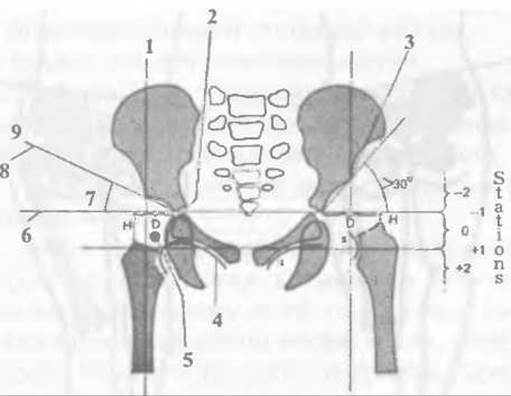
Fig. 133. Radiographic signs of hip dysplasia and congenital dislocation of the femur: 1 — vertical line from the center of the head; 2 — triradiate cartilage; 3 — acetabular fossa; 4 — Shenton's line; 5 — ossification center; 6 — Köhler's horizontal line; 7 — Wiberg angle = 27.5°; 8 — acetabular index <30°; 9 — line from the horizontal to the center of the acetabulum.

Fig. 134. Normal (a), increased (b), and decreased (c) Wiberg angle.
Although a neoarthrosis forms in the area of the dislocated femoral head beneath the iliac wing, it fails to meet anatomical and functional requirements. The deformed head lacks the necessary support, and the patient becomes disabled.
Late clinical signs of hip subluxation and dislocation.
Untreated children with hip dysplasia and subluxation begin to stand and walk somewhat later. If a child has not started walking by one year of age, an orthopedic evaluation is necessary. Children with unilateral congenital hip dislocation start walking a few months later, limp, and sway their torso toward the side of the dislocation. The lower limb is adducted, shortened, and externally rotated. The greater trochanter of the femur is palpable above Nelaton's line, and the additional "pistoneering" (telescoping) sign appears — when the femur is pulled upward and downward with a fixed pelvis, a gliding motion of the femoral head can be felt.
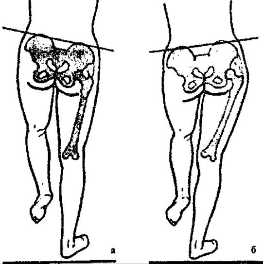
Fig. 135. Negative (a) and positive (b) Trendelenburg sign.
Due to the lack of support for the femoral head and the approximation of the attachment points of the gluteal muscles, muscle hypotonia and a positive Trendelenburg sign occur (Fig. 135). When standing on the single affected leg with the healthy knee bent, the pelvis tilts, and the gluteal fold on the opposite side drops below the fold of the loaded limb.
In bilateral hip dislocation, the child limps on both legs, walking with a waddling gait and swaying from side to side. Examination reveals lumbar hyperlordosis and an anterior pelvic tilt. In addition, all signs characteristic of unilateral or bilateral hip dislocation are pronounced.
The diagnosis of late-detected dysplasia and congenital hip dislocation is confirmed by sonography or radiography. However, when deciding on treatment tactics for a hip dislocation, it is sometimes necessary to use contrast radiography to clarify the condition of the acetabulum (the extent to which it is filled with a fat pad) and the ISTHMUS OF THE overstretched joint capsule, which may have an hourglass appearance in dislocation. This is required to determine whether the femoral head can be reduced conservatively. Contrast arthrography can be successfully replaced by magnetic resonance imaging.
Using Ozerov's special leg positioning technique, the degree of femoral neck anteversion and the neck-shaft angle (the degree of valgus deformity) should also be determined. However, Shevchenko S.D. and Polozov Yu.G. (1991) believe that in children aged 4–6 with subluxation and dislocation, epimetaphyseal relationships should also be determined using the inclination angle of the growth plate, as metadiaphyseal relationships are secondary in anteversion.
A detailed study of clinical and radiological examination data makes it possible to select the optimal treatment method individually for each patient.
Treatment. Looking at the treatment of children with hip dysplasia from a historical perspective, in the 1950s in Ukraine there were no widespread preventive screenings for newborns; consequently, orthopedic patients were typically older than one year with dysplastic hip dislocations. Treatment at that time followed Lorenz's method, performed after joint radiography without assessing their exact condition. Under anesthesia, the femoral head was forcibly reduced into the acetabulum, and the abducted thighs were fixed (according to Lorenz) in three "frog-leg" positions using a plaster cast. This caused pressure and Aseptic osteonecrosis of the femoral head in a significant number of children. Therefore, this reduction technique was later modified to be non-forceful. M.H. Zeleniy also proposed gradual leg abduction by changing the plaster cast every two weeks until the head was centered in the socket. Today, orthopedists have abandoned this method and, following a precise Assessment of the hip joint condition, employ gentle functional Methods starting from the first days of a child's life, as screening examinations for congenital musculoskeletal disorders are mandatory.
To prevent subluxation and dislocation of the femoral head in hip dysplasia, treatment should begin as early as possible, while the head is still located within the acetabulum.
The main goal of treatment is to create optimal conditions for the further development of all hip joint elements, especially the acetabular roof, during the period of rapid postnatal growth in the child.
Over 9 months (from conception to birth), the fetus grows by 50 cm, whereas in the first year of life a child grows by 25 cm, and from one to two years even less—meaning growth gradually slows down, which reduces the chances of successful treatment and prolongs its duration. Therefore, hip dysplasia should be detected in the maternity hospital, in the first days after the child's birth. Physicians, obstetricians, visiting nurses, district pediatricians, and rural paramedics must know the symptoms of dysplasia and check for them during the initial newborn examination. Overdiagnosis in this case is safer than missing pathology; therefore, if dysplasia is suspected, an orthopedic consultation is warranted.
During the examination, an orthopedist may identify Various Forms of hip joint pathology, which can present as:
1) tissue laxity;
2) tissue laxity with joint instability;
3) tissue laxity with a risk of femoral head subluxation;
4) tissue laxity with a risk of femoral head dislocation;
5) subluxation of the femoral head;
6) dislocation of the femoral head.
Depending on the identified pathological changes and the child's age, the orthopedist takes a differentiated approach to selecting the treatment method.
Treatment for infants from birth to 6 months of age.
If the orthopedist detects only soft tissue laxity in the hip joint area while the joint remains stable, they advise parents to provide the child with muscle massage, Therapeutic Exercises, wide swaddling (double or triple), use diapers, and visit the orthopedist for follow-up every two to three weeks (Fig. 136).
When hip instability combined with capsule laxity is detected, sonographic or radiographic examination is required to assess the degree of acetabular roof sloping and determine the acetabular index.
In such cases, treatment consists of shifting the femoral head from an unstable to a stable position. This is achieved by flexing and abducting the thigh—in other words, centering the femoral head within the acetabulum—which relieves the cartilaginous labrum from pressure and shifts the load to the bottom of the socket.
To keep the centered head stable within the acetabulum, the thighs are flexed to 90°, symmetrically abducted outward, and fixed in this position (the "frog" position).
Thigh abduction must be performed gently, gradually, without force, and within the permissible "safe zone." Irreducible hip adduction poses a risk of femoral head subluxation and dislocation, while maximum abduction beyond the "safe zone" can cause avascular Necrosis of the head due to excessive pressure.
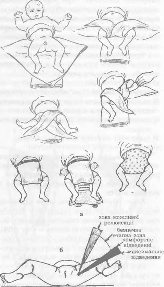
Fig. 136. Wide swaddling of an infant (a), safe zone, and comfortable degree of thigh abduction (b).
To abduct a severely adducted thigh in children aged three months and older, a tenotomy of the hip adductor muscles can be performed, which significantly reduces pressure on the head. The Procedure is carried out under anesthesia by puncturing the skin with the width of an eye scalpel while the Muscles of the abducted thigh are under tension. The proximal tendon of the adductor longus muscle is carefully transected near its attachment site, which is felt as an increased range of thigh abduction. Following the tenotomy, a single suture or simply an aseptic dressing is applied to the wound.
A variety of devices have been proposed to fix the legs in the "frog" position: the Frejka pillow, Pavlik harness, KITO pants (Kutsenok Ya.B.), Weickert devices, various splints, as well as rigid orthoses (Sitenko, Volkov, Szulz, etc.).
Nowadays, orthopedists have concluded that it is best to use devices that maintain the legs in the "frog" position while simultaneously allowing a certain range of motion in the hip joints, which promotes their development. Pavlik harnesses best meet these requirements (Fig. 137).
Most physicians believe that in Graf type I, when there is good bony Formation of the acetabulum and even when the cartilaginous labrum is short with a wide base (I B), but the femoral head is normally centered in the socket, normal hip development can be achieved through wide swaddling (for the first 3–4 weeks of life) followed by The Use of Pavlik harnesses (in three sizes).

Fig. 137. Pavlik harness—a device for hip abduction on an infant.
Lipczyk (Lipczyk Z., 1998) also considers it most appropriate to use dynamic treatment methods for hip dysplasia, as this reduces the risk of avascular necrosis of the femoral head, which is associated with rigid orthoses. He points out that when the head is normally centered in the acetabulum, clinical signs of dysplasia disappeared within 43–63 days of treatment with Pavlik harnesses in Graf types II A, II B, and even II C.
In all cases, it is crucial to unload the cartilaginous labrum of the socket, which—when the head is centered in an abducted thigh—hangs freely, embraces the head, ossifies, and provides normal support (Fig. 138). Creating treatment conditions that allow joint movement, applying thermal Procedures to improve Blood Circulation in the area, and muscle massage all contribute to the normal development of the hip joint.
In Graf groups D, III A, and III B, when a pronounced acetabular defect is detected, the bony roof is sloped and flat, and the cartilaginous labrum is deflected upward with decentration of the femoral head, there are clinically pronounced classic signs of dysplastic subluxation of the head. This manifests as significant limitation of abduction in a 90°-flexed thigh, along with the other previously mentioned symptoms.
In these children, femoral head subluxation is easily reduced by traction without anesthesia, centering it within the acetabulum. However, full thigh abduction is not always achievable due to adductor muscle contracture. In such cases, the required abduction should not be forced in a single stage; rather, the legs must be opened gradually to avoid excessive pressure on the head and subsequent avascular necrosis. Tenotomy of the hip adductor muscles is performed extremely rarely, followed by limb abduction.
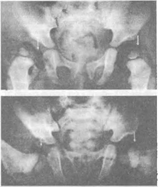
Fig. 138. Unloading the acetabular limbus by hip abduction using Pavlik harness (radiological view).
Hensinger (1985) considers simple closed reduction of the femoral head to be the most reliable approach, using a Frejka pillow or triple diaper for hip fixation to prevent resubluxation.
McKinnon et al. (1984) point out that the most severe forms of avascular necrosis of the femoral head occurred in infants treated within the first 6 months of life. Therefore, they adhere to THE PRINCIPLE OF gradual, functional femoral head reduction, which minimizes the risk of avascular necrosis.
Harcke (1992) also treats infants with developmental dysplasia of the hip and femoral head decentration using a Pavlik harness, monitoring treatment with weekly sonography until stabilization is achieved, and subsequently every three weeks. Once congruent reduction of the head into the acetabulum is attained, the hip joint develops normally and the dysplasia resolves spontaneously (Iwasaki, 1983).
Currently, orthopedic surgeons universally employ the functional method for treating developmental dysplasia of the hip using a Pavlik harness, which provides simultaneous centration of the femoral head in the acetabulum and gradual, non-forceful leg abduction via strap adjustment. The reduced head moves within the joint from a superior to a posteroinferior position and is maintained in the socket by the adductor muscles.
Race and Herring (1983) determined that the acetabular angle decreases by 1.6° for each month of treatment. They also indicate that when the acetabular angle is 25° or less, the Wiberg angle is greater than 20°, and Shenton's line is unbroken, aseptic necrosis of the femoral head never occurs during the treatment of hip dysplasia.
Alongside leg abduction, patients receive muscle massage, thermal therapy applied to the hip joint area, and passive therapeutic exercises.
Regarding the use of rigid splints/braces, Szulc (1981), for instance, utilizes them from the time of diagnosis until complete recovery. They are also indicated for delayed acetabular development during treatment with broad swaddling, as well as in cases of femoral head decentration (Burger et al., 1990; Gardiner et al., 1990; Herring, 1992).
In infants under six months of age, Graf type IV clinically presents with analogous signs. The bony Structure OF THE acetabulum is poor due to significant sloping and a flattened, upwardly displaced cartilaginous limbus, accompanied by marked subluxation or dislocation of the femoral head.
Radiological findings reveal a significantly increased acetabular angle, a disrupted Shenton's line, lateral displacement of the femur with an absent ossification center of the head, and occasionally displacement reaching the horizontal Köhler line.
In dysplastic dislocation, it is likewise necessary to lower the femoral head and center it within the acetabulum. This is achieved by flexing the hips to 90° and performing a very slow, gradual abduction within the "safe zone" without applying force.
Treatment takes considerably longer than in previous types of dysplasia, because following head centration in the acetabulum, time is required for the acetabular roof to develop to a normal state.
Lipczyk (1998) outlines varying treatment durations for children with different degrees of hip dysplasia accompanied by femoral head decentration (Graf types D, IIIA, IIIB, IV), i.e., in cases where the initial treatment stage requires head centration and stabilization of the femur in that position. During dynamic sonographic examination of the hip joints, the time required for Clinical Recovery varied across the specified Graf types: types IIA and IIB required 43–70 days, type IIC took 78–108 days, while femoral head decentration required 123–192 days. The longest treatment duration was observed in types IIIB and IV dysplasia with head decentration.
Treatment of hip dysplasia in children aged 6 to 18 months.
Occasionally, hip dysplasia is not detected at birth but is diagnosed at an older age. After six months, radiographic findings reveal all signs of dysplasia: a flat, sloped acetabulum, an absent or undersized ossification center of the femoral head, frequent lateralization and upward displacement of the femur, and a disrupted Shenton's line—all of which corroborate the clinical symptoms.
Since cartilage structures—especially the acetabular limbus—are not visible on standard radiographs, sonographic examination is much more informative, highly accessible, harmless to the child, and cost-effective.
In most infants under 12 months of age, and even in slightly older ones with femoral head subluxation, successful gradual reduction and centration of the femoral head within the acetabulum can be achieved using a Pavlik harness (Filippe & Carlioz, 1982). To reduce pressure on the head, tenotomy of contracted hip adductors is sometimes performed.
In cases of dysplastic hip dislocation, the thigh muscles—particularly the adductors—are contracted. Therefore, to relax them, lower the head, and center it into the acetabulum, traction with progressive abduction is currently employed. For children under 12 months, traction is typically applied using plaster casts over knees flexed to 90° (Fig. 139), whereas skeletal traction is used for older children. Abduction of the 90°-flexed thighs is performed gradually, increasing the angle by a few degrees daily while clinically monitoring reduction dynamics. Thus, without exerting pressure on the femoral head, centration in the acetabulum is achieved by the 16th–20th day of treatment, which is confirmed radiologically. Afterward, traction is removed, and the abducted thighs are stabilized for one month using a spica cast (Lorenz "frog" position).
Subsequently, the plaster cast is replaced by a Schneider splint, and two to three months later, by a Vienna splint, in which the child begins to walk. Following closed reduction, immobilization is required for no longer than six months.
However, reduction difficulties may arise in children older than one year with femoral head dislocation. The overstretched, thin joint capsule narrows at the isthmus, acquiring an hourglass shape that impedes or completely prevents reduction of the head into the acetabulum (Fig. 140).
To identify obstacles to reduction, diagnostic contrast arthrography of the hip joint is performed or, if necessary, magnetic resonance imaging. The contrast agent (such as cardiotrast, verografin, etc.) is injected into the hip joint by inserting the needle just below the inguinal ligament lateral to the femoral artery.
If an obstruction to closed reduction of the femoral head into the socket is identified, conservative management should not be pursued.
Following gradual centration of the femoral head in the acetabulum via skeletal traction, Volkov employed a specialized perforated polyethylene splint-orthosis. This rigid orthosis (available in four sizes) permits slight (5–8°) motion in the hip joints within the sagittal plane, which, in the author's view, positively influences their developmental process.
However, some orthopedic surgeons (Kahle et al., 1990; Fish, Herzenberg, & Hensinger, 1991; Quinn et al., 1994) do not advocate the traction method for dislocations, preferring femoral head reduction under general anesthesia (using the Ortolani method) without the application of force. If necessary, adductor tenotomy is performed, and the hips are fixed within the "safe zone" using a Pavlik harness. These harnesses are recommended provided that treatment does not exceed 8–9 months.
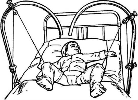
Fig. 139. Reduction of developmental hip dislocation via abduction and lowering of the thigh using traction with plaster spica casts.
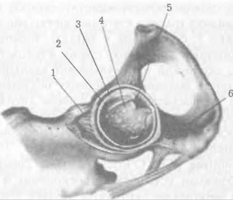
Fig. 140. Acetabular floor: 1. Iliofemoral ligament; 2. Acetabular cartilage; 3. Labrum; 4. Acetabular fat pad; 5. Transected ligamentum teres; 6. Ischiofemoral ligament.
Burger et al. (1990) apply a static orthosis until complete concentric reduction of the femoral head is achieved, followed by the dynamic Becker apparatus.
However, it should be noted that the treatment of developmental hip dislocation via traction remains optimal, as confirmed by our observations. If arthrography reveals obstacles to reduction, surgical intervention is indicated.
Treatment of children aged one and a half to three years.
At this age, children require a supplementary sonographic evaluation, and if necessary, radiography, contrast arthrography, or alternatively, magnetic resonance imaging (MRI) of the hip joints.
Plain radiographs reveal all classic signs of developmental dysplasia of the hip with subluxation or dislocation of the femur. Furthermore, positioning the leg according to Ozerov can sometimes demonstrate increased anteversion of the proximal femur, as well as coxa valga, which develop as the child grows.
Contrast arthrograms demonstrate an overstretched joint capsule in cases of femoral head subluxation, or a constricted, hourglass-shaped capsule in cases of hip dislocation. They also reveal a flat, sloped acetabular roof, the degree of acetabular filling by an overgrown fat "pad," and other signs of joint dysplasia and barriers to reduction. Arthroscopic studies are described in detail by Mitchell G.P. (1963), Renshaw T.S. (1981), and O'Sullivan M.E. & O'Brien T. (1994). Currently, these obstructions are addressed arthroscopically, after which traction is used to center the femoral head within the acetabulum.
Magnetic resonance imaging provides a detailed assessment of all osseous, cartilaginous, and soft-tissue structures within the hip joint region, though it is rarely used to avoid radiation exposure in children.
If examination reveals femoral head subluxation with no obstacles to reduction, a femoral traction treatment protocol may be employed to achieve reduction into the acetabulum. However, because muscle contractures tend to develop with the child's growth in such cases, it is advisable to perform an adductor and iliopsoas (m. iliopsoas) tenotomy prior to applying traction. Historically, M.I. Sitenko also successfully reduced the femoral head in children over two years of age using skeletal traction.
Between the second and third years of life, favorable conditions for hip joint development are still preserved in 19% of cases, whereas after three years of age, such conditions persist in only 7% of patients (Mardam-Bey T.N., McEwen G.D., 1982).
If obstacles to closed reduction of the femoral head into the acetabulum are identified (such as constriction of the capsular neck or the presence of transverse and elongated round ligaments), surgical intervention via arthroscopic or open procedures is required.
For children older than 3 years, conservative treatment methods are ineffective; therefore, it is currently standard practice to forgo these methods and proceed directly to surgery.
Open reduction ensures concentric placement of the head within the acetabulum and reduces mechanical pressure upon it, thereby eliminating the risk of avascular necrosis. If the head is not properly centered in the acetabulum, the surgery will be ineffective (Dhar S., Taylor J.F., 1990; Fairbank J.C. et al., 1986).
Most orthopedics specialists prefer open reduction because it carries fewer risks and does not cause damage to the epiphyseal cartilage or lead to avascular necrosis of the femoral head, complications sometimes associated with closed reduction. Williamson et al. (1989) report that following open femoral and pelvic osteotomy, the incidence of femoral head osteonecrosis decreases from 25% to 10%.
Surgical management of patients with hip dysplasia and dislocation is strictly individualized, depending on the child's age and the specific pathological changes present. Given that these conditions progress as an untreated child grows, earlier surgical intervention (at two to three years of age) is simpler, less traumatic, and yields superior functional outcomes by establishing proper conditions for the continued normal development of the hip joint.
When a constricted capsular neck precludes closed reduction, a straightforward open reduction of the femoral head into the acetabulum is performed.
Surgical technique. Under anesthesia, the joint capsule is exposed via the Smith-Petersen approach. The incision through the skin, subcutaneous tissue, and fascia lata is curvilinear, extending downward from the anterior superior iliac spine (spina iliaca anterior superior) and terminating below the greater trochanter of the femur. Muscles are separated bluntly. These minimal exposures help protect the lateral femoral cutaneous nerve (n. cutaneus). Meticulous hemostasis is performed during the procedure, as pediatric patients are highly sensitive to blood loss. The exposed capsule is incised in a T-shape over the femoral head (Fig. 141) at its constricted portion, taking care to preserve the cartilaginous labrum of the socket. The condition of the head and acetabulum is assessed. If a hypertrophied fat "pad" is found within the socket, it is excised. In a one-and-a-half to two-year-old child, the articular cartilage is normal, allowing the head to be reduced into the socket while flexing and abducting the thigh.
If adductor muscle contracture is felt during thigh abduction, a tenotomy is performed. The thighs are abducted within the "safe zone" without exerting excessive pressure on the femoral head. The joint is dried, and the capsule is closed hermetically. The wound is drained using a thin polyethylene tube and closed in layers. The limb is immobilized using a padded plaster spica cast extending to the tips of the toes, while the contralateral thigh is immobilized only down to the knee.
The following day, the plaster cast must be bivalved in the wound area, and blood-soaked sterile dressings replaced. The drainage tube is removed on the second or third day. Antibiotics are administered starting the day before surgery and continued until the wound has healed.
Three weeks later, the plaster cast is replaced with Pavlik harness straps, and control radiography is performed.
Most orthopedic surgeons favor simple open reduction because, as previously noted, pathological Changes in the hip joint progress as an untreated or improperly treated child grows, thereby increasing the complexity and extent of subsequent surgeries (Dhar S., Taylor J.F., Jones W.A., 1990).
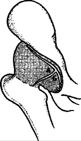
Fig. 141. Diagram of the hip joint capsule incision.
It is often necessary to perform extra-articular osteotomies of the ilium in various configurations, as shown in Fig. 142, to improve the inclination angle of the acetabular roof and head coverage, as well as femoral osteotomies to correct excessive anteversion and valgus deformity of its proximal end.
Indications for femoral osteotomy must be strictly defined. Shevchenko S.D. and Polozov Yu.G. (1991) point out that when planning corrective osteotomy, the main clinical landmarks should be epimetaphyseal relationships, while secondary ones should be metadiaphyseal; that is, the orientation of the physis is determined more by the head-neck relationship than by the neck-shaft angle.
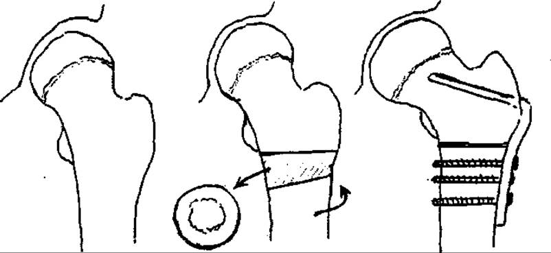
Fig. 142. Derotation and shortening-devalgizing osteotomy of the femur.
Therefore, radiographs should be taken with the thigh in internal rotation and abduction, and the angles of the growth plate and valgization must be determined. In children aged 4–6 years, Wiberg's angle reaches 20–30°, and the inclination angle of the growth plate relative to the horizontal does not exceed 30–35°. Derotation is required when, in unilateral dysplasia, the difference in parameters between the proximal ends of the femurs does not exceed 10–15°.
Regarding surgical approaches to the hip joint, they depend on The Scope of the operation. The Smith-Petersen incision is most commonly used, but nowadays, especially abroad, most pediatric orthopedists perform a transverse "bikini" incision, which leaves a less noticeable scar. This incision can also be extended proximally to perform Salter's pelvic osteotomy (Salter R., 1969) or extended medially to carry out a triple pelvic osteotomy.
The medial approach, described by Ludloff in 1913, has been successfully employed by some orthopedists. However, Kalamchi and McEwen (Kalamchi A. and McEwen G.D., 1982) indicate that injury to the circumflex artery and Veins is inevitable with this approach and occurs in 67% of cases.
The medial circumflex artery lies close to the tendon of the m. iliopsoas and is not a terminal artery, as it has anastomoses that supply blood to the femoral head and epiphysis. Mankey, Arntz, and Staheli (Mankey M.G., Arntz C.T., Staheli L.T., 1993) analyzed 66 cases of the Ludloff approach and concluded that it is safer and more effective in children operated on under two years of age.
The posterior approach to the hip joint is rarely used because it carries a higher risk of disrupting the Blood supply to the femoral head and epiphysis.
The posterior approach, performed for total hip arthroplasty in adults, is unsuitable for surgeries involving joint dysplasia. These hip approaches were comprehensively reviewed by Simmons (Simmons G.W., 1980).
Regarding the use of preoperative traction, A number of orthopedists (Schoenecker P.L., Strecker W.B., 1984) believe that it eliminates limb shortening, improves surgical conditions, reduces pressure on the head postoperatively, prevents any complications, and causes no harmful effects on the limb's Vessels and nerves. If traction is continued after surgery, it eliminates the conditions for femoral head reluxation. Thus, preoperative limb traction is beneficial, but it should not be used if a medial approach to the joint is planned (Roose P.E. et al., 1979).
We will not dwell on The history of the development and refinement of surgical intervention methods, starting from Hoffa, Lorenz, and subsequently Ludloff, Deutschlander, and Frejka, who attempted to help this category of children but failed due to the imperfection of the methods and the lack of adequate anesthetic support.
Credit should be given to Zahradníček, who in his time proposed a two-stage intervention not only on the acetabulum but also on the proximal end of the femur, eliminating increased anteversion and shortening the femur to achieve gentle, pressure-free reduction of the head into the socket. With proper anesthetic support and blood transfusion, this surgery began to be performed as a single-stage procedure.
The Zahradníček procedure is performed for dysplastic hip dislocation in children over 3 years of age. Prior to surgery, radiographs are used to determine the height of head displacement as well as the degree of acetabular obliquity, anteversion, and neck valgization.
Surgical technique. The incision of the skin, subcutaneous tissue, and fascia lata begins at the anterior superior iliac spine, curves along the lateral aspect of the thigh 3–4 cm below the greater trochanter, and terminates beyond the proximal region of the femur. On the lateral surface of the femur from this incision, Tissues are dissected down to the bone over a length of 5–8 cm. The periosteum is incised along the axis of the femur without damaging the epiphyseal cartilage, then elevated, and an osteotome is used to detach a very thin cortical plate from the greater trochanter, retracting it along with the attached muscles. It is important not to damage the growth epiphyseal cartilage, which actively influences the growth of both the greater trochanter and the femoral neck (Laurence G., 1958; Covanda M., Rott Z., 1960). Subperiosteal elevators are placed, and directly below the lesser trochanter, the femur is transected using a Gigli saw below the level of the lesser trochanter. Thereafter, a second oblique osteotomy shortens the femur by the length calculated from the radiograph, preserving the resected bone piece for acetabular roof plasty.
Soft tissues are separated from the capsule without damaging the iliopsoas muscle, and the capsule is incised above the head and obliquely across the isthmus toward the limbus, where it is further transected transversely to gain access to the socket. The socket is cleaned by removing the round and transverse ligaments and eliminating other obstacles that would hinder head reduction. Then, the resected round piece of bone is split longitudinally into two halves with an osteotome, and following a supra-acetabular marginal osteotomy of the ilium, bone grafts are wedged into the Koenig cleft, which lowers the cartilaginous limbus and forms the acetabular roof. Next, after eliminating excessive anteversion and valgus deformity through rotation and varization of the peripheral fragment, the head is reduced and centered in the socket. If angular deformities are not corrected immediately, a second operation will be required later.
To facilitate the surgical procedure, in our clinic in the 1960s, a linear score was made in the cortical layer of the exposed bone area so that, after double transverse osteotomy with a Gigli saw, anteversion could be corrected to the required angle more precisely using the score line. Due to the transverse osteotomy, the femoral fragments were fixed intramedullary with a triangular bone peg from the supra-acetabular fossa to prevent potential rotation of the peripheral fragment. If neck varization is required, the femur is resected at the appropriate angle. The fragments are fixed with a metal L-shaped device (Ter-Yegizarov et al.).
The joint capsule is closed hermetically, reinforcing its anterior wall. The detached muscles are sutured to the greater trochanter, and the wound is drained and closed in layers. A hip spica cast is applied with the thighs in flexion, moderate abduction, and internal rotation. Medial rotation is required by a congruently reduced head.
Postoperative management of the patient does not differ from that described above after open simple head reduction.
The plaster cast is removed after one month, replacing it with adhesive traction (the Eloesser method / pull) with the thigh abducted, and gentle joint mobilization exercises are initiated. After another two months, joint radiography is performed, and the patient is discharged home with permission to walk using crutches.
For an oblique and shallow acetabulum, Colonna proposed deepening it combined with capsuloplasty of the narrowed isthmus. He considered the indications for surgery to be high dysplastic hip dislocation in children aged 3–6 years, but he also performed this procedure in slightly older children up to ten years of age. It should not be used in cases of subluxation and marginal dislocation of the femoral head, nor when the capsule is fused to the ilium and transformed into cartilage, as capsuloplasty is impossible in these cases.
Before surgery, skeletal traction is used to lower the femoral head to the level of the socket, sometimes accompanied by adductor tenotomy if necessary.
Colonna's surgical technique. Through an approach similar to the previous Zahradníček operation, the hip joint and femur are exposed. Then, via a longitudinal incision closer to the cartilaginous limbus, the hip joint cavity is opened and the condition of the socket is assessed. The socket is deepened in its posterosuperior portion using a reamer while protecting the cartilaginous limbus. Hemostasis is performed, and the socket is packed.
The capsule is resected transversely from within the joint cavity in such a way that it is sufficient to cover the head. The head is sometimes slightly smaller but retains its round shape. The capsule over the head is sutured tightly so that the joint cavity remains hermetic and blood does not enter it. Prior to this, however, a standard derotational and shortening subtrochanteric osteotomy is performed with osteosynthesis of the fragments (using a bone peg, Ter-Yegizarov's angular plate, screws, etc.).
The femoral head, completely covered by the capsule, is reduced into the acetabulum, centered, and, with the thigh abducted, the wound is drained and closed in layers. With the thighs abducted within the "safe zone" and flexed, the lower limbs are immobilized with a plaster cast.
The reduced femoral head within the deepened acetabulum, while the thighs are abducted, presses the capsule against the spongy bone wall, with which it eventually fuses. Following the removal of the plaster cast, the synovial membrane of the capsule—under the influence of femoral Head movements in accordance with Wolff's law—metaplasizes into cartilage-like tissue, which fully meets the functional demands of the hip joint.
The Colonna procedure was widely used by orthopedic surgeons in this age group of children, occasionally with minor modifications to the classical technique. For instance, I. Movshovych and Mitrofanova (1959), Ter-Yegizarov (1963), as well as surgeons at our clinic, performed a transverse subtrochanteric osteotomy rather than an oblique one; specifically, Movshovych detached the gluteal muscle group from the greater trochanter by transecting their tendons.
M. V. Volkov (1963) treated congenital hip dislocation in children older than eight years using a method of his own design. In The First stage, he lowered the gluteal muscle group by detaching them along with the periosteum of the iliac wing, and performed subspinous myotomy and adductor tenotomy. After closing the wounds, skeletal traction was applied to the thigh.
Three weeks later, the Second Stage of the surgery was performed—open reduction of the dislocation, which involved a subtrochanteric resection (by 3–5 cm) of the femur. After correcting the anteversion and valgus of the femoral neck, the fragments were fixed with a bone peg. The acetabulum was then deepened, the femoral head covered with an amniotic cap, and reduced. Volkov noted that normal joint mobility is restored following this procedure, and recommended it for bilateral congenital hip dislocation. However, in clinical practice, the use of caps did not yield the desired results.
We analyzed the medical histories of over 100 patients who underwent surgery in the postwar years and returned for a follow-up examination in 1968. Clinical and radiological evaluation of different age groups revealed discrepancies in surgical outcomes. Despite radiologically good CHARACTERISTICS OF THE hip joint, one-third of the patients exhibited unsatisfactory joint range of motion and pain upon limb overloading. Conversely, many patients maintained a fairly satisfactory limb function despite significant radiologically detected periarticular ossifications.
The best surgical outcomes were observed in patients operated on before the age of six who, according to surgical logs, underwent the Zahradníček procedure or, notably, simple reduction during the second year of life. The primary causes of unsatisfactory treatment outcomes were forcible reduction by the Lorenz method, avascular necrosis of the femoral head during conservative treatment, and operating on children older than 6 years.
According to B. Frejka (1968), good functional surgical results were achieved in 84% of children operated on before the age of six, and when the Zahradníček procedure was performed in cases of ineffective conservative treatment.
F. R. Bogdanov (1959) believed that the optimal age for congenital hip dislocation surgery is two to three years, as capsule resection and acetabular deepening are unnecessary at this stage. Since intraoperative procedures did not always yield favorable functional results, extra-articular osteotomies were developed.
To provide adequate coverage of the femoral head within a dysplastic acetabulum, Chiari, Salter, and Pemberton iliac osteotomies, as well as double and triple osteotomies, are now widely employed. These are extra-articular procedures performed under appropriate anesthesia in children older than 18 months.
Undoubtedly, these operations are most effective when the femoral head is contained within the acetabulum. Establishing reliable head coverage promotes the subsequent normal development of the hip joint and prevents angular deformities at the proximal end of the femur. If increased femoral anteversion and valgus are detected, they must be corrected simultaneously with the iliac osteotomy.
In 1953, Chiari described the technique of supra-acetabular pelvic osteotomy (Fig. 143).
Chiari surgical technique. Using the Smith-Petersen surgical approach, tissues are dissected in layers and muscles are bluntly retracted to expose the hip joint area. To ensure free access to the supra-acetabular region, the tendons of the gluteal muscles are transected, or the cortical plate of the greater trochanter is osteotomized and reflected upward. During the subperiosteal dissection of muscles in the supra-acetabular area, it is crucial to avoid damaging the superior gluteal artery and sciatic nerve, the joint capsule, and the cartilaginous labrum of the acetabulum. Next, the muscle is detached from the anterior superior iliac spine and, by stripping the iliacus muscle, a tunnel is created toward the greater sciatic notch, proceeding strictly along the inner surface of the ilium. Special care must be taken not to injure the sciatic nerve and the superior gluteal artery, which exits the pelvis near the upper margin of the greater sciatic notch, where it is anchored to the bone by fascia and surrounded by a venous plexus.
Injury to the artery results in significant Hemorrhage because the vessel has a relatively large diameter and does not readily collapse. To arrest the bleeding, ligation of its posterior trunk within the pelvic cavity becomes necessary.
To prevent such complications and simultaneously facilitate the passage of the Gigli saw through the sciatic foramen, our clinic designed a specialized raspatory (Fig. 144). After a bone-level tunnel is created with this instrument, a thread tied to the Gigli saw is threaded through the loop of the raspatory groove, and both are guided along the bone through the greater sciatic notch. The gluteal muscles are then retracted, the thread is grasped with forceps, and the Gigli saw is introduced. Proper positioning of the saw is essential so that the bone transection runs above the labrum, directly adjacent to the acetabular roof. Following bone transection, abduction of the thigh shifts the lower portion of the pelvis—and consequently the acetabulum—medially, while the cut surface of the iliac wing overlaps the displaced cartilaginous labrum over the femoral head.
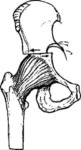
Fig. 143. Schematic drawing of the Chiari procedure.
To immobilize the iliac bone fragments in this position, they are frequently fixed with one or two Kirschner wires inserted percutaneously from the gluteal region. Subsequently, the gluteal muscle group is sutured to the greater trochanter, and the sartorius muscle to the anterior superior iliac spine; the wound is drained and closed in layers. With the thigh abducted, a spica plaster cast is applied, including a leg extension on the healthy thigh.
A wound inspection and dressing change to replace blood-soaked Sponges are mandatory on the following day. Active wound drainage is maintained for 1–2 days. The pins are removed after two to three weeks by grasping their ends protruding above the skin.
Antibiotics are prescribed the day before surgery and throughout the postoperative period, alongside supportive symptomatic treatment.
Some orthopedic surgeons perform this procedure for dysplastic coxarthrosis. Judet and others report favorable long-term outcomes following Surgical treatment in young patients.
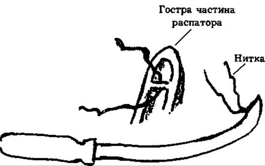
Fig. 144. Schematic drawing of the raspatory for passing the Gigli saw through the sciatic notch.
To correct superior acetabular deficiency, Salter (Salter R., 1961) proposed performing a supra-acetabular pelvic osteotomy with redirection of the acetabular roof using a bone graft wedged into the osteotomy site (Fig. 145). He reoriented the acetabulum by tilting its roof, avoiding more complex osteotomies. He performed this operation in children older than 18 months, predominantly between two and three years of age.
The femoral head must be properly seated within the acetabulum; otherwise, pelvic osteotomy will be ineffective.
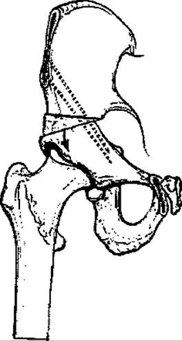
Fig. 145. Salter's operation principle with bone wedge insertion into the osteotomized cleft.
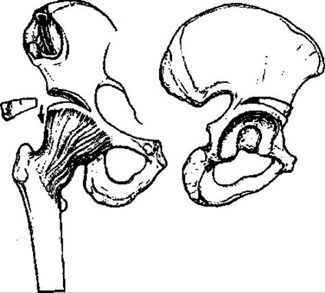
Fig. 146. Schematic drawing of Pemberton's procedure.
Salter believed that in children older than one and a half years, open reduction of the femoral head can be combined simultaneously with pelvic osteotomy. As Salter (1969) pointed out, it is crucial that the surgery is performed strictly According to the rules described by the author.
Obviously, it is unwise to perform Salter's procedure in children younger than 18 months, as such children still possess excellent potential for the normal restoration of the acetabular roof.
Technique of Salter's operation. Using an iliofemoral approach (Smith-Petersen) — which is essentially identical to that described for Chiari's osteotomy — the hip joint and iliac bone areas are exposed at the planned site of osteotomy.
THE ORIGIN OF m.sartorius is detached, and hemostasis is performed. Following the osteotomy of the iliac bone directly above the acetabular labrum, a triangular-shaped bone wedge, harvested from the region of the anterior superior iliac spine (spina iliaca anterior superior), is impacted into the created cleft (Fig. 145). It is important that the distal fragment is slightly rotated to cover the femoral head superiorly and anteriorly. The correction is then secured using two Kirschner wires driven through both iliac fragments and the inserted wedge. The detached muscles are sutured back into place, and the wound is closed in layers.
The wound is drained using an active suction drain, and a hip spica cast is applied, including the thigh on the healthy side.
Pemberton's operation is similar, involving an incomplete supra-acetabular osteotomy into which a bone graft harvested from the iliac wing is wedged (Fig. 146).
To restore the normal relationship between the femoral head and the acetabulum, various modifications of pelvic osteotomy have been proposed, as illustrated in Figure 147.
Triple pelvic osteotomy was first performed by Le Coeur. In 1973, H. N. Steel described a more complex triple osteotomy involving supra-acetabular transection of the ilium along with osteotomies of the pubic and ischial bones. A very similar, yet significantly easier, double pelvic osteotomy was later proposed by D. H. Sutherland and Greenfield (1977).
Other types of osteotomies are also aimed at spatial redirection of the acetabulum to ensure congruence and stability within the hip joint.
An effective yet rather complex osteotomy procedure deserves attention, as it provides optimal correction of the acetabular orientation. Performing this surgery requires advanced anesthesia management and specialized osteotomy instruments (straight osteotomes and those angled at 45°, as well as 40° angled osteotomes with up to 35° of anteversion).
Surgical technique. Under endotracheal anesthesia, a slightly modified Smith-Petersen incision is made, extending along the PROJECTION OF THE iliac wing and the anterior superior iliac spine (spina iliaca ant. sup.), curving downward toward the anterolateral aspect of the thigh. The fascia lata, along with the gluteus maximus and medius muscles, is stripped from the iliac crest, leaving the m.gluteus min. in place. The muscles are retracted to expose the joint capsule and acetabular labrum. Next, a thin cortical plate from the anterior superior iliac spine is osteotomized, the m.sartorius is retracted, and the m.rectus femoris is detached from the anterior inferior iliac spine (spina iliaca ant. infer.). These muscles are retracted inferiorly, and the iliacus and psoas muscles (mm.iliacus et psoas) are elevated from the ilium, thereby exposing its internal (pelvic) surface.
Following this, the m.iliopsoas is carefully mobilized all the way up to the iliopectineal eminence (eminentia ileopectinea). To relax this muscle and improve access, the hip must be flexed to 45°. The required level of tissue dissection is determined by palpation, and the surgical field is exposed using retractors. The sites of the osteotomies around the acetabulum are identified visually. Figure 148 schematically illustrates the location and direction of the pelvic osteotomies viewed from the acetabular and internal pelvic Perspectives. It is essential that both surfaces are clearly visible during the osteotomy and that the bone-cutting plane is properly oriented. If the osteotomy is directed too posteriorly, a fracture of the ischium may occur; if directed too anteriorly, intra-articular damage to the acetabulum is possible.
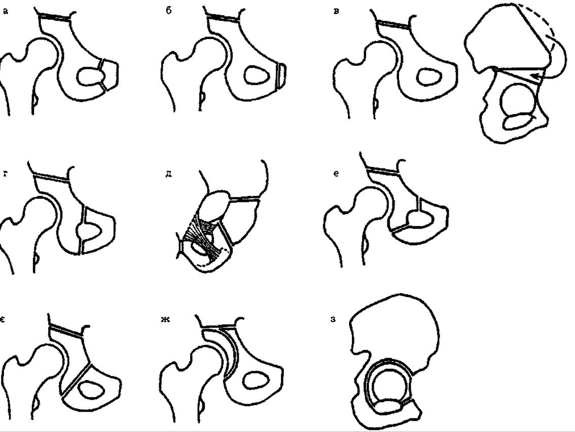
Fig. 147. Schematic drawings of pelvic osteotomy procedures:
a - triple osteotomy by Le Coeur; b - double osteotomy by Sutherland and Greenfield; c - Salter's osteotomy (anteroposterior and lateral views); d - triple osteotomy by Steel; e - triple osteotomy by Toennis (lateral view); f - triple juxta-acetabular osteotomy by Carlioz; g - double osteotomy by Hopf; h - spherical osteotomy by Wagner; i - translocation osteotomy by Kuzmenko.
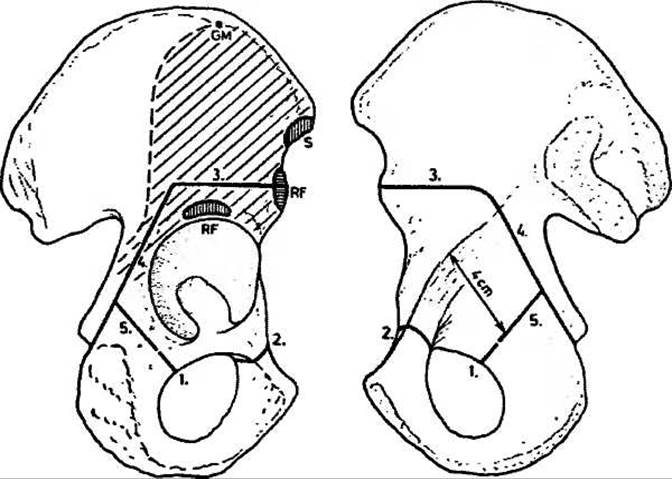
Fig. 148. Osteotomy lines and sequence of execution, shown schematically from the acetabular and pelvic cavity perspectives (Ganz R., Klaue K., et al., 1993). Muscle attachment sites: GM - gluteus medius, RF - rectus femoris head, S - sartorius head.
Using an osteotome angled 20° proximally, the bone is transected starting approximately 4 cm posterior to the anterior inferior iliac spine (spina iliaca ant. infer.) in such a way that the cutting line terminates anterior to the posterior inferior iliac spine (spina iliaca post. infer.), leaving the ischium uninjured. This osteotomy must be performed under visual control from both sides of the bone.
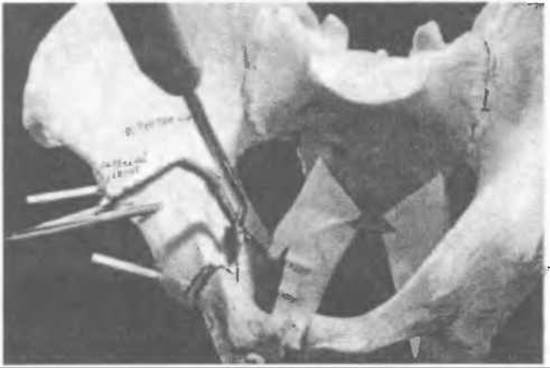
Fig. 149. Diagram of pelvic osteotomy on a skeletal model.
Subsequently, a threaded pin is inserted into the "Schantz pin placement site" (Fig. 148) parallel to the created bone cut, allowing the acetabular fragment to be manipulated and corrected. Guided by the anterior margin of the acetabulum, the pubic bone is osteotomized. Extreme caution must also be exercised here to avoid injuring the obturator artery and nerve.
Next, guided by the inferior margin of the acetabulum, the bone is transected across using an osteotome bent at the desired angle (Fig. 149). The mobilized acetabular fragment is shifted into the required position using a threaded pin screwed into the bone.
To correctly orient the planned displacement, two Kirschner wires are first inserted into both the acetabular fragment and the iliac bone above the osteotomy site in the frontal plane. Once the acetabulum is set in the proper position, the fragment is temporarily fixed with Kirschner wires, the range of motion in the hip joint is assessed, and control radiography is performed.
Following this, the acetabular fragment is secured with two screws and the wires are removed (Fig. 150); any gaps around the fragment can be packed with bone grafts. Hemostasis is then verified. The detached muscles are sutured back into place. The wound is drained using three active drains and closed in layers.
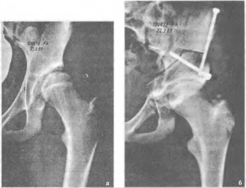
Fig. 150. Radiographs of pelvic osteotomy before surgery (a) and after surgery (b) according to Clane K. and Ganc R. (1993).
Postoperative patient care does not differ from the protocol described above.
The Sytenko Institute in Kharkiv developed the Korzh-Miteleva procedure (Fig. 151), which is indicated for secondary subluxations of the femoral head and is recommended for children older than six years, as the acetabular roof constructed in this manner can withstand greater loads (Shevchenko S.D., Polozov Yu.G., 1991).
The surgical technique is relatively straightforward and involves overlapping the femoral head with the outer cortical plate of the supra-acetabular region of the ilium. This plate is deflected downward and secured in position using one or two triangular allografts wedged into the iliac bone. When necessary, a derotational and varus intertrochanteric osteotomy is performed. Postoperatively, a hip spica cast extending down the thigh of the unaffected side is applied and kept in place until the graft integrates.
Loskutov O.Ye., Golovakha M.L. et al. (2001) note that Surgical Treatment of children with hip dysplasia and dislocation yields good outcomes; however, in bilateral cases, the postoperative period is often marked by a phase of "apparent well-being," during which the compensatory mechanisms of the growing child's body ensure satisfactory joint function for quite some time.
Often, after childbirth, operated women experience a breakdown in compensatory mechanisms, as also reported by Guryev V.N. (1975) and Mikhelman M.D. et al. (1975). This leads to rapidly progressive coxarthrosis. Patients suffer from pain and, notably, impaired hip joint function that hinders normal walking and standing. Clinical examination reveals a pathological position of the thigh, which is flexed, adducted, and externally rotated. As a rule, this is accompanied by a valgus deviation of the lower leg of up to 10°. The range of motion in the hip joints is limited, which, in bilateral cases, fails to adequately compensate for lower limb function.
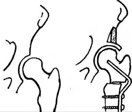
Fig. 151. Diagram of the surgical procedure according to Korzh O.O. and Miteleva Z.V.
Gait disturbances and pain associated with bilateral dysplastic coxarthrosis are a direct indication for joint replacement, staged six months apart.
Last update: 10/08/2026
Editorial and Educational Adaptation: This material has been compiled based on the primary/original source text. The project team performed an editorial review, corrected technical inaccuracies, structured sections, and adapted the content for an educational format.
What was processed:
- elimination of formatting defects (OCR errors, structural breaks, corrupted characters);
- editorial organization of content;
- standardization of terminology in accordance with academic sources;
- verification of factual statements against the original source text.
All mentions of the author, publication year, and origin of the primary text have been preserved in accordance with the source.