Medical Radiology - Lazar A.P. 2008
Radiological examination of the urogenital system
The Kidneys are retroperitoneal, bean-shaped Organs located on either side of THE Vertebral Column at the level of the last thoracic and the upper two lumbar vertebrae. The right Kidney sits about 1/2 vertebra lower than the left because of the Liver above it. The twelfth rib crosses the posterior surface of the left kidney slightly above its middle, and the right kidney at its upper third, or at the junction of its upper and middle thirds. Each kidney weighs 120-200 g, with a length of 11-12 cm, width of 5-6 cm, and thickness of 3-4 cm. At the renal hilum, located on the medial border of the kidney, the renal artery lies anterior to the vein.
Urine secreted through the papillae of the renal pyramids first enters the funnel-shaped extensions of the renal pelvis, known as the calyces. From the minor calyces, urine collects into the major calyces. Two or three major calyces merge to form the renal pelvis. The renal pelvis is situated within the renal sinus at the level of the L1-L2 vertebrae, posterior to the renal vessels. The tapered portion of the pelvis is directed inferiorly and medially, transitioning into the Ureter at the level of the lower border of the renal hilum.
The ureter is a tube approximately 30 cm long and 3-7 mm in diameter that connects the renal pelvis to the Urinary Bladder, running retroperitoneally. It descends inferiorly and medially from the renal pelvis, enters the pelvic cavity, and courses along its lateral wall. The ureter then turns medially, reaches the Base of the urinary bladder, and obliquely pierces its wall. Based on its topography, the ureter is divided into abdominal, pelvic, and intramural parts.
The urinary bladder is an unpaired, hollow, egg-shaped organ with an average capacity of about 500 mL. When empty, the urinary bladder is located within the pelvic cavity posterior to the Pubic Symphysis; when full, it extends 4-5 cm above the level of the symphysis into the Abdominal cavity AND comes into contact with the anterior abdominal wall. The empty urinary bladder is extraperitoneal in relation to the Peritoneum, whereas the distended bladder is mesoperitoneal.
Imaging Methods for examining the kidneys and Urinary Tract include ultrasound, CT, MRI, radiography, and radionuclide imaging.
Ultrasound imaging is currently the primary method for assessing the Morphology of the kidneys and urinary bladder. On a sonogram, a normal kidney appears oval or rounded, surrounded by an echo-negative (hypoechoic) adipose capsule and an echo-positive (hyperechoic) fibrous capsule (Fig. 235).
Beneath the 1-1.5 mm thick fibrous capsule lies a low- echogenicity layer of renal cortex and medulla, approximately 2 cm thick. The calyceal-pelvic system, together with the major renal vessels, forms a unified hyperechoic complex. When the calyces are filled with urine, rounded echo-negative fluid collections up to 0.5 cm in size are visualized against the Background of the central echogenic complex. The ratio of the transverse dimension of the central renal complex to the surrounding parenchyma at the level of the hilum is 1:2, and in children, 1:2.5. The renal artery and vein are most readily identified in their medial segments—near their origin from the aorta and entry into the INFERIOR VENA CAVA. Intrarenal Blood flow is evaluated using Doppler Ultrasonography. The normal ureter is not visualized on ultrasound.
Class="center">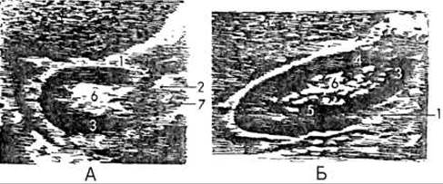
Fig. 235. Renal sonogram. A - transverse section; B - longitudinal section.
1 - capsule; 2 - renal artery and vein; 3 - renal cortex; 4 - renal medulla; 5 - pyramid; 6 - pelvicalyceal complex; 7 - psoas major Muscle.
On sonograms, the urinary bladder appears as a rounded, echo-negative Structure devoid of any internal echoes. The bladder wall, up to 1 cm thick (depending on the degree of distension), has well-defined contours.
For computed tomography of the kidneys, axial slices are obtained from the level of the XI thoracic to the III-IV lumbar vertebrae. The kidneys appear as rounded structures with a densitometric density of +30 to +40 HU, surrounded by a fat capsule with a density of -80 to -100 HU. On an axial slice, the ureter presents as a dense, rounded structure with a diameter of 0.3-0.7 cm.
Magnetic Resonance imaging differentiates the renal cortex and medulla by signal intensity (medium in the cortex, low in the medulla on T1-weighted images). The adipose body and renal capsule produce a high-intensity signal, whereas the renal pelvis and renal fascia produce a low-intensity signal. During intravenous renal contrast enhancement, arteriographic, corticomedullary, nephrographic, urographic, and excretory phases of enhancement are distinguished (Fig. 236). Calyces are normally not visualized. In the normal anatomical position, the long axes of the kidneys converge superiorly, and their transverse axes converge anterior to the vertebral column (Fig. 237).
The urinary bladder is evaluated on CT and MRI scans when it is distended with urine.
Radiological examination of the kidneys begins with a plain abdominal radiograph (KUB), which reveals the bean-shaped renal shadow in 2/3 of patients.
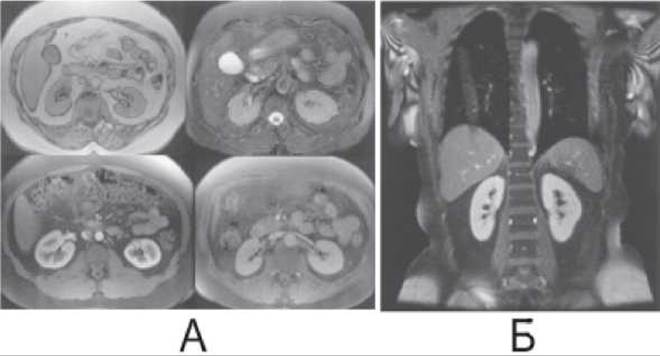
Fig. 236. Magnetic resonance images of the kidneys.
A - Axial MRI; top scans show unenhanced T2- and T1-weighted images; bottom scans show gadolinium-enhanced T1-weighted images, demonstrating the corticomedullary and nephrographic enhancement phases.
B — Contrast-enhanced coronal MRI.
Non-contrast radiography provides insight into the shape, size, and position of the kidneys, as well as the presence of calcifications, calculi, or gas.
Contrast examination of the urinary tract involves intravenous (excretory, or descending) urography and retrograde (ascending) pyelography. Excretory urography is performed by intravenous administration of 20–60 mL (depending on body weight) of body-Temperature-warmed urotropic iodinated ionic (Triombrast, Urografin, Hypaque) or non-ionic radiopaque agents (Omnipaque, Ultravist). Maximum Filling of the calyces, pelvis, and Ureters occurs within 10–20 minutes; therefore, radiographs are taken as follows: the first at 5–7 minutes, the second at 15–20 minutes, the third at 25–30 minutes, and an additional one at 1–2 hours if one of the renal pelves was not visualized on previous scans. Normally, opacification of the calyces and pelves occurs simultaneously on both sides. In unilateral renal disease, a delay in contrast excretion from the affected side of up to 30 minutes or more is frequently observed.
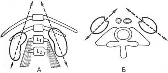
Fig. 237. Renal position on tomograms (diagram).
A - in the sagittal plane;
B - in the horizontal (axial) plane.
To obtain high-quality radiographs, the patient must be carefully prepared—the bowel should be cleansed to remove gas, just as on the eve of an irrigation Procedure (barium enema). Prior to intravenous urography, a sensitivity test to the contrast agent must also be performed.
Retrograde pyelography (retrograde urography) is performed using urinary catheters inserted under cystoscopic guidance. 10 ml of a contrast agent is injected into the renal pelvis through the catheter. Retrograde pyelography provides significantly better contrast and clarity of the renal pelvis, calyces, and ureters compared to the intravenous method. It provides insight into the morphological state of the urinary tract organs, such as filling defects, deformations, displacements, pathological cavities, etc.
Thus, intravenous urography is used to evaluate the functional state of the kidneys, whereas retrograde urography is used to detect morphological Changes in the urinary tract.
Infusion urography is indicated in cases of reduced renal function and combines the diagnostic capabilities of both intravenous and retrograde urography. Because of the high concentration of the radiopaque agent in the urine, it allows for the evaluation of both the Excretory Function of the kidneys and the morphological state of the urinary tract. During infusion urography, a triiodinated radiopaque agent (e.g., a 76% triombrast solution) diluted 1:1 with a 5% glucose solution or isotonic sodium chloride solution is administered by intravenous drip over 5–7 minutes, at a dose of 1 ml per 1 kg of the patient's body weight (60–80 ml). Radiography is performed immediately after the infusion and again after 15–20 minutes. On infusion urograms, the kidneys produce a more intense shadow than on excretory urograms, which facilitates the detection of tumors, cysts, developmental anomalies, and other conditions.
On urograms, the kidneys typically reveal three major calyces: upper, middle, and lower, from each of which minor calyces branch out (Fig. 238). The capacity of the renal pelvis is approximately 6–10 ml, and its shape can vary. The most common type is the dendritic (arborescent) branching of a narrow renal pelvis into elongated calyces, whereas the ampullary type of renal pelvis with poorly developed calyces is less common. The smooth and distinct contours of the renal pelvis seamlessly transition into the similar contours of the ureter, which appears as a narrow shadow 3–10 mm wide. Two distinct curvatures of the ureter are visible in the frontal plane: the abdominal segment curves medially, and the pelvic segment curves laterally.
On the urogram, the urinary bladder appears as an oval shadow of medium intensity with smooth contours, the lower edge of which is located at the level of the pubic symphysis or slightly above it. On a cystogram performed after filling the bladder with a contrast agent via a catheter, the urinary bladder produces an intense shadow. Tumors, calculi, and foreign bodies within the bladder cavity appear as filling defects.
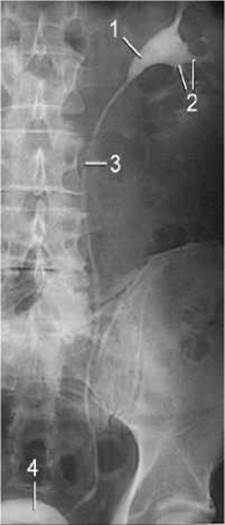
Fig. 238. Normal retrograde urogram.
1 - renal pelvis;
2 - calyces;
3 - ureter;
4 - urinary bladder.
Voiding cystography refers to the radiography of the Urethra performed during the urination of radiopaque urine from the bladder. Such a urethrogram can reveal strictures, tumors, diverticula, and INJURIES OF THE urethra.
Renal angiography is performed by puncture and catheterization of the femoral artery using the Seldinger technique. During general renal arteriography, the tip of the catheter is placed in the aorta above the Water/144.html">Origin of the renal Arteries (at the level of the first lumbar vertebra or slightly below), 40–60 ml of a water-soluble radiopaque agent is injected under pressure, and a high-speed series of radiographs is acquired. In selective arteriography, the catheter tip is inserted directly into one of the renal arteries, providing a more contrasted image of the renal artery than general arteriography. Renal angiography is indicated: 1) when renal artery stenosis is suspected and subsequent Surgical Treatment (balloon angioplasty) is planned; 2) in the Treatment of renal Hemorrhage by embolization of the bleeding Branches of the renal artery.
Radionuclide diagnostic methods make it possible to detect functional Disorders of the kidneys.
Renography (or dynamic scintigraphy) records, as a curve on a graph, the changes in radioactivity over each kidney and over the cardiac region where the collimators of the radiochronograph are positioned. 131I-labeled hippuran, with an activity of 2 to 5 kBq/kg administered intravenously, is most commonly used as the nephrotropic radiopharmaceutical (RP). The radioactivity curve recorded over The Heart region reflects blood clearance. The renographic curve recorded over the kidney is conventionally divided into three segments: 1) the vascular segment, lasting the first 20–25 s, characterized by a rapid rise in radioactivity caused by the passage of the RP through the renal vasculature; 2) the secretory segment, which is more gradual, with Tmax (secretion phase) = 3.2 min (2–5 min); T1/2 (half-elimination time) = 7.2 min (6–12 min); blood half-clearance time up to 20 min; renogram amplitude = 83.4±3.0 counts/s; the ratio of these parameters in the left and right kidneys is approximately equal.
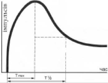
Fig. 239. Normal renogram.
continuing until the peak of the curve is reached at 2–5 minutes, associated with The excretion of the radionuclide into the lumen of the nephron tubules; 3) the excretory segment, where, as the radionuclide is eliminated, its concentration in the kidneys begins to decrease exponentially with a half-life of 6–12 minutes (Fig. 239).
Obstructive renograms are characterized by a decreased amplitude, flattening and delayed peak relative to THE START OF the study, a distorted shape of the excretory segment of the curve, and a slowed excretory phase. Obstructive-type curves are observed in Hydronephrosis, ureteral obstruction by a calculus, or external compression of the ureter by a mass lesion. The parenchymal-type curve is marked by a reduced curve height, the absence of a pronounced peak, a rightward shift of the maximum radioactivity, and a moderate slowing of the excretory segment decline. A parenchymal-type renogram occurs in inflammatory renal diseases (Pyelonephritis, Glomerulonephritis), urolithiasis, and arterial Hypertension. In cases of severe impairment of renal function, the renogram takes the form of a two-segment isosthenuric curve. Over a non-functioning kidney, the renogram will resemble the blood clearance curve, but will be significantly lower. The isosthenuric renogram is found in chronic renal failure. In vesicoureteral reflux, a second peak is observed on the renogram. A non-functioning kidney is characterized by an afunctional curve type, which occurs in the absence of one kidney (congenital or post-nephrectomy), renal artery occlusion, and primary or Secondary contracted kidney (Fig. 240).
Renoscintigraphy (or static scintigraphy) is used to obtain a radionuclide image of the kidneys. Rapidly cleared radiopharmaceuticals such as 99mTc-DMSA and 99mTc-gluceptonate are used. Maximum renal accumulation is observed 2 hours after radionuclide administration, at which point scintigraphy should be performed. Scanning is typically performed using a gamma camera in the posterior projection. If necessary, lateral, anterior, and oblique projections of the kidney can be obtained. Areas of non-functioning renal parenchyma caused by a pathological lesion (tumor, cyst, abscess) are characterized by the presence of a "cold" spot on the scintigram. Renoscintigraphy is also used to determine the volume of functioning renal parenchyma (especially in the presence of renographically "silent" kidneys) and to detect renal developmental anomalies (Horseshoe kidney, hypoplastic kidney).
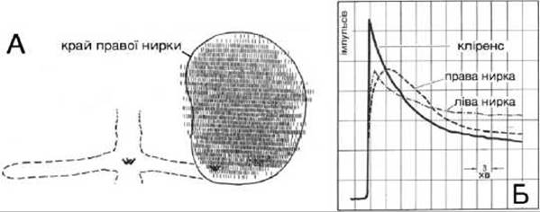
Fig. 240. Atherosclerotic occlusion of the left renal artery (radionuclide study).
A - scanogram. The right kidney is enlarged; the distribution of the radiopharmaceutical is even. Functioning parenchyma of the left kidney is not visualized.
B - renogram. The curve of the right kidney is of a normal type (Tmax = 4.5 min, T1/2 = 16.5 min), with initial impairment of excretory function. The curve of the left kidney is of an afunctional type (T1/2 is not determinable).
Last update: 08/08/2026
Editorial and Educational Adaptation: This material has been compiled based on the primary/original source text. The project team performed an editorial review, corrected technical inaccuracies, structured sections, and adapted the content for an educational format.
What was processed:
- elimination of formatting defects (OCR errors, structural breaks, corrupted characters);
- editorial organization of content;
- standardization of terminology in accordance with academic sources;
- verification of factual statements against the original source text.
All mentions of the author, publication year, and origin of the primary text have been preserved in accordance with the source.