TEXTBOOK PEDIATRIC GYNECOLOGY - 2013
Chapter 4. METHODS OF EXAMINATION IN PEDIATRIC GYNECOLOGY
FEATURES OF GYNECOLOGICAL EXAMINATION IN GIRLS AND ADOLESCENTS
A gynecological examination of girls differs significantly from that of adult women and has A number of specific features.
1. The examination environment must be calm and reassuring, as children often resist out of fear, which hinders the collection of data. It is unacceptable to have unauthorized persons in the room where the examination is performed, or to conduct examinations of other patients simultaneously.
2. The internal genitalia of girls are difficult to access for manual and instrumental examination. This compels the physician to avoid Procedures that may cause pain to the girl, even at the expense of obtaining valuable information about the child's condition.
3. The pelvic floor in girls is firm, spatial relationships are severely limited, and the genital Organs are small and lacking in distinct contours.
4. Girls lack the fixed position, shape, and dimensions of the internal reproductive organs that are characteristic of adult women.
Therefore, it must be emphasized that examinations of girls should be performed exclusively by physicians with specialized training in PEDIATRIC AND ADOLESCENT gynecology.
The examination should be performed with an empty bladder and bowel.
The patient's position during the examination may be supine, squatting, or knee-chest. Excessive thigh abduction should be avoided. Grasping the Labia Majora with a pinch-like grip using the thumb and index fingers of both hands, the examiner parts the pudendal cleft, inspects the external genitalia, and assesses their degree of development and the condition of the vaginal vestibule. During the inspection of the hymen, its integrity and type are assessed (annular, crescent, labiate, with one or multiple openings, imperforate septate, etc.), as well as its thickness, elasticity, and distensibility.
In cases of defloration, additional findings may include scarring deformity of the hymenal remnants, excessive distensibility, hyperpigmentation of the Tissues near the vaginal introitus, signs of inflammation, and The Nature of any discharge.
Subsequently, smears for bacteriological and bacterioscopic examination are taken from the vaginal vestibule, Urethra, paraurethral ducts, excretory ducts of the greater vestibular glands, and the rectum. When necessary, vaginal secretions are collected for cytologic examination.
Various instruments are used to collect smears: special pediatric Glass catheters, pipettes, capillary tubes, grooved probes, curettes of various shapes, or cotton-tipped wooden applicators. The sampling technique ranges from gently touching the mucosa to light scraping. Collecting secretions using a Braun syringe is also permissible. The obtained material is placed on a glass Microscope slide, dried, fixed, stained, and examined microscopically. The microscope slides are labeled accordingly.
The internal reproductive organs of girls are examined bimanually via the rectum.
Rectal examination in girls under 6 years of age is performed using the fifth (little) finger!
As an exception, a combined rectovaginal examination is permitted (if a vaginal or cervical tumor is suspected), strictly with the permission of parents or guardians and in the presence of a third party.
A specialized examination of a girl is performed in the following sequence:
— inspection and Assessment of the degree of secondary sexual characteristics development;
— inspection, Palpation, and Percussion of the abdomen;
— inspection of the external genitalia and hymen, taking into account age-related changes, along with inspection of the Perineum and anus;
— rectoabdominal examination;
— vaginoscopy (if a foreign body in the Vagina is suspected).
Prior to the examination, the lower bowel (via cleansing enema) and the bladder must be emptied.
The examination of younger girls (under 3 years of age) is performed on a changing table, while older girls are examined on a pediatric gynecological chair, which differs from adult chairs by having a special mechanism to adjust its depth, as well as a step for the girl to climb onto the chair independently.
The stage of sexual development is determined using the Ma, Ax, P, Me formula, where Ma stands for Mammary Glands (mammae), Ax for axillary Hair, P for pubic hair (pubes), and Me for menarcheal age.
When examining young girls, as well as during the initial evaluation, the presence of a mother or legal guardian is required.
During the examination of the external genitalia, the hair distribution pattern is assessed (female type with a horizontal hair line, or male type forming a triangle extending toward the Linea Alba of the abdomen and the inner thighs), along with the Water/140.html">Anatomical Structure of the Clitoris, labia majora and minora, hymen, color of the vaginal mucosa, and the nature of any vaginal discharge.
Rectoabdominal examination is performed on all girls with gynecological conditions to evaluate the state of the vagina (presence of foreign bodies, tumors, or Blood accumulation) and to assess the Uterus, appendages, parametrium, and adjacent organs. Palpation of the uterus determines its position, mobility, tenderness, the ratio between the sizes of the cervix and the uterine body, and the angle formed between the uterine body and cervix.
Depending on the Nature of the girl's condition, Additional Diagnostic Methods in pediatric gynecology are employed.
Vaginoscopy is a method for examining the vaginal walls and the cervix using an optical device. Vaginoscopy can be performed on girls of any age and helps determine the condition of the vaginal mucosa, the shape and size of the cervix, the shape of the external os, the presence of the "pupil" sign, pathological Changes in the cervical and vaginal areas, foreign bodies, and Congenital Malformations of the vagina and cervix.
Endoscopic diagnostic methods include laparoscopy and hysteroscopy. Laparoscopy is performed to rule out congenital Malformations of the internal genitalia, tumors, tumor-like masses of the pelvic organs, and sclerocystic Ovaries (Fig. 1).
Class="center">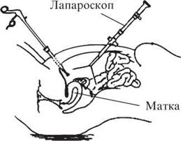
Fig. 1. Diagram of laparoscopy Procedure
Indications and contraindications for laparoscopy are standard. During the laparoscopic procedure, when necessary, gonadal biopsy, electrocoagulation of endometrioid heterotopias, and lysis of adhesions can be performed (Fig. 2).
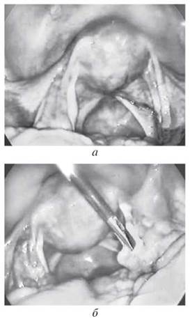
Fig. 2. Diagnostic laparoscopic view of the internal genitalia of patient S., 17 years old. Diagnosis: secondary Amenorrhea (a, b)
Hysteroscopy is a highly informative method for detecting intrauterine pathology, including endometrial hyperplasia and polyps, adenomyosis, and uterine developmental anomalies (Figs. 3, 4).
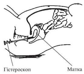
Fig. 3. Diagram of hysteroscopy procedure
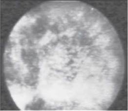
Fig. 4. Hysteroscopic view of the uterine cavity endometrium. Micrograph, ×20
Ultrasonography (US) is widely used in pediatric gynecological practice due to its safety, non-invasiveness, feasibility of dynamic monitoring, and high diagnostic accuracy. In pediatric gynecology, ultrasound allows for the DIAGNOSIS OF GENITAL malformations, ovarian tumors, and other gynecological conditions.
An ultrasonic wave has The ability to propagate in a specific direction while simultaneously transferring energy. The impact of an ultrasound wave on a portion of a medium causes it to oscillate and induces vibration in adjacent particles. This generates an ultrasonic wave that propagates at a specific speed, which depends on the Physical Properties of the medium.
In childhood, the internal reproductive organs (uterus, ovaries) are very small, which complicates Ultrasound examination. Careful preparation of the patient is required: 1–2 days prior, a gas-eliminating diet is prescribed, and an enema is administered the evening before.
Under normal conditions, pelvic organs are inaccessible for examination due to significant ultrasound reflection from gas-containing bowel loops. Therefore, ultrasound scanning is performed with a sufficiently filled Urinary Bladder (the patient should drink 1 liter of fluid 1–1.5 hours prior). When adequately filled, the bladder displaces the intestinal loops out of the pelvis and acts as an acoustic window, as fluid is the optimal medium for ultrasound propagation. In addition, the image of the urinary bladder serves as a reference for a cystic structure, which is necessary for comparing solid and fluid-filled structures.
Along with transabdominal scanning, transvaginal scanning is widely used in adult gynecology and in sexually active young women. This method does not require special preparation (diet, cleansing enema, bladder filling). However, it cannot be used to examine girls and adolescents who are not sexually active.
Echography makes it possible to assess the growth of internal reproductive organs during the prepubertal period, as well as to monitor the dynamic growth of the uterus and ovaries during physiological and pathological sexual development. Furthermore, it reveals a clear correlation between The Development of secondary sexual characteristics, anthropometric data, echographic dimensions of the uterus and ovaries, and their degree of maturity at a given age.
The uterus is easily visualized during ultrasound examination. In physiological development, it appears as a dense structure with numerous linear and punctate echogenic formations, located in the center of the pelvis behind the urinary bladder.
The uterus grows slowly and progressively. Between the ages of 1 and 5 years, the uterus has a tubular appearance, with its length changing from 2.8 cm at age 1 to 3.2 cm at age 5. Meanwhile, the width and anteroposterior dimensions of the uterus remain virtually unchanged. From age 5 until the onset of menarche, further enlargement of the uterus occurs in both length and width. Over the period from age 1 to menarche, the linear growth of the uterus increases by 3 cm, and its width by 2.3 cm. Significant uterine enlargement begins at ages 8–9, reflecting the onset of ovarian hormonal activity.
A particularly intensive growth of the internal reproductive organs, including the uterus, is observed with the onset of menarche. During this period, the uterus grows in length, width, and thickness, and an angle forms between the uterine body and its cervix. Furthermore, with the onset of menstrual function, the shape of the uterus changes to a pear-like form, and variations in uterine size occur throughout the Menstrual cycle: In the second phase of the cycle, the dimensions of the uterus are larger than in the first. By the age of 17, the dimensions of the uterus approach those of adult women, although, as a rule, they do not fully reach them.
Endometrium. Dynamic monitoring carried out throughout the menstrual cycle helps study the characteristics of endometrial changes (M-echo) in girls. In the majority of healthy girls at the beginning of the cycle, the M-echo is either undetectable or visualized as a narrow echopositive strip 1–2 mm thick, located centrally. Subsequently, an increase in endometrial thickness is observed, reaching 8–10 mm by the end of the menstrual cycle. In older girls with a biphasic menstrual cycle, endometrial thickness in the second phase of the cycle can reach 10–15 mm. In The first phase, the endometrial structure is homogeneous. Starting from the middle of the menstrual cycle, a rejection zone appears at the uterotubal/myometrial boundary in the form of an echonegative rim, the manifestations of which increase as menstruation approaches. By the end of the second phase, the entire endometrium becomes hypoechoic. A clearer median intrauterine M-echo is determined during menstruation.
The cervix begins to differentiate after 4 years of age, but as a rule, it is visualized from 8–9 years, when it accounts for 2/3 of the uterine body and is slightly wider than the body itself. At the age of 10–11, the cervix is clearly differentiated, an angle begins to form between the uterine body and the cervix, and The ratio of the length of the uterine body to the cervix is approximately 1:1. The uterocervical angle is clearly defined with the onset of menarche. At the age of 17, Puberty is completed, and the echogram reveals the following changes: the angle between the uterine body and the cervix is well expressed, and the ratio of the length of the uterine body to the length of the cervix is 2:1 (a decrease in this ratio at an older age more frequently indicates sexual infantilism and less commonly represents an individual anatomical variation of the uterus). The cervical canal is generally not visualized; however, during ultrasound examination during menstruation, remnants of the exfoliating endometrium may be detected within the cervical canal.
The vagina is represented on the echogram by two parallel lines that connect at an angle to the uterus and are located posterior to the urinary bladder.
The ovaries are the most challenging object for echographic examination, especially in young children. They are defined as ellipsoid structures with a delicate texture compared to The structure of the uterine wall. In childhood, the ovaries are located high in the pelvic cavity or at the boundary of the pelvic inlet, near its walls. On a transverse scan echogram, they are identified near the lower edge of the transverse section of the obturator internus/pelvic Muscle. At the age of 2–7 years, the average dimensions of the ovaries are 1.7×1.3×1.6 cm. At 8–9 years of age, with the onset of puberty, the ovaries are the first to begin enlarging, with dimensions of 1.9×1.4×1.8 cm. The ovaries are positioned high near the pelvic walls. At 10–11 years of age, they descend slightly into the pelvic cavity and are located 2–4 cm above the uterine angles. The ovarian dimensions are 2.1×1.5×2.0 cm. By the time of menarche, further growth of the ovaries occurs up to 3.0×2.12×3.0 cm. They are located near the uterine angles, which, combined with a pronounced uterocervical angle and a uterine body-to-cervix length ratio of 2:1, is a sign of maturity of the internal reproductive organs. At the age of 17, when puberty is completed in girls, the ovaries are located adjacent to the uterus and abut its walls, and follicles are identifiable within the Gonads.
Ovulatory cycles in girls appear 10 months after the onset of menstrual function, and from this time on, the development of dominant follicles can be tracked using ultrasound scanning. There is no significant difference between the sizes of the right and left ovaries in the First and Second Phases of the cycle, which indicates the equivalent participation of both ovaries in a normal menstrual cycle during puberty. As a rule, in healthy girls on the 10th day of an ovulatory menstrual cycle, a follicle with a diameter of 8–10 mm appears in one of the ovaries; subsequently, the follicle grows by 2 mm per day, reaching 18–20 mm on the 15th day, with a maximum of 25 mm. Girls exhibit a slower follicular growth rate, and ovulation may occur in follicles of a smaller diameter than in women of reproductive age. Immediately prior to ovulation, the follicle resembles a cystic formation of one of the ovaries. In the following days, the follicle is no longer detectable, indicating ovulation, which is also confirmed by functional diagnostic tests.
Ultrasound Diagnostics has the highest informational value in cases of amenorrhea, hypomenstrual syndrome, juvenile uterine bleeding (JUB), Developmental anomalies of the internal reproductive organs in girls, ovarian and uterine tumors, as well as Inflammatory Diseases of the internal reproductive organs. In conditions of hypomenstrual syndrome and amenorrhea, ultrasound reveals that the dimensions of the uterus lag behind age norms, the ovaries predominantly have a solid structure with isolated small echonegative inclusions (follicles), and the median uterine structure (M-echo) is thinned. The ultrasound method makes it possible to diagnose pubertal uterine bleeding, correctly select the Treatment method, and monitor its effectiveness dynamically (Fig. 5).
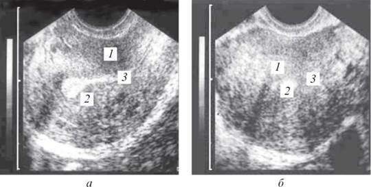
Fig. 5. Ultrasound appearance of the female genitalia in patient T., aged 15: 1 — uterus; 2 — endometrium; 3 — synechiae; a — in longitudinal transvaginal scan; b — in transverse transvaginal scan
Ultrasound examination allows for the differentiation of ovarian retention cysts from true tumors requiring surgical intervention, as well as monitoring the effectiveness of ongoing therapy. Of particular note are developmental Anomalies of the internal reproductive organs in girls, especially their diagnosis and ultrasound evaluation, which presents certain difficulties.
Uterine duplication is often clinically asymptomatic and in some cases is discovered as an incidental finding during ultrasound. The most challenging cases for ultrasound diagnosis are hemi-genital atresias, which most frequently manifest after menarche, when, despite normal menstruation, menstrual blood accumulates in the second, blind vagina. With this pathology, erroneous surgical intervention is frequently performed by mistaking hematocolpos for an ovarian cyst. Differential diagnostic features in this case include the Location of an echonegative mass below one of the duplicated uteri, the presence of ovaries on both sides in the Abdominal cavity, and renal aplasia on the affected side, which occurs in 100% of cases (Table 10).
Table 10. Age-related changes in the dimensions of the uterus and ovaries in adolescent girls
Age, years |
Length of uterine body (cervix), cm |
Uterine width, cm |
Anteroposterior uterine dimension, cm |
Ovarian volume, cm3 |
2-7 |
3.19±0.06 |
1.50±0.05 |
0.9±0.7 |
1.70±0.25 |
8-9 |
3.50±0.06 |
1.70±0.07 |
1.10±0.04 |
2.5±0.3 |
10-11 |
4.9±0.2 |
2.0±0.1 |
1.5±0.1 |
3.3±0.2 |
14-16 |
4.25±0.10 (2.57±0.03) |
3.8±0.8 |
2.82+0.10 |
6.9±0.3 |
17-19 |
4.80±0.11 (2.6±0.1) |
4.1±0.1 |
3.30±0.04 |
8.8±0.4 |
Ultrasound diagnostics of inflammatory processes of the internal reproductive organs reveals signs of uterine displacement and the presence of fluid in the pelvic cavity.
Probing of the vagina and uterine cavity is performed to diagnose malformations, in the presence of a foreign body, and when hematometra or pyometra is suspected.
Aspiration biopsy is performed in girls with uterine bleeding during the juvenile period to clarify the cause of the bleeding (neoplasms, endometrial hyperplastic process).
Curettage of the mucous membrane of the uterine body (with preceding and subsequent hysteroscopy) is indicated to stop uterine bleeding for diagnostic purposes in cases of prolonged, scanty spotting in girls with a disease duration of more than two years and in the absence of an effect from symptomatic and hormonal therapy.
Radiographic and radiopaque imaging methods (pneumopelvioradiography, hysterosalpingography, vaginography) make it possible to detect genital malformations, tumor-like formations of the small pelvis, and sclerocystic ovaries.
Radiological methods hold a special place in the practice of pediatric gynecologists. To diagnose neuroendocrine disorders, delayed or absent sexual development, radiological examinations of the shape, size, and contours of the sella turcica—the bony bed of the Pituitary Gland—are utilized. On a targeted lateral radiograph, its sagittal and vertical dimensions, as well as the ratio of the height to the length of the sella turcica (sellar index), which changes with age, are measured.
The sellar index in childhood is greater than or equal to 1, while in puberty it is less than 1. A pituitary tumor can cause Osteoporosis of the sellar walls, thinning and irregularity of the bony contours of the sellar walls, and other changes.
In pediatric gynecology, the radiological method is used to determine bone age, which characterizes the biological maturity of the Organism. It correlates closely with the age of menarche, and in cases of delayed sexual development, it correlates better with the onset of secondary sexual characteristics than with chronological age. The main indicators of skeletal bone differentiation are ossification centers and The formation of synostoses. To determine a child's bone age, obtained radiographs are compared with bone maturation standards using special radiological tables.
Girls outpace boys in bone age values by 12–18 months. During puberty, sexual development according to this indicator reaches 18–24 months.
In cases of impaired sexual development, the dynamics of ossification undergoes changes, which are taken into account in diagnostics and in determining the stage of sexual development.
Based on specially developed tables indicating the timing and sequence of ossification development, along with the synostoses between the metaphyses and epiphyses of long tubular bones, the timing of skeletal and limb ossification in children is determined according to age (Table 11).
Table 11. Timing of Ossification of the hand Skeleton and distal forearm in children and adolescents (M. A. Zhukovsky et al., 1980)
Site of ossification and synostosis |
Earliest term |
Latest term |
Average term |
|||
boys |
girls |
boys |
girls |
boys |
girls |
|
Capitate and hamate bones |
1 mo. |
1 mo. |
10 mo. |
8 mo. |
3-4 mo. |
2-3 mo. |
Distal epiphysis of the radius |
6 « |
4 « |
2 yrs |
1 S yrs |
10-12 « |
8-10 « |
Epiphyses of the proximal Phalanges and Metacarpal bones |
10 « |
8 « |
3 « |
2 S yrs |
15-18 « |
10-12 « |
Epiphyses of the middle and distal phalanges |
1 yr |
10 « |
3 « |
3 « |
20-24 « |
12-15 « |
Triquetral bone |
1 S yrs |
1 yr |
5 yrs |
4 « |
3-3 S yrs |
2-2 S yrs |
Lunate bone |
2 « |
2 yrs |
6 « |
4 S « |
3 S-4 « |
2 S-3 « |
Trapezium and scaphoid bones |
4 « |
3 S « |
8 « |
6 « |
5 S-6 yrs |
4-4 S « |
Distal epiphysis of the ulna |
6 yrs |
5 yrs |
10 « |
8 « |
7-7 S « |
6-6 S « |
Styloid process of the ulna |
7 « |
6 « |
12 « |
10 « |
9 S-10 « |
7 S-8 « |
Pisiform bone |
10 « |
7 « |
13 « |
11 « |
11-12 « |
8 S-9 « |
Sesamoid bones in the I metacarpophalangeal joint |
11 « |
9 « |
15 « |
13 « |
13 S-14« |
11-11 S « |
Synostosis in the I metacarpal bone |
14 « |
12 « |
17 « |
15 « |
15 S-16 « |
12 S-13 « |
Synostoses in the distal phalanges |
14 « |
12 « |
18 « |
16 « |
16-16 S « |
13 S-14 « |
Synostoses in the proximal phalanges |
14 « |
12 « |
19 « |
17 « |
16 S-17 « |
14-15 « |
Synostoses in the middle phalanges |
14 « |
12 « |
19 « |
17 « |
16 S-17 « |
15 S-16 « |
Synostoses in the II—V metacarpal bones |
14 « |
12 « |
19 « |
17 « |
16 S-17 « |
15 S-16 « |
Synostosis of the distal epiphysis of the ulna |
16 « |
13 « |
19 « |
17 « |
17-18 « |
15 S-16 « |
Synostosis of the distal epiphysis of the radius |
16 « |
14 « |
20 « |
18 « |
18-19 « |
16 S-17 S « |
An extremely important diagnostic method in girls is radiography of The Skull and sella turcica, which provides insight into the STRUCTURE OF THE cranial vault bones as well as the shape and size of the sella turcica as an indicator of pituitary gland size.
Among other X-ray Examination methods, adrenal radiography is used (when a tumor is suspected).
Hysterosalpingography (HSG) is performed according to clear indications: suspected Genital Tuberculosis, adenomyosis, and developmental anomalies of the internal genitalia in girls after 14–15 years of age.
In addition to the aforementioned methods, cytogenetic testing (Determination of Sex Chromatin, and karyotyping when indicated) is widely used to diagnose a range of gynecological disorders, which is particularly important in cases of impaired somatosexual development (sexual differentiation, delayed puberty, etc.).
Last update: 08/08/2026
Editorial and Educational Adaptation: This material has been compiled based on the primary/original source text. The project team performed an editorial review, corrected technical inaccuracies, structured sections, and adapted the content for an educational format.
What was processed:
- elimination of formatting defects (OCR errors, structural breaks, corrupted characters);
- editorial organization of content;
- standardization of terminology in accordance with academic sources;
- verification of factual statements against the original source text.
All mentions of the author, publication year, and origin of the primary text have been preserved in accordance with the source.