Human Anatomy and Physiology - I. V. Gaivoronsky 2011
Cardiovascular System
Lymphatic System
An integral component of The Vascular System is The Lymphatic system (Fig. 12.20). It is a network of Lymphatic vessels and nodes through which Lymph flows from the Tissues into the venous bed. Lymph is a clear or milky-white fluid whose chemical composition closely resembles that of Blood Plasma. It consists of interstitial fluid that has filtered into the lymphatic capillaries, along with lymphocytes. A significant portion of dietary fat is absorbed directly into the lymphatic Circulation. Lymphatic vessels can also transport toxins, microorganisms, and malignant tumor Cells, with metastasis primarily occurring along lymphatic drainage pathways. Lymph flow is facilitated by Muscle contractions, arterial pulsation, external pressure (such as massage), and other factors.
Lymph moves much more slowly than blood. Its circulation is aided by the Structural Features of the drainage pathways: capillaries, postcapillaries, lymphatic vessels, trunks, and ducts. Lymphatic pathways originate as blind-ended lymphatic capillaries (i.e., lacking an initial opening). The diameter of lymphatic capillaries exceeds that of blood capillaries, and their walls feature gaps between endothelial cells that allow interstitial fluid to pass into the capillary lumen. The next component of the lymphatic system is the lymphatic postcapillaries. Their walls feature Valves formed by the inner lining of the vessels, which prevent the backflow of lymph. Lymphatic capillaries and postcapillaries together form the lymph microcirculatory bed. From there, lymph flows into lymphatic vessels, along the course of which Lymph Nodes are situated.
Lymph nodes are aggregates of lymphoid tissue ranging in size from a pea to a bean. They are enveloped in a Connective Tissue capsule from which inward projections called trabeculae extend (Fig. 12.21). In cross-section, a lymph node exhibits a darker outer cortical substance and a lighter central medullary substance. As lymph flows through the lymph nodes, it becomes enriched with lymphocytes and Antibodies. The nodes carry out the phagocytosis of Bacteria and foreign particles, as well as the specific differentiation of T AND B lymphocytes. Consequently, the lymph draining from a lymph node contains a higher concentration of white Blood Cells than the lymph entering it.
In the HEAD, neck, trunk, and limbs, superficial and deep lymphatic vessels and nodes are distinguished. In the upper and lower extremities, as well as the head and trunk, the course of superficial lymphatic vessels largely mirrors that of the subcutaneous Veins in those regions. Deep lymphatic vessels, which drain lymph from joints, Muscles, and bones, travel alongside major blood Vessels and nerves as part of neurovascular bundles.
Class="center">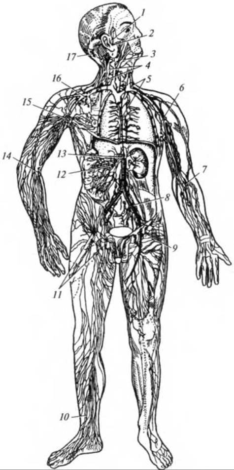
Fig. 12.20. The lymphatic system (diagram):
1 — auricular lymph nodes; 2 — parotid lymph nodes; 3 — submandibular lymph nodes; 4 — cervical lymph nodes; 5 — Thoracic duct; 6 — lateral axillary nodes; 7 — deep cubital lymph nodes; 8 — iliac lymph nodes; 9 — deep inguinal lymph nodes; 10 — lymphatic Vessels of the leg; 11 — superficial inguinal lymph nodes; 12 — mesenteric lymph nodes; 13 — cisterna chyli; 14 — superficial cubital lymph nodes; 15 — central axillary lymph nodes; 16 — subclavian lymph nodes; 17 — occipital lymph nodes
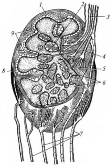
Fig. 12.21. Diagram of lymph node Structure:
1 — cortex; 2 — trabeculae; 3 — efferent lymphatic vessels; 4 — hilum; 5 — anastomosis between afferent and efferent lymphatic vessels; 6 — medulla; 7 — afferent lymphatic vessels; 8 — capsule; 9 — connective tissue trabeculae
Lymph nodes are predominantly arranged in groups, categorized into superficial and deep nodes. Nodes that drain lymph from specific Regions of the body are referred to as regional nodes. Clusters of lymph nodes are found in the bronchial region, the pulmonary hilum, and the Abdominal cavity. Major groups are located in the axilla, the cubital fossa, the popliteal fossa, the inguinal region, the neck, and beneath the Mandible. In these areas, they lie superficially, directly beneath the Skin, making them readily palpable. The following lymph node groups can be palpated: occipital, parotid, submandibular, submental, cervical, axillary, cubital, inguinal, and popliteal.
Lymph nodes act as biological barriers that trap foreign cells present in the lymph, such as tumor cells and microorganisms. Thus, the lymphatic system fulfills a barrier function by neutralizing foreign particles and microorganisms that enter the body. In addition, it unloads the Venous system by removing excess fluid from tissues into the lymphatic circulation. When an infection occurs, lymph nodes become tender and enlarged. Therefore, when an infectious disease is suspected, the regional superficial lymph nodes should be palpated first—for instance, submandibular nodes in dental conditions, cervical nodes for a sore throat, and inguinal nodes in cases of lower limb trauma and infected wounds.
The largest lymphatic vessel is the thoracic duct. It originates at the level of the first lumbar vertebra (L1), ascends through the thoracic cavity posterior to the aorta, runs up the right side of THE Vertebral Column into the neck, and empties into the left venous angle (Fig. 12.22). The initial segment of the thoracic duct is dilated and known as the cisterna chyli. It receives the right and left lumbar trunks, which drain lymph from the lower extremities, pelvis, and abdominal wall. In 40% of cases, the unpaired intestinal trunk, which collects lymph from the intestines, also empties into the cisterna chyli. Lymphatic vessels running within the mesentery of the intestines are called lacteals; they get their milky-white appearance from the fats absorbed in the digestive tract. Immediately before emptying into the left venous angle, the thoracic duct receives the left jugular trunk (which drains the left side of the Head and Neck), the left subclavian trunk (from the left upper extremity), and the left bronchomediastinal trunk (from the left lung and the left side of the Thorax).

Fig. 12.22. Lymphatic trunks and ducts (diagram):
1 — thoracic duct; 2 — Internal jugular vein; 3 — left jugular trunk; 4 — left subclavian trunk; 5 — subclavian vein; 6 — left bronchomediastinal trunk; 7 — left brachiocephalic vein; 8 — intestinal trunk; 9 — left lumbar trunk; 10 — right lumbar trunk; 11 — cisterna chyli; 12 — right bronchomediastinal trunk; 13 — right subclavian trunk; 14 — Right lymphatic duct; 15 — right jugular trunk
Thus, the thoracic duct collects lymph from three-quarters of the body: the lower extremities and abdominal cavity, the left side of the head and neck, the left upper extremity, the left half of the thorax, and the left lung.
The second major lymphatic vessel is the right lymphatic duct. It drains lymph from the right upper extremity and the right sides of the head, neck, and thorax. The right lymphatic duct is formed by the confluence of the right jugular, subclavian, and bronchomediastinal trunks, and it empties into the right venous angle.
The Central Nervous system (Brain AND SPINAL cord) lacks lymphatic vessels and lymph nodes. Lymphatic vessels are likewise absent in the epithelium of the skin and mucous membranes, Cartilage, the lens of the eye, and the sclera.
Knowledge of the Location of major lymphatic vessels and nodes is essential for the proper application of massage, which promotes the rapid evacuation of lymph from specific areas of the body. In doing so, massage helps remove Metabolic waste products that accumulate in tissues As a result of physical exertion and can exert adverse effects.
1. Provide a Classification of the vascular system.
2. What is the Structural Organization of the walls of Arteries and veins?
3. Characterize the SYSTEMIC AND PULMONARY circulations.
4. What is the Internal Structure of The Heart chambers?
5. List the layers of the heart wall.
6. Describe the borders of the heart.
7. Describe The properties of the heart muscle.
8. List and characterize the Phases of the cardiac cycle.
9. What is the Conduction system of the Heart?
10. Which vessels are involved in the Blood supply to the heart?
11. List the Branches of the External Carotid Artery.
12. Name the major Arteries of the upper limb.
13. How are the Branches of the Thoracic Aorta classified?
14. List the Branches of the Abdominal Aorta.
15. Name the main arteries of the lower limb.
16. What are intra-systemic and inter-systemic anastomoses?
17. Name the sites for digital compression of major arteries.
18. List the roots and tributaries of the superior and inferior venae cavae.
19. Which vessels form the roots of the portal vein?
20. How are the VEINS OF THE upper and lower limbs classified?
21. Name the superficial veins of the upper and lower limbs.
22. List the vessels of the Hemomicrocirculatory bed.
23. Describe the formation, topography, and drained areas of the thoracic duct.
24. What specific features characterize FETAL Blood Circulation?
Last update: 08/08/2026
Editorial and Educational Adaptation: This material has been compiled based on the primary/original source text. The project team performed an editorial review, corrected technical inaccuracies, structured sections, and adapted the content for an educational format.
What was processed:
- elimination of formatting defects (OCR errors, structural breaks, corrupted characters);
- editorial organization of content;
- standardization of terminology in accordance with academic sources;
- verification of factual statements against the original source text.
All mentions of the author, publication year, and origin of the primary text have been preserved in accordance with the source.