Human Anatomy, Part 1 - K. A. Dyubenko, A. K. Kolomiysev, Y. B. Chaikovsky 2002
Special Part
Splanchnology, splanchnologia [the study of viscera] — Major Digestive Glands
Pancreas — Peritoneum
The Peritoneum is a thin serous membrane that lines the inner surface of the abdominal walls and the Internal Organs located within the Abdominal cavity. Its total surface area averages 2 m2, which is roughly equivalent to the total surface area of human Skin. The peritoneum has a glistening appearance because its Connective Tissue base is covered by a single layer of squamous epithelium (mesothelium), which secretes a small amount of serous fluid, liquor peritonei, to moisten its surface. In males, the peritoneum forms a closed serous cavity, whereas in females, this cavity communicates with the external environment through the openings of the uterine tubes. The uterine part of the tube opens into the uterine cavity, which in turn communicates with the external environment via the Vagina. The peritoneum performs several vital physiological Functions:
- it provides smooth surfaces within the abdominal cavity, thereby facilitating easy and appropriate displacement of abdominal organs;
- it acts as a sero-hemato-lymphatic barrier regulating fluid movement from Blood Vessels into the abdominal cavity, as well as from the abdominal cavity into Blood and Lymphatic vessels;
- it maintains the Circulation of fluid within the abdominal cavity, which is continuously renewed through simultaneous processes of transudation and resorption;
- it plays a major role in maintaining systemic Homeostasis and Water-electrolyte balance. It is capable of absorbing up to 70 L of fluid per day (O. O. Shalimov et al., 1983).
The peritoneum consists of two layers: 1) the layer lining the inner surface of the abdominal cavity is called the parietal peritoneum, peritoneum parietale; 2) the layer covering the internal organs is called the visceral peritoneum, peritoneum viscerale (Fig. 270). By fusing with the surfaces of internal organs, the second layer forms their serous coats. Between the parietal and visceral layers lies the peritoneal cavity, cavum peritonei, which represents an intricate labyrinth of clefts, recesses, and spaces. The peritoneal cavity contains a small amount (30 cm3) of serous fluid, liquor peritonei, produced by the membrane itself. This serous fluid moistens the surfaces of the internal organs and the abdominal walls, facilitating peristalsis and mutual organ movement. Pathological conditions (such as Peritonitis and other acute inflammatory processes in the abdominal cavity) lead to an increased fluid volume, causing the peritoneal cavity to expand.
The parietal layer, or parietal peritoneum, peritonium parietale, transitions into the organ peritoneum, peritonium viscerale, which covers the internal organs. A connective tissue layer containing adipose tissue, tela subserosa, lies between the parietal peritoneum and the abdominal walls; this is the subserous fascia (tela subserosa), which varies in development across different regions. Specifically, it is absent in the diaphragmatic region, yet pronounced on the posterior abdominal wall, where it surrounds the Kidneys, Adrenal Glands, Ureters, Abdominal Aorta, INFERIOR VENA CAVA, and their branches. In the anterior abdominal wall, the subserous layer is poorly developed, increasing only in the regio pubica, where the peritoneum connects loosely to the abdominal wall. Consequently, when the Urinary Bladder is distended, it displaces the peritoneum away from the anterior abdominal wall, creating an area approximately 4-5 cm above the Pubic Symphysis that remains uncovered by the peritoneum.
Class="center">
Fig. 270. Sagittal section of the trunk.
Relationships between the peritoneum and abdominal organs (diagram) (after V. P. Vorobyov)
Tracing the further course of the peritoneum along the anterior and superior abdominal walls reveals that it passes onto the diaphragmatic surface of the Liver, forming the falciform ligament, lig. falciforme hepatis, within the free margin of which lies the round ligament of the liver, lig. teres hepatis. From the Inferior surface of the Diaphragm posterior to the falciform ligament, the peritoneum reflects onto the diaphragmatic surface of the liver, forming the coronary ligament of the liver, lig. coronarium hepatis, which terminates at its margins in triangular sheets known as the right and left triangular ligaments, lig. triangulare dextrum et sinistrum.
From the diaphragmatic surface of the liver, the peritoneum extends over its inferior margin onto the visceral surface, from which it gives rise to several ligaments passing to internal organs: to the Kidney, lig. hepatorenale; to the lesser curvature of The Stomach, lig. hepatogastricum; and to the duodenum, lig. hepatoduodenale. Together, lig. hepatoduodenale, lig. hepatogastricum, and lig. phrenicogastricum form the lesser omentum, omentum minus. The hepatoduodenal ligament, lig. hepatoduodenale, connects the porta hepatis to the initial segment of the duodenum. From right to left, this ligament transmits the common Bile duct (ductus choledochus), portal vein (v. portae), and proper hepatic artery (a. hepatica propria) — memorized via the anatomical mnemonic DVA (ductus, vena, arteria) — along with Lymphatic vessels and nerves. This is a crucial consideration during surgical interventions on extrahepatic biliary ducts.
At the lesser curvature of the stomach, the layers of the lesser omentum diverge to cover the anterior and posterior surfaces of the stomach. At the greater curvature, these layers converge and descend in front of the transverse colon and Small Intestine, forming the anterior layer of the greater omentum, omentum majus (see Fig. 270). The layers of the greater omentum descend and then loop upward to form its posterior layer. Thus, the greater omentum is composed of four peritoneal layers. Upon reaching the transverse colon and its mesentery, the posterior layer of the greater omentum fuses with them and extends dorsally toward the Pancreas, where its layers diverge. One layer covers the pancreas and ascends toward the diaphragm, while the other covers the inferior surface of the gland and transitions into the mesentery of the transverse colon, colon transversum.
The organs located within the abdominal cavity have varying relationships with the peritoneum (Fig. 271).

Fig. 271. Transverse section of the trunk. Organ-peritoneum relationships (diagram):
1 - extraperitoneal position of the kidney; 2 - peritoneum viscerale; 3 - peritoneum parietale; 4 - intraperitoneal position of the small intestine; 5 - mesenterium; 7 - cavitas peritonei; 8 - spatium retroperitoneale
An organ is said to have an intraperitoneal position if it is covered by the peritoneum on all sides; a meso-peritoneal position if it is covered on three sides; and an extraperitoneal position if it is covered on only one side (Table 7).
Table 7. Relationships of internal organs to the peritoneum
|
Intraperitoneal organs |
Mesoperitoneal organs |
|
|
Stomach Jejunum Ileum Transverse colon Sigmoid colon Upper third of the rectum Uterine tubes |
Liver Ascending colon Descending colon Middle third of the rectum Urinary bladder (distended) |
Pancreas Duodenum (ascending and descending parts) Adrenal glands Ureters Lower part of the rectum Urinary bladder (empty) |
As the visceral peritoneum transitions from one organ to another, onto the parietal wall, or vice versa, it forms ligaments, folds, omenta, as well as grooves, recesses, fossae, sinuses, compartments, and mesenteries. The latter attach to the posterior abdominal wall. Consequently, the peritoneum forms the following mesenteries:
- mesenterium - for the jejunum and ileum;
- mesocolon transversum - for the transverse colon;
- mesocolon sigmoideum - for the sigmoid colon;
- mesoappendix - for the appendix.
The abdominal cavity, cavum abdominis, is conventionally divided into three floors: upper, middle, and lower.
1. The upper floor is bounded superiorly by the diaphragm, and inferiorly by the transverse colon and its mesentery. It contains the stomach, liver, and spleen.
2. The middle floor occupies the area from the mesentery of the transverse colon, mesocolon transversum, to the pelvic inlet. It contains the jejunum, ileum, ascending colon, descending colon, and cecum with the appendix.
3. The lower floor extends from the pelvic inlet to the pelvic diaphragm, deepening its cavity. It contains the rectum, urinary bladder, ureters, prostate, and Seminal Vesicles in males, and the Uterus and Ovaries in females.
1. For the Organs of the upper floor, the peritoneum forms three recesses, bursae (D. M. Zernov): the hepatic bursa, bursa hepatica; the pregastric bursa, bursa pregastrica; and the omental bursa, bursa omentalis.
The hepatic bursa, bursa hepatica, is located beneath the diaphragm and is separated from the pregastric bursa by the falciform ligament; posteriorly, it is bounded by the coronary ligament, lig. coronarium hepatis. The hepatic bursa contains the right lobe of the liver, and deep within the bursa, beneath the liver, the upper pole of the right kidney and the Adrenal gland can be palpated.
The pregastric bursa, bursa pregastrica, is located in front of The Liver and spleen beneath the diaphragm. It contains the left lobe of the liver, the spleen, and the anterior surface of the stomach, featuring a deep extrasplenic space.
The omental bursa, bursa omentalis (Fig. 272), is located posterior to the stomach and the lesser omentum, omentum minus, which is formed by three peritoneal ligaments: the hepatogastric ligament, lig. hepatogastricum, extending from the porta hepatis to the lesser curvature of the stomach, and the hepatoduodenal ligament, lig. hepatoduodenale, connecting the porta hepatis to the pars superior duodeni. The omental bursa is bounded by the anterior wall formed by the lesser omentum, the posterior wall of the stomach, and the gastrocolic ligament, lig. gastrocolicum.

Fig. 272. Horizontal section of the trunk at the level of the XII thoracic vertebra (after R. D. Sinelnikov)
The posterior wall is formed by the parietal layer of the peritoneum; the superior wall by the inferior surface of the caudate lobe of the liver and the diaphragm; and the inferior wall by the mesocolon transversum et colon transversum. The left wall of the omental bursa is formed by the splenic ligaments: lig. gastrolienale et colon transversum and lig. phrenicosplenicum.
The omental bursa communicates with the peritoneal cavity through the omental foramen, foramen omentale [epiploicum] (Winslow) (see Fig. 272), which is bounded: anteriorly by the lig. hepatoduodenale, posteriorly by the lig. hepatorenale, inferiorly by the lig. duodenorenale, and superiorly by the caudate lobe of the liver. The omental bursa features a vestibule, as well as superior and inferior splenic recesses. During surgical interventions, a surgeon may access the omental bursa through the omental foramen for inspection.
The greater omentum, omentum majus, hangs like an apron covering the loops of the small intestine anteriorly (see Fig. 270). It is formed by four layers of peritoneum fused together in the form of plates. The anterior plate is formed by two layers of peritoneum descending from the greater curvature of the stomach and, passing anterior to the transverse colon, fusing with it to form the gastrocolic ligament, lig. gastrocolicum. The anterior plate descends to the level of the pubic bones and then folds back, forming the posterior plate of the greater omentum. A slit-like cavity lies between the layers of the anterior and posterior plates of the omentum, which communicates with the cavity of the omental bursa, although in adults the omental cavity is partially obliterated.
2. The middle floor of the peritoneal cavity can be examined by lifting the greater omentum and the transverse colon upward. The middle floor comprises four compartments: the right and left lateral channels, canalis lateralis dexter et sinister, which run between the lateral abdominal walls and the ascending and descending colons, as well as two mesenteric sinuses, sinus mesentericus dexter et sinister, formed by the division of the middle floor by the mesentery of the small intestine, which runs obliquely from top to bottom and from left to right. The left and right sinuses are separated from each other by the ROOT of the mesentery of the small intestine and communicate with the lesser pelvis.
The mesentery, mesenterium, is a fold formed by two layers of peritoneum that attaches the small intestine to the posterior abdominal wall. The posterior edge of the mesentery is its root, radix mesenterii, which originates on the left side of the II lumbar vertebra and runs obliquely toward the right iliac fossa. Along its path, the root of the mesentery crosses the terminal part of the duodenum, the aorta, the inferior vena cava, the right Ureter, and the psoas major Muscle.
Between the serous layers of the mesentery lie adipose tissue, Lymph Nodes, blood and lymphatic vessels, and nerves. On the posterior parietal peritoneum, There is a series of recesses of practical significance, as they can sometimes give rise to internal retroperitoneal hernias: duodenojejunal hernias of Treitz, intersigmoid, cecal, and subfascial. At the transition of the duodenum into the jejunum, small recesses are formed, recessus duodenalis superior et inferior. In the region where the small intestine transitions into the Large Intestine, above and below the ileocecal fold, plica ileocecalis, There are two recesses: recessus ileocecalis superior et inferior. At the site of the cecum, the parietal peritoneum forms a depression known as the retrocecal recess, recessus retrocecalis. Sometimes, an opening leading into the retrocecal recess is present in the retrocecal fossa. On the opposite side, on the left surface of the sigmoid colon mesentery, lies the intersigmoid recess, recessus intersigmoideus (a site for The formation of retroperitoneal hernias).
3. The peritoneum of the lower floor covers its walls and the organs located within it, depending on sex. The sigmoid colon and the initial part of the rectum are covered by the peritoneum on all sides (intraperitoneally) and possess their own mesentery. The middle section, rectum, is covered by the peritoneum anteriorly and laterally, while its posterior surface remains uncovered; further inferiorly, at a distance of 7.5–8 cm from the anus, the peritoneum transitions from the anterior surface of the rectum to the posterior surface of the urinary bladder, forming the rectovesical pouch, excavatio rectovesicalis. A feature of the male peritoneum is that a portion of the serous sac is isolated within the Scrotum, enclosing each Testis separately. During development, a finger-like sac—the processus vaginalis—evaginates through the Inguinal Canal into the scrotum, which obliterates in 99% of cases except for its distal part. Thus, two serous sacs remain in the scrotum, in which serous fluid accumulates during testicular inflammation (orchitis).
In females, the uterus is located between the urinary bladder and the rectum. It is covered by the peritoneum on all sides; therefore, the female pelvic cavity contains two pouches: the rectouterine pouch, excavatio rectouterina (between the rectum and the uterus), and the vesicouterine pouch, excavatio vesicouterina (between the uterus and the urinary bladder). The excavatio rectouterina, or pouch of Douglas (Douglasi), is of practical significance: blood, pus, or serous fluid accumulates within it during inflammatory conditions and intra-abdominal hemorrhages; hence, in clinical (gynecological) practice, puncture of this pouch is performed for diagnostic purposes. On the sides of the uterus, the peritoneum extends onto the walls of the lesser pelvis, forming the broad ligament of the uterus, lig. latum uteri, which lies transversely in the pelvic cavity and divides it into anterior and posterior compartments.
In both sexes, in the pubic region, regio pubica, lies the prevesical cellular space, spatium prevesicale (cavum Retzii), bounded anteriorly by the transversalis fascia, fascia transversalis, and posteriorly by the urinary bladder and peritoneum. The space is filled with adipose tissue, the venous plexus of the urinary bladder and prostate, and Arteries supplying the urinary bladder. The prevesical space provides access to the urinary bladder during the operation of high suprapubic cystostomy. The prevesical space is a site where blood accumulates (in fractures of the pubic bones) and urine escapes (in injuries to the urinary bladder). All of this is of great importance in clinical (urological) practice. Therefore, when the urinary bladder is distended, the peritoneum is displaced upward, and the bladder closely abuts the anterior abdominal wall, allowing for cystocentesis (using a Veress needle or trocar) above the symphysis in clinical (surgical) practice.
Within the lower floor of the abdominal cavity, the peritoneum forms folds and fossae. On the posterior surface of the anterior abdominal wall, five umbilical folds extend from the umbilicus to the urinary bladder: the median umbilical fold, plica umbilicalis mediana; two medial umbilical folds, plicae umbilicales mediales; and two lateral umbilical folds, plicae umbilicales laterales (Fig. 273). The median umbilical fold contains the obliterated urachus; the medial folds contain the obliterated umbilical arteries; and the lateral folds contain the inferior epigastric arteries, aa. epigastricae inferiores (Branches of the external iliac artery, a. iliaca externa). Lateral to the median umbilical fold lie the supravesical fossae, fossae supravesicales. Between the medial and lateral folds on each side are the medial inguinal fossae, fossae inguinales mediales, and lateral to the lateral folds are the lateral inguinal fossae, fossae inguinales laterales. The lateral inguinal fossa corresponds to the deep inguinal ring, and the medial one to the superficial ring; inguinal hernias may protrude through these fossae, which is clinically significant in surgical practice (see the chapter "Myology", Inguinal Canal).
Blood supply to the peritoneum is provided by branches (arteries) of the abdominal aorta: the inferior phrenic, superior and inferior mesenteric, anterior and задняя (anterior and posterior) cecal, suprarenal, renal, and lumbar arteries. Venous blood drains into the superior and inferior vena cava systems and into the portal vein.
Lymphatic drainage from the peritoneum occurs via lymphatic capillaries of the superficial and deep lymphatic networks (L. V. Chernyshenko, A. M. Synytska, 1982), after which lymph penetrates through Stomata into the lymphatic Vessels of the peritoneum.
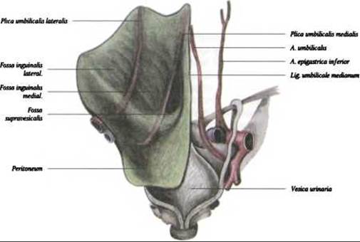
Fig. 273. Folds and fossae on the posterior surface of the anterior abdominal wall. (On the right side, the parietal peritoneum has been removed)
Innervation of the peritoneum is provided by a superficial nerve plexus located in the peritoneum above the elastic networks, and a deep nerve plexus located within the deep reticular Collagen-elastic layer.
Last update: 08/08/2026
Editorial and Educational Adaptation: This material has been compiled based on the primary/original source text. The project team performed an editorial review, corrected technical inaccuracies, structured sections, and adapted the content for an educational format.
What was processed:
- elimination of formatting defects (OCR errors, structural breaks, corrupted characters);
- editorial organization of content;
- standardization of terminology in accordance with academic sources;
- verification of factual statements against the original source text.
All mentions of the author, publication year, and origin of the primary text have been preserved in accordance with the source.