Human Anatomy Part 1 - K. A. Diubenko, A. K. Kolomiitsev, Iu. B. Chaikovsky 2002
Special Part
Splanchnology, splanchnologia [the study of viscera] - Major digestive glands
Pancreas - Structure of the gland
The Pancreas has an tubuloalveolar Structure. Externally, it is covered by a Connective Tissue capsule, from which trabeculae extend into the parenchyma, dividing it into lobules, known as lobuli pancreatici. Blood Vessels, ducts, and nerves run between these lobules. The pancreatic lobules comprise both exocrine and endocrine components.
The exocrine portion of the pancreas, pars exsocrina pancreatis, is represented by pancreatic acini, acinus pancreaticus (Fig. 268), which serve as the Structural and functional units of the gland. They consist of 8–12 exocrine pancreatocytes, or acinar Cells, that synthesize digestive Enzymes. The exocrine part includes intralobular and interlobular ducts, as well as the main pancreatic duct, ductus pancreaticus (Wirsung). It originates in the tail region of the gland, runs from left to right, receiving smaller tributaries. In its terminal section, it merges with the common Bile duct, and together they empty into the duodenum through a common opening on the major duodenal papilla, papilla duodeni major (see Figs. 262, 263). Various anatomical variations of the pancreatic duct opening into the duodenum are known; notably, the ducts may open separately, either with or without The formation of a major papilla. These variations in the opening of the BILE AND PANCREATIC ducts into the duodenum must be considered in surgical practice. In its terminal segment, the duct features the sphincter of the pancreatic duct, m. sphincter ductus pancreatici, which regulates the flow of pancreatic juice into the duodenum. In addition to the main pancreatic duct, the gland often contains an accessory pancreatic duct, ductus pancreaticus accessorius, which opens into the duodenum at the minor duodenal papilla. Cases of an accessory pancreas, pancreas accessorius, may also occur.
The Endocrine Part of the gland, pars endocrina pancreatis, is represented by pancreatic islets, insulae pancreaticae, which are scattered among the acini and have a spherical or ovoid shape (see Fig. 268). These islets consist of hormone-producing endocrine cells (insulocytes). The highest concentration of pancreatic islets is found in the tail of the gland, with their total number reaching 1–2 million. (For a more detailed Discussion of the endocrine portion, see the chapter "Endocrine glands".)
Ultrasonography (US) of the pancreas. The pancreas is visualized in 85% of cases following special preparation. It is well-delineated during scanning through the Liver while the patient holds their breath. Transverse scanning is performed first, followed by longitudinal scanning. Transverse scanning is conducted at an angle of 10–20° along a conventional line drawn from the hilum of the right Kidney to the hilum of the Spleen or the upper pole of the left kidney, moving the transducer from the xiphoid process toward the umbilicus. Longitudinal scans are performed from the right midclavicular line toward the anterior axillary line.
Class="center">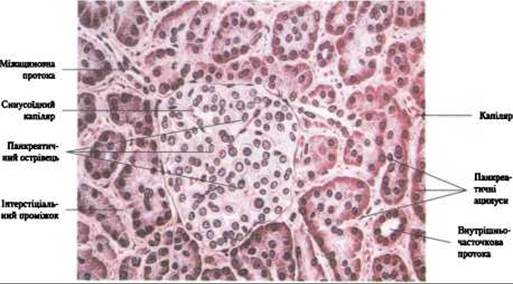
Fig. 268. Microscopic STRUCTURE OF THE pancreas (pancreatic islet and acini)
Normally, the HEAD of the pancreas is located directly beneath the right lobe of the liver, while the body and tail lie beneath the left lobe of The Liver and The Stomach. The tail of the pancreas is identified in the region of the splenic hilum or the upper pole of the left kidney. Posteriorly, the pancreatic head is adjacent to the INFERIOR VENA CAVA. On sonograms, it appears as a large echo-negative structure situated to the right of THE Vertebral Column. To the left and anterior to it lies the superior mesenteric vein, which serves as the primary landmark for identifying the pancreatic head. The splenic vein serves as a landmark for identifying the body of the gland, with the splenic artery running slightly superior to it. Normally, the superior mesenteric artery can be detected between the splenic vein and the aorta.
Topographic studies of the gland on sonograms have established that it initially runs from right to left and inferiorly to superiorly, and then sharply turns posteriorly at the junction of the body and tail. The head of the gland lies to the right of the vertebral column, the neck lies superficial to it, and the body and tail lie to its left. During transverse scanning, the head of the pancreas appears oval or circular, whereas the body and tail are identified as cylindrical structures of approximately uniform thickness.
Dimensions of the pancreas: the anteroposterior diameter of the head ranges from 2.2 to 3 cm, the body from 1 to 2 cm, and the tail from 1.8 to 2.8 cm. The width of the body and tail is approximately 1.5 times greater than their thickness. The pancreatic duct is detected in less than one-third of observations, typically in a fragmented manner, with a diameter not exceeding 1 mm. Determining the dimensions and structure of the gland is of great diagnostic importance for identifying pathologies (such as tumors, cysts, and Chronic Pancreatitis).
Computed tomography (CT) of the pancreas. Computed tomography is a vital, highly informative diagnostic method that provides accurate information on the state of the pancreas, while also allowing for a better Assessment of the morphofunctional status of adjacent Organs (Kidneys, spleen, biliary tract, mesentery, as well as the small and large intestines).
Optimal imaging of the pancreas is achieved by scanning at the level of ThXII–LII. On tomograms, the head of the pancreas is located approximately at the level of LI, while the body and tail are situated 1–2 vertebrae higher. Landmarks that allow for the assessment of pancreatic topography and its spatial relationships with surrounding organs (syntopy) include the outlines of the superior mesenteric artery and the vascular pedicle of the spleen (Fig. 269). On the posterior surface of the gland, posterior to the artery, lies the splenic vein, whose diameter is twice that of the corresponding artery and which is more clearly visualized on CT scans. The pancreatic structure is homogeneous in young individuals, whereas in elderly individuals, it decreases in size and exhibits a lobular architecture. The retroperitoneal adipose tissue surrounding the organ allows for clear visualization of the pancreas.
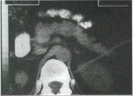
Fig. 269. Computed tomogram of the retroperitoneal space at the level of the pancreas, kidneys, and Adrenal Glands (according to O. Ivankov):
1 - pancreas; 2 - head of the pancreas; 3 - body of the pancreas; 4 - tail of the pancreas; 5 - Abdominal Aorta; 6 - kidneys; 7 - duodenum; 8 - inferior vena cava; 9 - vertebra
Blood supply to the pancreas is provided by Branches of the superior pancreaticoduodenal artery (from the gastroduodenal artery) and the inferior pancreaticoduodenal artery (from the superior mesenteric artery), as well as branches of the splenic artery. Venous blood drains via corresponding Veins into the portal vein.
Lymphatic drainage flows into the pancreatosplenic Lymph Nodes.
The Innervation of the pancreas is complex and derives from multiple sources: the hepatic, splenic, superior mesenteric, and celiac nerve plexuses, as well as branches of the Vagus nerve. Nerve trunks extend from these neural structures into the parenchymal tissue, accompanying blood vessels and forming plexuses around them. Parasympathetic innervation is mediated primarily by the right vagus nerve trunk (D. M. Golub, 1939). Sympathetic innervation is supplied by the celiac plexus through its ganglia. Sensory innervation is predominantly spinal, mediated by Spinal Nerves arising from the V–XII thoracic segments (V. Yu. Pervushin, 1956, 1957).
Last update: 08/08/2026
Editorial and Educational Adaptation: This material has been compiled based on the primary/original source text. The project team performed an editorial review, corrected technical inaccuracies, structured sections, and adapted the content for an educational format.
What was processed:
- elimination of formatting defects (OCR errors, structural breaks, corrupted characters);
- editorial organization of content;
- standardization of terminology in accordance with academic sources;
- verification of factual statements against the original source text.
All mentions of the author, publication year, and origin of the primary text have been preserved in accordance with the source.