MEDICAL BIOLOGY, HUMAN ANATOMY, PHYSIOLOGY AND PATHOLOGY - Ya.I. Fedoniuk 2010
ANATOMY, PHYSIOLOGY, PATHOLOGY
SECTION 3. ANATOMICAL AND PHYSIOLOGICAL ASPECTS OF BODY FUNCTION SELF-REGULATION
FUNCTIONAL ANATOMY OF THE PERIPHERAL NERVOUS SYSTEM
CRANIAL NERVES
The Brain gives rise to 12 pairs of cranial
Spinal Nerves, which are mixed; some Cranial Nerves are purely motor (IV, VI, XI, XII), some are sensory (I, II,
VIII pairs), and some are mixed (III, V, VII, IX, X pairs).
The cranial nerves also contain parasympathetic fibers that innervate smooth Muscles and glands (III, VII, IX, and X pairs).
Pair I – olfactory nerves (nn. olfactorii) – constitute a collection of fine olfactory filaments, which are the processes of Olfactory nerve Cells located in the mucous membrane of the superior nasal meatus, superior nasal concha, and nasal septum. They pass through the cribriform plate of the Ethmoid bone and reach the olfactory bulb, where The Cell bodies of the second Neurons of the olfactory pathway are located. From here, nerve impulses are transmitted along the olfactory pathway to the Cerebral Cortex (Fig. 3.44).
Class="center">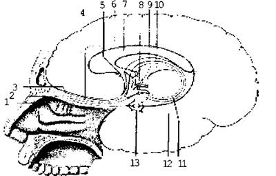
Fig. 3.44. Pathway of the Olfactory Organ:
Pair II – Optic nerve (n. opticus) is formed by the processes of nerve Cells of the retina, exits the Orbit through the optic canal, and forms an incomplete chiasm in the region of the sella turcica.
Only the medial fibers decussate, passing into the optic tract (Fig. 3.45).
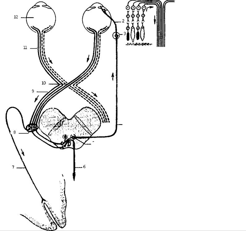
Fig. 3.45. Optic pathway
Fibers of the latter approach the lateral geniculate bodies, superior colliculi of the Midbrain, and the pulvinar of the thalamus, where the subcortical centers of Vision are located. From here, fibers extend to the cerebral cortex, ending along the banks of the calcarine sulcus in the occipital lobe. This is the Location OF THE cortical visual analyzer.
Pair III – Oculomotor nerve (n. oculomotorius); its motor and parasympathetic nuclei are located on the floor of the cerebral aqueduct at
the level of the superior colliculi of the midbrain. Passing through the superior orbital fissure into the orbit, its motor fibers innervate the superior, inferior, and medial rectus Muscles of the eye, the inferior oblique Muscle, and the levator palpebrae superioris muscle. The parasympathetic fibers provide innervation to the sphincter pupillae and ciliary muscles.
Pair IV – Trochlear nerve (n. trochlearis); its motor Nucleus is located on the floor of the cerebral aqueduct at the level of the inferior colliculi of the midbrain. The nerve passes through the superior orbital fissure into the orbit and innervates the superior oblique muscle of the eye.
Pair V – Trigeminal nerve (n. trigeminus), mixed (Fig. 3.46). It has one motor and three sensory nuclei. The processes of the cells of the sensory nuclei form the three Branches of the trigeminal nerve: the ophthalmic, maxillary, and mandibular nerves. The processes of the motor nucleus cells run within the mandibular nerve. The ophthalmic nerve passes into the orbit through the superior orbital fissure and innervates the membranes of the Eyeball, the Skin of the forehead, the mucosa of the Nasal cavity, the lacrimal gland, the frontal and sphenoid sinuses, and the ethmoid air cells.
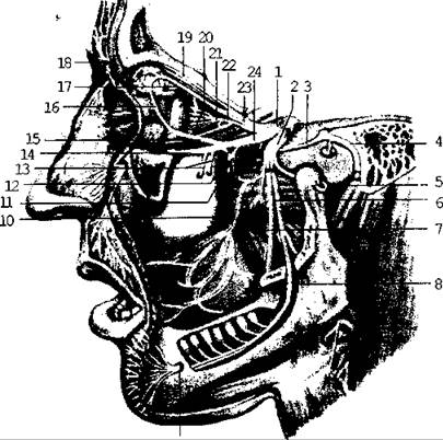
Fig. 3.46. Trigeminal nerve
The maxillary nerve exits the Skull through the foramen rotundum, enters the pterygopalatine fossa, and gives off branches that innervate the Teeth of the upper jaw, the mucosa of the nasal cavity, the soft and hard palate, and the skin of the face from the medial angle of the eye to the angle of the Mouth. The parasympathetic pterygopalatine ganglion is associated with this nerve, and its postganglionic fibers provide secretory innervation to the lacrimal gland.
The mandibular nerve exits the skull through the foramen ovale and gives off branches that innervate the teeth of the lower jaw, the buccal mucosa, the anterior 2/3 of the Tongue, the Cytology/practical/97.html">Parotid salivary gland, the skin of the chin, the temporal region and auricle, the Muscles of Mastication, the anterior belly of the digastric muscle, the mylohyoid muscle, the tensor veli palatini muscle, and the tensor tympani muscle.
The parasympathetic ganglia associated with this nerve are: the otic, sublingual, and submandibular ganglia.
Postganglionic fibers from the otic ganglion provide secretory innervation to the parotid salivary gland, whereas those from the sublingual and submandibular ganglia supply secretory innervation to the corresponding Salivary Glands.
The VI pair is the Abducens nerve (n. abducens), which is a motor nerve originating from cell nuclei located in the Rhomboid fossa on the dorsal surface of the Pons. It enters the orbit via the superior orbital fissure, where it innervates the lateral rectus muscle of the eye.
The VII pair is the Facial Nerve (n. facialis), a motor nerve (Fig. 3.47); however, it incorporates the so-called XIII pair of cranial nerves (the intermediate nerve, n. intermedius), which is mixed. Consequently, some authors consider the VII pair to be of a mixed type.
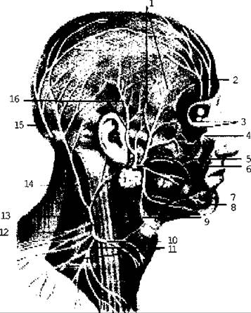
Fig. 3.47. Superficial NERVES OF THE HEAD and Neck
The facial nerve originates from cell nuclei located in the pons, which is also the site of the sensory and parasympathetic nuclei of the intermediate nerve. Both nerves pass through the internal acoustic meatus into the facial canal. The facial nerve exits the skull via the stylomastoid foramen and innervates the facial muscles. The sensory fibers of the intermediate nerve, together with the mandibular nerve, supply the mucous membrane of the anterior two-thirds of the tongue, while its parasympathetic secretory fibers innervate the lacrimal, submandibular, and sublingual salivary glands, as well as the Glands of the nasal cavity.
The VIII pair is the Vestibulocochlear nerve (n. vestibulocochlearis), which consists of two parts: vestibular and cochlear. The vestibular part transmits sensations from the static apparatus located in the vestibule and semicircular canals of the Inner ear labyrinth, whereas the cochlear part conveys auditory sensations from the Spiral organ of Corti situated within the cochlea (Figs. 3.48 and 3.49). Both parts feature nerve ganglia composed of bipolar cells lying within the pyramid of the Temporal bone. Within the internal acoustic meatus, these two parts unite to form the vestibulocochlear nerve. The latter enters the brain and terminates in nuclei located in the pons at the border with the Medulla Oblongata.
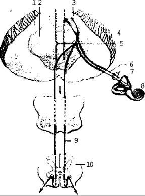
Fig. 3.48 Diagram of the vestibular pathways
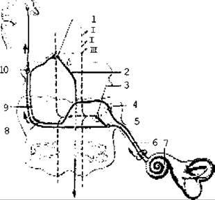
Fig. 3.49. Auditory Analyzer pathway
The IX pair is the Glossopharyngeal nerve (n. glossopharyngeus), a mixed nerve containing motor, sensory, and parasympathetic fibers. Its nuclei are located in the medulla oblongata. The nerve exits the skull through the jugular foramen. Its sensory fibers innervate the posterior third of the mucous membrane of the tongue, the pharyngeal mucosa, Tonsils, and Middle ear. The motor fibers supply the stylopharyngeus muscle and the pharyngeal muscles (along with the X pair). The parasympathetic secretory fibers provide innervation to the parotid salivary gland (Fig. 3.50).
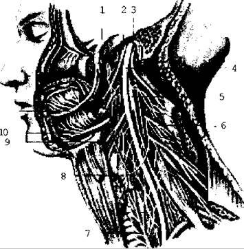
Fig. 3.50. Nerves of the head and neck; left lateral view (muscles, Blood Vessels, lateral wall of the skull base, and left half of the Mandible removed)
The X pair is the Vagus nerve (n. vagus), a mixed nerve and the longest of the cranial nerves. Its motor, sensory, and parasympathetic nuclei lie within the medulla oblongata. The vagus nerve exits the skull through the jugular foramen and, in the neck region, courses alongside the Internal jugular vein and common carotid artery. Through the superior thoracic aperture, it enters the thoracic cavity, where the left nerve lies on the anterior surface of the Esophagus and the right on its posterior surface; anastomosing with each other, they form the esophageal plexus.
Passing through the Diaphragm into the Abdominal cavity, the vagus nerves form the anterior (left) and posterior (right) vagal trunks. The terminal branches of the vagus nerves in the abdominal cavity extend to the ganglia of the celiac (solar) plexus and, together with sympathetic nerves, form plexuses along the Arteries to supply the Organs.
The motor fibers of the vagus nerves innervate the striated muscles of the Pharynx, soft palate, and Larynx; the parasympathetic fibers supply the smooth muscle of the Internal Organs, The Heart muscle, and glands; while the sensory fibers transmit general visceral sensation from the internal organs, the posterior cranial fossa dura mater, and the External ear.
Thus, the vagus nerve comprises four divisions: cranial, cervical, thoracic, and abdominal. From each of these divisions, branches arise that innervate the respiratory organs, the greater part of the digestive tract (extending down to the descending colon), the Liver, Pancreas, and Kidneys, as well as branches to The Heart and the ROOT of the aorta.
The XI pair is the Accessory nerve (n. accessorius), which is a motor nerve. Its nuclei lie in the medulla oblongata and the upper cervical segments of the Spinal Cord. It exits the skull via the jugular foramen alongside the IX and X pairs. The accessory nerve innervates the sternocleidomastoid and trapezius muscles.
The XII pair is the hypoglossal nerve (n. hypoglossus), a motor nerve. Its nucleus is located in the medulla oblongata. The nerve exits the skull through the hypoglossal canal of the Occipital bone and innervates the muscles of the tongue. Together with fibers from the Cervical plexus, it forms the ansa cervicalis, the branches of which supply the infrahyoid group of Neck Muscles.
Last update: 08/08/2026
Editorial and Educational Adaptation: This material has been compiled based on the primary/original source text. The project team performed an editorial review, corrected technical inaccuracies, structured sections, and adapted the content for an educational format.
What was processed:
- elimination of formatting defects (OCR errors, structural breaks, corrupted characters);
- editorial organization of content;
- standardization of terminology in accordance with academic sources;
- verification of factual statements against the original source text.
All mentions of the author, publication year, and origin of the primary text have been preserved in accordance with the source.