LEHNINGER PRINCIPLES OF BIOCHEMISTRY - VOL. 1. THE FOUNDATIONS OF BIOCHEMISTRY: STRUCTURE AND CATALYSIS - 2011
PART I. STRUCTURE AND CATALYSIS
3. AMINO ACIDS, PEPTIDES, AND PROTEINS
3.2. Peptides and Proteins
We now turn our attention to the polymers of Amino acids: Peptides and Proteins. Polypeptides of vastly different sizes exist in nature, ranging from just two or three to thousands of amino acid residues. Here, we focus on the fundamental Chemical properties of these polymer molecules.
Peptides are chains of Amino Acids
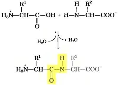
Two identical or different Amino acids can be covalently joined through a substituted amide linkage, termed a peptide bond, to yield a dipeptide. This bond forms by the removal of the elements of Water from the α-carboxyl group of one Amino Acid and the α-amino group of another (Fig. 3-13). Peptide bond formation is a Condensation reaction, widely prevalent in living Cells. Under standard biochemical conditions, the equilibrium of the reaction shown in Fig. 3-13 favors the amino acids rather than the dipeptide. To make the reaction thermodynamically favorable, the carboxyl group must be modified or activated to facilitate the departure of the hydroxyl group. The chemical approach to this problem is discussed later in this chapter, whereas biological strategies are the primary subject of Chapter 27.
Class="center">Fig. 3-13. Formation of a peptide bond via a condensation reaction. The α-amino group of one amino acid (with its R2 group) acts as a nucleophile, displacing the hydroxyl group of another amino acid (with its R1 group) to form a peptide bond (highlighted in yellow). Although Amino acids are good nucleophiles, the hydroxyl group is a poor leaving group and is not readily displaced. Consequently, under physiological pH conditions, the reaction shown here virtually does not proceed.
Three amino acids can be joined by two peptide bonds to form a tripeptide; similarly, tetrapeptides, pentapeptides, and so forth can be formed. A few amino acid residues joined in this fashion constitute an oligopeptide. When many amino acids are linked, the product is a polypeptide. Proteins may contain thousands of amino acid residues. Although the terms "protein" and "polypeptide" are sometimes used interchangeably, molecules with molecular weights below approximately 10,000 are generally referred to as polypeptides, while larger molecules are called proteins.
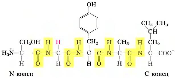
Figure 3-14 illustrates The Structure of a pentapeptide. As noted earlier, amino acid units in peptides are commonly called residues because they represent what remains of an amino acid molecule after the loss of a hydrogen atom from the amino group and a hydroxyl group from the carboxyl group. The amino acid residue at the end of the peptide with a free α-amino group is called the amino-terminal (or N-terminal) residue; the residue at the opposite end, which carries a free carboxyl group, is the carboxyl-terminal (or C-terminal) residue.
Fig. 3-14. The pentapeptide seryl-glycyl-tyrosyl-alanyl-leucine (Ser-Gly-Tyr-Ala-Leu). Peptide names are derived from the Amino Acid Sequence, starting with the N-terminal residue, which is conventionally written on the left. Peptide bonds are highlighted in yellow, and R groups are shown in red.
Key Convention.
By convention, the amino acid sequence of a peptide, polypeptide, or protein is written with the N-terminus on the left and the C-terminus on the right. The sequence is read from left to right, starting from the N-terminus. ■
Although peptide bond Hydrolysis is an exergonic process, it occurs very slowly due to a high activation energy barrier (see p. 48). As a result, peptide bonds in proteins are quite stable, having an average half-life (t1/2) of about seven years under standard intracellular conditions.
Peptides Vary in Their Ionization Behavior
A peptide molecule has only one free α-amino group and one free α-carboxyl group, located at opposite ends of the peptide chain (Fig. 3-15). These groups ionize in peptides much as they do in free amino acids, although with different ionization constants because the oppositely charged groups are no longer attached to the α-carbon atom. The α-amino and α-carboxyl groups of all other amino acid units, linked by covalent peptide bonds, do not contribute to the acid-base Properties of the peptide because they are no longer ionizable. However, the R groups of Certain amino acids can be ionized (Table 3-1), contributing to the acid-base CHARACTERISTICS OF THE entire molecule (Fig. 3-15). Thus, the acid-base properties of a peptide can be predicted from the presence of a single free α-amino group, a single free α-carboxyl group, and The Nature and number of its ionizable R groups.
Fig. 3-15. Alanyl-glutamyl-glycyl-Lysine. This tetrapeptide features one free α-amino group, one free α-carboxyl group, and two ionizable R groups. Groups ionized at pH 7.0 are highlighted in red.

Like amino acids, peptides have characteristic titration curves and isoelectric points—the pH values at which they do not migrate in an electric field. These properties are exploited in various Methods for the Separation of peptides and proteins, as discussed later in this chapter. It is worth emphasizing once again that the pKa values of ionizable groups in a free amino acid can differ from those of the same amino acid incorporated into a peptide. Within a peptide, pKa values are influenced by the loss of charge at the α-amino and α-carboxyl groups, interactions with neighboring R groups, and other environmental shifts. The pKa values of R groups listed in Table 3-1 should be used only as a guide to the pH range over which these groups ionize, rather than as precise pKa values for peptides.
Biologically Active Peptides and Polypeptides Vary Widely in Size and Composition
There is no fixed correlation between the size of peptides and proteins and their biological activity. Naturally occurring peptides range in size from two to several thousand amino acid residues, and even small protein molecules can possess biological activity. A prime example is the artificially synthesized dipeptide L-aspartyl-L-phenylalanine methyl ester, widely used as a non-nutritive sweetener under the trade name aspartame or NutraSweet.

Many small peptides exert potent biological effects at very low concentrations. For instance, Vertebrate Hormones (Chapter 23) are typically small peptides. These include oxytocin (9 amino acid residues), secreted by the posterior Pituitary Gland, which stimulates uterine contractions; and thyrotropin-releasing hormone (3 residues), produced in the Hypothalamus, which stimulates the release of another hormone, thyrotropin, from the anterior pituitary gland. Other Examples of small peptides include highly toxic fungal poisons (such as α-amanitin) and numerous Antibiotics.
What is the length of peptide chains in proteins? As seen in Table 3-2, this parameter varies over a wide range. Human cytochrome c consists of 104 residues joined in a single chain; bovine chymotrypsinogen consists of 245 residues. Titin, a giant Muscle protein found in vertebrates, contains about 27,000 amino acid residues and has a molecular mass of roughly 3,000,000. The vast majority of natural proteins are much smaller than titin, typically containing fewer than 2,000 amino acid residues.
Table 3-2. Molecular Characteristics of Selected proteins
Molecular mass |
Number of residues |
Number of chains |
|
Cytochrome c (human) |
12 400 |
104 |
1 |
Ribonuclease A (bovine Pancreas) |
13 700 |
124 |
1 |
Lysozyme (hen egg white) |
11 300 |
129 |
1 |
10 700 |
153 |
1 |
|
Chymotrypsin (bovine pancreas) |
25 200 |
241 |
3 |
Chymotrypsinogen (bovine) |
25 700 |
245 |
1 |
Hemoglobin (human) |
64 500 |
574 |
4 |
Serum albumin (human) |
66 000 |
609 |
1 |
Hexokinase (Yeast) |
107 900 |
972 |
2 |
RNA polymerase (E. coli) |
450 000 |
4 158 |
5 |
Apolipoprotein B (human) |
513 000 |
4 536 |
1 |
Glutamine Synthetase (E. coli) |
619 000 |
5 628 |
12 |
Titin (human) |
2 993 000 |
26 926 |
1 |
Some proteins consist of a single polypeptide chain, whereas others, known as multisubunit proteins, contain two or more chains held together by noncovalent interactions (Table 3-2). The chains in multisubunit proteins may be identical or different. If at least two chains in a protein are identical, the protein is called oligomeric, and the identical subunits (each consisting of one or more chains) are referred to as protomers. For example, hemoglobin is composed of four polypeptide subunits: two identical α chains and two identical β chains, held together in a single complex by noncovalent interactions. Both α subunits pair identically with the β subunits, so hemoglobin can be viewed either as a tetramer consisting of four polypeptide chains or as a dimer of αβ protomers.
In certain proteins, polypeptide chains are linked by covalent bonds. For example, the two polypeptide chains in an Insulin molecule are interconnected by disulfide bridges. In such cases, the individual polypeptides are considered simply as polypeptide chains rather than as subunits.
The Amino Acid Composition of proteins is highly diverse. The twenty standard amino acids are almost never found in proteins in equal proportions. Some amino acids may occur only once or not at all in a given type of protein, whereas others may be present in large amounts. Table 3-3 shows the amino acid composition of bovine cytochrome c and bovine chymotrypsinogen, the inactive precursor of the digestive enzyme chymotrypsin. These two proteins, which have completely different Functions, differ markedly in their amino acid composition.
Table 3-3. Amino acid composition of two proteins
Number of residues |
per protein molecule* |
|
Amino acid |
Bovine cytochrome c |
Bovine chymotrypsinogen |
Ala |
6 |
22 |
Arg |
2 |
4 |
Asn |
5 |
14 |
Asp |
3 |
9 |
Cys |
2 |
10 |
Gin |
3 |
10 |
Glu |
9 |
5 |
Gly |
14 |
23 |
His |
3 |
2 |
lie |
6 |
10 |
Leu |
6 |
19 |
Lys |
18 |
14 |
Met |
2 |
2 |
Phe |
4 |
6 |
Pro |
4 |
9 |
Ser |
1 |
28 |
Thr |
8 |
23 |
Trp |
1 |
8 |
Туг |
4 |
4 |
Val |
3 |
23 |
Total |
104 |
245 |
* In Analytical Methods such as acid hydrolysis, Asp and Asn are virtually indistinguishable and are therefore grouped together and designated as Asx (or B). Similarly, Glu and Gln cannot be distinguished and are designated as Glx (or Z). In addition, Trp is completely destroyed during acid hydrolysis. Determining the exact amino acid composition requires supplementary analytical methods.
To estimate the number of amino acid residues in a protein that contains no non-protein components, its molecular mass should be divided by 110. Let us explain where this number comes from. The average molecular mass of the twenty standard amino acids is 138, but lighter amino acids predominate in proteins. When the contributions of various amino acids to protein chains are taken into account (Table 3-1), the average molecular mass of an amino acid residue in a protein turns out to be approximately 128. Because The formation of each peptide bond releases one molecule of water (Mr = 18), the average molecular mass of an amino acid residue is 128 - 18 = 110.
Some proteins contain other groups In addition to amino acid residues
Many proteins, such as the Enzymes ribonuclease A and chymotrypsinogen, contain only amino acid residues and no other chemical components; these are called simple proteins. However, some proteins contain other chemical groups besides amino acids; these are known as Conjugated Proteins. The non-protein part of a conjugated protein is called a prosthetic group. Conjugated proteins are classified According to the Chemical Nature of their prosthetic groups (Table 3-4). For example, Lipoproteins contain Lipids, Glycoproteins contain sugars, and Metalloproteins contain specific metals. Some proteins contain multiple prosthetic groups. Typically, the prosthetic group plays a vital role in the biological function of the protein.
Table 3-4. Conjugated proteins
Class |
Prosthetic group |
Example |
Lipoproteins |
Lipids |
Blood β1-lipoprotein |
Glycoproteins |
Immunoglobulin G |
|
Phosphate groups |
Milk casein |
|
Heme (iron-protoporphyrin complex) |
Hemoglobin |
|
Flavin NUCLEOTIDES |
Succinate dehydrogenase |
|
Metalloproteins |
Iron |
Ferritin |
Zinc |
||
Calcium |
Calmodulin |
|
Molybdenum |
||
Copper |
Plastocyanin |
Summary of Section 3.2 Peptides and Proteins
■ Amino acids can be linked by covalent peptide bonds to form peptides and proteins. Cells typically contain thousands of different proteins, each possessing characteristic biological activity.
■ Some Proteins are very long polypeptide chains containing anywhere from 100 to several thousand amino acid residues, whereas Natural peptides consisting of just a few amino acids also exist. Certain proteins are made up of multiple noncovalently linked polypeptide chains called subunits. Hydrolysis of simple proteins yields a mixture of amino acids. Conjugated proteins also contain components of other chemical structures, such as Metal Ions or organic prosthetic groups.
■ The amino acid sequence is an essential characteristic of a protein and is referred to as its Primary Structure. This is the first of the four Levels of Protein molecular Organization.
Last update: 06/08/2026
Editorial and Educational Adaptation: This material has been compiled based on the primary/original source text. The project team performed an editorial review, corrected technical inaccuracies, structured sections, and adapted the content for an educational format.
What was processed:
- elimination of formatting defects (OCR errors, structural breaks, corrupted characters);
- editorial organization of content;
- standardization of terminology in accordance with academic sources;
- verification of factual statements against the original source text.
All mentions of the author, publication year, and origin of the primary text have been preserved in accordance with the source.