Biochemistry and Molecular Biology - Belyasova N.A. 2002
Molecular Foundations and Mechanisms of Heredity
Organization of the Cellular Genetic Apparatus
Nucleic Acid Replication
The primary function of DNA is its ability to self-duplicate (Replication). Replication is an extremely precise mechanism with an almost zero error rate. DNA itself (or RNA in certain Viruses) encodes information regarding the METABOLISM/9.html">Structure of Enzymes responsible for nucleic acid duplication, the synthesis of new NUCLEOTIDES—the Building Blocks of replication—correction of replication errors, and the Repair of DNA Damage caused by various factors. Ultimately, the very structure of DNA, specifically its double-stranded composition, facilitates the copying process because each strand can act as a template for synthesizing new DNA molecules. This hypothesis was proposed by James Watson and Francis Crick as early as 1953 and was subsequently confirmed experimentally. This mode of DNA copying, where each strand Functions as a template and the newly synthesized molecules are hybrid (consisting of one old and one new strand), is termed semi-conservative.
Besides the semi-conservative model, two other Replication Models were proposed: conservative and dispersive. The characteristics of these DNA replication models are as follows. According to the dispersive model, the parental DNA helix is cleaved at each half-turn through multiple fragmentation during duplication, and new strands are synthesized on these fragments (Fig. 1.9). In the conservative model, the DNA helix does not unwind at all; instead, it serves as a template for two entirely new strands, resulting in a parental helix composed entirely of old material and a daughter helix composed of new material. The validity of the semi-conservative DNA Replication Mechanism was proven by Meselson and Stahl in 1958 through cesium chloride density gradient ultracentrifugation experiments using isotopically labeled bacterial DNA.
The Essence of these experiments was as follows: E. coli DNA was labeled with the radioactive isotope 15N and then allowed to undergo one round of replication by growing the Cells for ~50 min in a nutrient medium containing the normal nitrogen isotope 14N. DNA extracted from the cells was subjected to cesium chloride density gradient ultracentrifugation. Under these conditions, CsCl molecules form a density gradient in the centrifuge tube, causing molecules of other substances to distribute according to their density. E. coli DNA grown on a 15N-containing medium has a density of 1.724 g/cm3, whereas DNA from cells grown on normal medium with the 14N isotope has a density of 1.710 g/cm3. Thus, a mixture of these Two Types of DNA is easily separated by density gradient centrifugation. DNA localization in the CsCl gradient tube can be determined by ultraviolet absorbance (DNA absorbs radiation at a wavelength of 260 nm). Consequently, DNA appears in the tube as distinct "bands"—the "light" band near the top and the "heavy" band near the bottom. In this experiment, the cesium chloride gradient tube formed only a single band of intermediate "weight," corresponding in position to hybrid DNA containing both nitrogen isotopes, 15N and 14N. This finding ruled out one specific DNA replication model: the conservative model. To distinguish between the two remaining replication models, Meselson and Stahl allowed the Bacteria, whose DNA contained both isotopes, to undergo another Cell Division in 14N medium. Their DNA was then subjected to ultracentrifugation once again. This time, two DNA bands formed in the tube—a "light" band and an intermediate "weight" band—thus confirming the validity of the semi-conservative mechanism of DNA replication.
Class="center">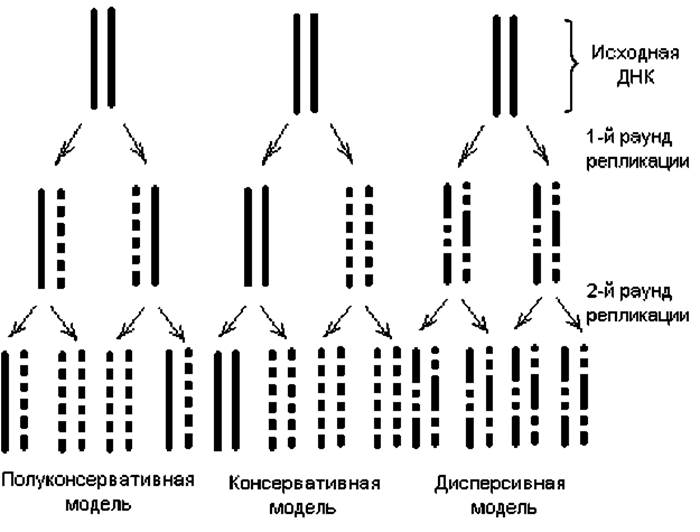
Fig. 1.9. Proposed models of duplex DNA replication. Solid lines represent the original ("heavy," 15N-containing) DNA strands, and dashed lines indicate the new ("light," 14N-containing) DNA strands generated As a result of replication.
Thus, all Nucleic Acid Replication pathways studied to date rely on a semi-conservative mechanism, whereby after each round of replication, one strand in each of the two daughter molecules is parental (i.e., conserved) and the other is newly synthesized. The replication of single- and double-stranded Nucleic Acids representing the genomes of various organisms proceeds according to specific regularities across different mechanisms, as discussed below. Common features of all these processes include: 1) the involvement of a complex enzymatic machinery driving replication; 2) three main stages: initiation, elongation, and termination; 3) adherence to THE PRINCIPLE OF complementarity during the construction of new strands, using the parental strand as a template; 4) high fidelity; and 5) The ability to correct replication errors via proofreading.
Replication of double-stranded DNA. Double-stranded DNA constitutes the genomes of all cellular organisms, both PROKARYOTES AND EUKARYOTES. The Mechanism of DNA replication is best understood in Prokaryotic Cells, particularly the bacterium E. coli. Experiments with prokaryotes have shown that under conditions limiting Protein Synthesis, DNA replication halts, implying that the process requires protein factors. It has now been established that the products of more than 10 genes participate in DNA replication. These primarily include DNA polymerases, as well as topoisomerases, helicases, and ligases. Accumulating evidence points to the involvement of a highly organized multi-enzyme complex—the replisome—which includes the primosome-primase complex, helicases, the Pol III holoenzyme, and gyrases.
DNA polymerases are the Key Enzymes of the replicative process, directly responsible for elongating polynucleotide chains based on the principle of complementarity. The DNA polymerases of E. coli have been studied most thoroughly. Three distinct types of DNA polymerases (Pol I, Pol II, and Pol III) have been identified in these bacteria, differing primarily in catalytic speed and nuclease activity. DNA polymerase I (Pol I) is a single polypeptide consisting of approximately 1,000 amino acid residues. An E. coli cell contains about 400 molecules of this enzyme. Pol I exhibits the following activities: polymerase activity—adding deoxynucleotides complementary to the template strand to the free 3'-OH group of a primer in the 5' to 3' (5'→3') direction of the nascent DNA molecule; and exonuclease activity—hydrolyzing phosphodiester bonds (cleaving nucleotides) within a single DNA strand or at the unpaired end of a duplex DNA, starting from either the 3' end (3'→5') or the 5' end (5'→3'). Exonuclease activities play a crucial role in the Replication and Repair of E. coli chromosomal DNA. The 3'→5' exonuclease activity monitors The addition of each nucleotide and removes erroneous nucleotides from the growing chain terminus (proofreading), whereas the 5'→3' exonuclease activity is utilized to remove pyrimidine dimers and the RNA primers of Okazaki fragments.
DNA polymerase II (Pol II) is present in significantly fewer copies per E. coli cell and carries out polymerization much slower than Pol I (accounting for only 5% of DNA polymerase I activity). Unlike Pol I, this enzyme lacks 5'→3' exonuclease activity. The exact role of this polymerase in replication remains fully unresolved. It is generally believed that this enzyme is not essential for DNA replication but can substitute for certain Pol I functions when Pol I is damaged.
DNA polymerase III (Pol III) is the primary enzyme responsible for replicating the chromosomal DNA of E. coli. Each cell contains only 10–20 molecules of this enzyme, yet it operates approximately 60 times faster than DNA polymerase I. Furthermore, Pol III displays a higher affinity for the template and ensures superior copying efficiency. Like Pol II, this enzyme lacks 5'→3' exonuclease activity. Consequently, replicating the lagging strand requires the participation of Pol I to remove the RNA primers at the 5' ends of Okazaki fragments.
A greater variety of DNA polymerases has been identified in Eukaryotic cells, though their functions are less well understood.
The function of topoisomerases is to resolve mechanical and topological constraints encountered during the unwinding of The Double Helix at the Replication fork. These enzymes alter the degree of supercoiling and create a "swivel" that allows the replication fork to move continuously. Two MAIN TYPES OF topoisomerases have been identified across various organisms: Type I topoisomerases nick one of the two strands, allowing the terminal region of the double helix to rotate around the intact strand before resealing the cut strand. Type II topoisomerases introduce transient double-strand breaks in both complementary strands, alter the supercoiling state, and subsequently rejoin the severed ends.
Helicases facilitate the formation and progression of the replication fork—the region of the molecule where the strands are unzipped—along the DNA helix. These enzymes utilize The energy released from ATP Hydrolysis to unwind the strands. To ensure a high unwinding rate, multiple helicases act in concert with single-strand DNA-binding Proteins, which attach to the single-stranded regions and thereby stabilize the unzipped duplex.
Finally, DNA ligases catalyze the rejoining of DNA strand fragments by forming covalent bonds (phosphodiester bridges) between the 5'-P and 3'-OH groups of adjacent deoxyribonucleotides. These enzymes also harness the energy of high-energy bonds derived from ATP or GTP hydrolysis.
The Mechanism of double-stranded DNA replication is best understood in the bacterium E. coli and will be examined using this model.
Initiation of DNA replication. The replication of E. coli DNA begins at a strictly defined site called the origin (ori), located at 85 min on the Genetic Map of the bacterial chromosome. At the replication ori, enzymes (topoisomerases, helicases) act on the DNA to form a replication fork, where strand copying actually takes place. Replication requires: a DNA template in the form of a single-stranded DNA region, a mixture of deoxynucleoside triphosphates, a replisome (the enzymatic assembly involved in replication), and a nucleic acid 3'-OH group acting as a primer to which the DNA polymerase attaches the next nucleotide. Notably, none of the DNA polymerases can initiate nucleotide polymerization de novo. This function is performed by RNA polymerases, which recognize the replication ori within the replication fork and synthesize short (10–60 ribonucleotide) sequences known as RNA primers. Primer synthesis proceeds in the 5' to 3' direction, yielding a free 3'-OH terminus that DNA polymerase can use to extend the chain during the elongation stage of replication (Fig. 1.10).
Elongation of DNA replication. The synthesis of new DNA strands proceeds in accordance with the principle of complementarity: each nucleotide incorporated into the growing chain must be complementary to the corresponding (opposite) nucleotide in the template strand.
Because all DNA polymerases polymerize nucleotides exclusively in the 5'→3' direction, while the replication fork moves along the DNA in both directions, only one strand—designated the leading strand—can be synthesized continuously in each direction. The opposite (second) strand is synthesized in short fragments (Okazaki fragments) and is called the lagging strand (Fig. 1.10). Okazaki fragments consist of approximately 1,000 nucleotides in prokaryotes and 100–200 nucleotides in eukaryotes.
Aside from chain polymerization, which is carried out primarily by DNA polymerase III, several other key events occur during DNA replication:
— excision of RNA primers from the leading strand and from each Okazaki fragment, a function performed by Pol I via its 5'→3' exonuclease activity;
— Filling of the "gaps" left after RNA primer removal, a task also executed by DNA polymerase I utilizing the free 3'-OH group of the adjacent Okazaki fragment;
— joining of DNA fragments in the lagging strand by DNA ligase: once the growing 3'-hydroxyl end of each Okazaki fragment reaches the 5'-deoxynucleotide end of the neighboring fragment, DNA ligase steps in to establish a continuous lagging strand;
— correction of replication errors via proofreading, a mechanism inherent to both Pol I and Pol III and based on their 3'→5' exonuclease activity. DNA polymerase checks the complementarity of each incoming nucleotide by measuring the geometry of the putative new nucleotide pair within its Active Site, engaging its polymerase activity only when proper complementarity is verified. Furthermore, each newly incorporated nucleotide is subsequently checked for correct pairing within the enzyme's active site. If the dimensions of the newly formed nucleotide pair deviate from the norm (indicating mismatched bases), the enzyme uses its 3'→5' exonuclease activity to excise the non-complementary nucleotide and replace it. DNA Repair serves as an additional error-minimizing mechanism. Consequently, the frequency of nucleotide misincorporation into the newly synthesized DNA strand is extremely low (10-8–10-10).

Fig. 1.10. Replication in E. coli. Arrows indicate the direction of new DNA strand synthesis. Unshaded thick lines represent RNA primers initiating synthesis; solid bold lines denote elongating DNA strand fragments. Vertical lines indicate Hydrogen Bonds between complementary nucleotides.
Termination of replication. During Bidirectional Replication of a circular genome (such as in Escherichia coli), replication forks meet at a distance of 180° from THE ORIGIN OF replication, where replication is completed. The circular DNA molecules are joined together by ligase at the meeting point, resulting in catenated pairs that are subsequently separated into individual genomes by topoisomerase type II.
The rate of DNA replication in E. coli is approximately 1,500 nucleotide pairs per second. Thus, the complete genome of the bacterium (4∙106 bp) is replicated in about 40 min. However, E. coli cells divide faster—every 20 min—which means that at the same copying speed, the frequency of initiation events at the same origin of replication increases. That is, even before the first round of genome replication is completed at the ori site, a second round of replication is initiated. The rate of replication fork movement in eukaryotic cells is significantly lower (10–100 bp per second), but the completion of replication within a reasonable time is ensured by simultaneous initiation at multiple sites. As a result, the Drosophila chromosome, for example, containing 6.5∙107 bp, is replicated in just a few minutes.
In general, the patterns of replication identified in prokaryotes are also characteristic of most Eukaryotic Genomes. The differences lie primarily in the presence of multiple replication initiation sites on each chromosome in eukaryotes, different mechanisms for correcting replication errors compared to prokaryotes, and the enzymatic machinery involved in the replication process. A schematic representation of the replication processes of circular genomes (forming Prokaryotic Genomes and Plasmids) and linear (eukaryotic) genomes is shown in Fig. 1.11.
In linear DNA, strand unwinding occurs by the rotation of one strand around the other. In circular DNA, unwinding and replication lead to The formation of a structure resembling a ring with an internal loop. It is called a theta loop because its shape resembles the Greek letter Θ. Such loops can be observed on autoradiographs of replicating bacterial DNA, as was first demonstrated by Cairns for E. coli DNA.
The mechanism of bidirectional DNA replication described above is the most common, but not the only one. The DNA of phages P22, 186, P2, as well as phages T4 and $\lambda$ at late Stages of the lytic cycle, replicates via a unidirectional mechanism (the rolling-circle type). In this case, double-stranded Circular DNA is nicked by a specific enzyme at a unique site in one strand (the rolling-circle origin). The resulting 5' end of the nicked strand binds to the enzyme that made the nick. DNA Synthesis begins with the Displacement of the enzyme-bound 5' end into the solution, allowing DNA polymerase to add nucleotides to the 3'-OH end. Semi-conservative replication takes place, during which the 5' end of the broken strand is displaced as a free tail of increasing length, with the intact closed strand serving as the template. This replicating structure (Fig. 1.12) is called a rolling circle because the unwinding of the free single strand is accompanied by the Rotation of the double-stranded template around its axis.
If this mechanism is used for the replication of double-stranded DNA, the 5'-terminal tails serve as templates for the synthesis of small DNA fragments that are immediately joined together by DNA ligase. As a result, the growing tails soon acquire a double-stranded structure after their formation. Tail elongation sometimes results in their length significantly exceeding the total length of the initial circular molecule. This mode of replication is used, for example, by phage $\lambda$. During DNA packaging into capsids, nicks are formed at specific sites called cos-sites, which are separated by a distance equal to the viral genome length; as a result, the long duplexes of repeatedly replicated phage DNA are cleaved into fragments corresponding in size to the mature DNA found in bacteriophage $\lambda$ virions.

Fig. 1.11. Types of bidirectional replication of double-stranded DNA. Solid lines indicate the parental DNA strands; dashed lines indicate the newly synthesized strands.
Rolling-circle replication is also characteristic of the formation of copies of the bacterial chromosome of E. coli Hfr and the F+ factor transferred during conjugation into a recipient cell.
Replication of single-stranded DNA. In phages M13 or $\phi$X174, whose mature genomes are single-stranded circular DNAs, replication occurs via the rolling-circle mechanism (Fig. 1.12). This takes place at late stages of the infection process, after the infecting DNA is converted into a double-stranded circular form. In this case, replication of the 5'-terminal regions does not occur, unlike the replication of phage $\lambda$ genomes (Fig. 1.12, pos. 5); therefore, the replication products are long single DNA strands that continuously detach from the "rolling spool." These strands are cleaved at each replication origin and circularized to form mature circular forms that are packaged into capsids.

Fig. 1.12. Rolling-circle replication of double-stranded DNA. Solid lines indicate the parental DNA strands; dashed lines indicate the strands newly synthesized during replication. Position 1 — initial circular DNA; 2 — nick formation in one of the strands, exposing the 3' and 5' ends; 3, 4 — replication of the circular strand at the replication fork; 5 — initiation of 5'-tail replication; 6, 7 — replication products.
RNA replication. The production of Introduction/7.html">RNA-containing Viruses occurs through the replication of their RNA, whereas all cellular RNAs are formed as a result of DNA Transcription. With the exception of Retroviruses, RNA replication largely mimics the DNA replication process. As in DNA replication, the order of nucleotides is determined by complementary template copying, in this case necessarily an RNA strand. The enzymes carrying out this process are called RNA-dependent replicases. The RNAs of bacterial viruses R17 and MS2, as well as polioviruses and Sindbis virus infecting animals, are always designated as plus (+)-strand, since The sequence of their RNA genomes is identical to that of mRNA. Thus, The Genome of the infecting virus can serve as mRNA and contains information for the synthesis of some, if not all, viral proteins. A specific replicase encoded by the viral genome and produced shortly after infection binds to one or more host cell proteins and initiates the copying of the (+)-strand from its 3' end, producing a complete (-)-strand associated with the template (+)-strand. Then, the same replicase synthesizes numerous copies of the RNA (+)-strand, using the newly synthesized (-)-strand as a template. The genomes of some viruses (vesicular stomatitis virus, Influenza virus) are represented by one or more (-)-strands. In this case, they serve as templates for the synthesis of (+)-strands, which act as mRNA and are used in the synthesis of progeny (-)-strands.
A distinctive feature of retroviral genome replication is that after their RNA penetrates the host cell, the viral genome undergoes reverse transcription. Initially, an RNA-DNA duplex is formed, followed by double-stranded DNA. The enzyme that catalyzes the complementary copying of RNA to form DNA is called Reverse Transcriptase (revertase). It is contained in retroviral particles (virions) and is activated upon entering The Cell. Accumulating evidence suggests that reverse transcription occurs in A wide variety of eukaryotic cells, and reverse transcriptase plays an important role in genome rearrangement processes. Replication of the double-stranded form of retroviral DNA does not begin until it integrates into the cellular DNA. The mechanism of recombinational integration is not yet fully understood. Following integration, retroviral DNA replicates as part of the cellular DNA. The RNA of progeny virions is produced through the transcription of integrated copies of the viral DNA.
Last update: 06/08/2026
Editorial and Educational Adaptation: This material has been compiled based on the primary/original source text. The project team performed an editorial review, corrected technical inaccuracies, structured sections, and adapted the content for an educational format.
What was processed:
- elimination of formatting defects (OCR errors, structural breaks, corrupted characters);
- editorial organization of content;
- standardization of terminology in accordance with academic sources;
- verification of factual statements against the original source text.
All mentions of the author, publication year, and origin of the primary text have been preserved in accordance with the source.