Principles of Biochemistry Volume 3 - A. Lehninger 1985
Molecular mechanisms of genetic information transfer
DNA: structure of chromosomes and genes
Circular DNA is supercoiled
Careful and gentle isolation of circular viral DNAs reveals that they are supercoiled, or superhelical. It appears as though The Double Helix was partially unwound before the ends of its strands joined to form a ring. This compensatory twisting exerts a torsional stress on the circular DNA molecule, causing it to twist back upon itself (Fig. 27-18). If such a supercoiled DNA, which stores extra Free energy, is treated with an endonuclease that cleaves a single strand, the twist-induced strain is relieved, and the DNA relaxes into its standard low-energy state (Fig. 27-18). Supercoiled viral DNA is significantly more compact than its relaxed circular counterpart.
Analysis of DNA isolated from E. coli under very careful conditions reveals that it is organized into numerous loops held together by Proteins. Each of these loops is, in turn, supercoiled (Fig. 27-19). This loop Organization and supercoiling help package exceptionally large circular DNA molecules within small, non-membrane-bounded cellular volumes. Linear DNAs cannot form supercoils unless both ends of the molecule are anchored. Supercoiling plays a critical role in many DNA-dependent processes. In fact, certain Proteins and Enzymes fail to bind to DNA unless it is in a supercoiled conformation. Enzymes known as topoisomerases can regulate the degree of supercoiling by increasing or decreasing the number of superhelical turns.
Class="center">
Fig. 27-18. Circular supercoiled DNA, twisted As a result of underwinding—that is, rotations in the direction opposite to the coiling of the double helix itself. A single-strand break in this type of DNA relieves the supercoiling, converting the circular DNA into a relaxed form.
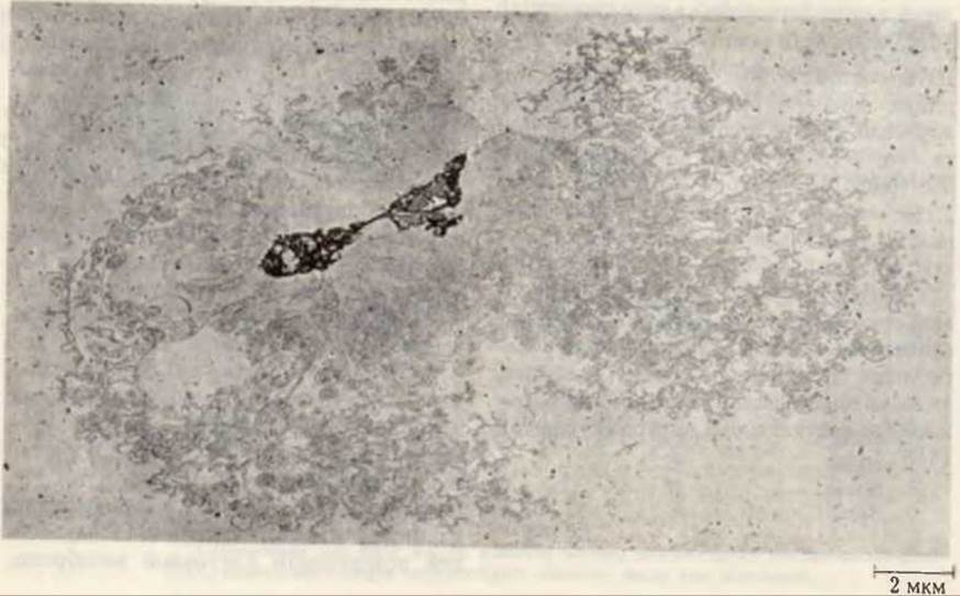
Fig. 27-19. Electron micrograph of an intact supercoiled chromosome isolated from a single E. coli Cell. The integrity of the chromosome was verified using a highly magnified photograph. Fragments of The Cell membrane are visible slightly to the left of the center.
Last update: 06/08/2026
Editorial and Educational Adaptation: This material has been compiled based on the primary/original source text. The project team performed an editorial review, corrected technical inaccuracies, structured sections, and adapted the content for an educational format.
What was processed:
- elimination of formatting defects (OCR errors, structural breaks, corrupted characters);
- editorial organization of content;
- standardization of terminology in accordance with academic sources;
- verification of factual statements against the original source text.
All mentions of the author, publication year, and origin of the primary text have been preserved in accordance with the source.