INVERTEBRATE ZOOLOGY IN THREE VOLUMES - BOOK 3 - H.I. Shcherbak - 1997
DEUTEROSTOMIA
PHYLUM ECHINODERMATA
SUBPHYLUM ASTEROZOA
This subphylum comprises two classes: Sea Stars (Asteroidea) and Brittlestars, or Serpent Stars (Ophiuroidea).
CLASS ASTEROIDEA (SEA STARS)
Representatives of this Class are exclusively marine inhabitants of seas and oceans, and are never found in waters with a salinity below 30 ‰.
Only a single species, Marthasterias glacialis, occurs in the Black Sea near the Bosphorus (see Fig. 229, b). Over 300 fossil and about 1,500 extant species are known.
Body sizes range from one to 80 centimeters or more. Sea stars are often brightly colored in A wide variety of hues, sometimes mottled.
The body of sea stars is more or less flattened along the oral-aboral axis. With few exceptions, one can distinguish a central disc that gradually transitions into radial rays, or arms. The arms may be very short, as, for example, in Patiria pectinifera, which is frequently found in the Sea of Japan. They may project only slightly along the edges of the disc, giving the body a pentagonal shape, or they may be longer, with the body radius (the distance from the center to the tip of the ray) significantly exceeding the interradius length (the distance from the center of the disc to its margin between the rays). Sea stars always face the substrate with their oral side. A five-rayed (pentamerous) Structure is typical for sea stars, although quite a few species possess six or even more (up to 50) rays.
The body surface is covered by a thin cuticle, beneath which lies a single-layered ciliated epithelium containing glandular Cells In addition to epithelial cells (Fig. 211). The bulk of the body wall (integument) consists of Connective Tissue, within the thickness of which lie calcareous skeletal plates and Muscle fibers. From the inside, the Skin is lined with peritoneal epithelium that bounds the coelom.

Fig. 211. Cross-section through the outer body wall of Asterias rubens:
1 — cuticle; 2 — glandular cells; 3 — epithelial cells; 4 — Nerve Tissue layer
Unlike sea urchins, the body of sea stars is not enclosed in a solid test. Their Skeleton consists of numerous calcareous plates interconnected by connective tissue and Muscles. The Skeleton of the oral side is developed more strongly than the opposite, aboral side. Along the oral and partially lateral sides of each ray, there are eight rows of skeletal plates (Fig. 212). Along the margin of each ray, there are one or two rows of marginal plates, medial to these are a row of adambulacral plates, and centrally lie two rows of ambulacral plates positioned at an angle to each other, forming a gabled roof over the ambulacral groove. Each ambulacral plate bears a small depression through which the canal of the ambulacral podium passes.

Fig. 212. Skeleton of sea stars:
a — diagram of skeletal plate arrangement in a transverse section of a ray; b — external view of the skeletal plates of the ambulacral groove; c — skeletal plates around the oral region; d — aboral skeleton (top view);
1 — paxillae; 2 — aboral skeletal plates; 3 — upper marginal plate; 4 — lower marginal plate; 5, 6 — adambulacral and ambulacral plates; 7 — pores for ambulacral podia; 8 — first ambulacral (oral) plate; 9 — first ambulacral plate with a tooth-like process; 10 — spines
The structure of the skeletal plates around the Mouth region is somewhat different. Here, the first ambulacral plates are significantly widened and transformed into oral plates, while the first adambulacral plates, also widened, bear pointed so-called tooth-like processes directed toward the oral opening. The degree of Development of the aboral skeleton varies among different species. The skeleton consists of narrow calcareous plates that may form an irregular meshwork or cover the entire surface.
Among the aboral skeletal plates, a large madreporic plate stands out.
The skeletal plates of both the oral and aboral sides bear numerous skeletal appendages on their surface (except for the plates of the ambulacral series), mostly in the form of needles and spines. Spines may be simple or in the form of so-called paxillae — calcareous pillars with small spinelets and needles at the apex. As in sea urchins, some of the spines are modified into pedicellariae of various shapes (Fig. 213), which perform the same Functions as in urchins — sanitary and protective.

Fig. 213. Paxillae (a) and pedicellariae (b, c) of sea stars: 1 — open pedicellaria; 2 — closed pedicellaria
The ambulacral system of sea stars is similar to that of sea urchins, though with minor differences. The madreporic plate leads into an ampulla and further into the stone canal, the walls of which are lined with calcareous rings. The canal may be tortuous or even spirally wrap around the axial organ. The oral end of the canal connects to the circumoral ring canal surrounding the mouth opening. Positioned dorsally on the ambulacral ring in pairs are Tiedemann's bodies, typically numbering nine; in one of the interradiuses, the stone canal opens in place of a Tiedemann's body. It is believed that Tiedemann's bodies function as Lymph Nodes AND produce amebocytes, much like the lymphoid part of the axial organ. Polian vesicles may open into the ring canal (sometimes they are absent). Their number varies, and there may occasionally be several even within a single interradius.
Radial ambulacral canals originate from the ring canal and extend to the tip of each arm along the bottom of the ambulacral grooves, protected by the ambulacral ossicles of the oral skeleton. Numerous lateral branches depart from both sides of the radial canals, passing through the pores between the ambulacral ossicles to connect with the tube feet. Inside the body, an ampulla—a thin-walled muscular vesicle—extends from each lateral canal. The tip of each tube FOOT features a well-developed sucker (Fig. 214), except for those located at the very tip of the arm, where the tube feet lack suckers and instead perform a tactile function.
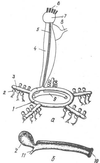
Fig. 214. Ambulacral system of a starfish:
a — structural diagram; b — tube foot; 1 — ring canal; 2 — tube foot ampulla; 3 — radial canal; 4 — axial sinus; 5 — stone canal; 6 — madreporite; 7 — ampulla; 8 — genital stolon; 9 — oral perhemal ring; 10 — sucker; 11 — lateral branch of the radial canal
The primary function of the tube feet is locomotion. The feet can contract or extend in any direction. Contraction is driven by the longitudinal muscles in their walls, while extension results from the hydrostatic pressure of the ambulacral fluid pumped into the FOOT FROM THE ampulla when it contracts. By extending a group of tube feet, the starfish attaches its suckers to the substrate, an adhesion enhanced by adhesive mucus secreted by the glandular Cells of the sucker epithelium. The feet then contract, the fluid returns to the ampullae, and the starfish pulls its body forward. Next, the feet release from the substrate, extend, and the cycle repeats. Through the synchronized action of numerous tube feet, the starfish moves along.
The Digestive System of sea stars is straight and very short. The mouth is surrounded by a soft peristomial membrane that allows it to expand widely. A short Esophagus leads from the mouth into a Stomach, which is divided by a constriction into two regions. The first is the cardiac stomach, which can be everted outwards and bears five radial pouches; the second is the pyloric stomach, from which five radial tubular outgrowths extend into the arms, where they branch to form paired ducts of branched digestive glands, sometimes referred to as hepatic caeca (Fig. 215). These glands produce a series of Enzymes that accelerate food Digestion, and this is also the primary site of nutrient absorption. The pyloric stomach continues into a short, slender intestine, from which two branched blind pouches extend; their function remains unknown. This is followed by a barely perceptible rectum that terminates in a microscopic anus in the center of the aboral surface. In some sea stars, the STRUCTURE OF THE digestive system may be simplified, with the hindgut and anus being absent. In all sea stars, indigestible food remains are expelled through the mouth.
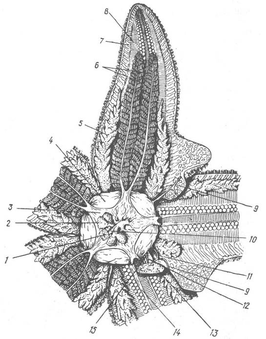
Fig. 215. Dissection fragment of the sea star Asterias rubens (aboral view):
1 — skin region with the anus; 2 — rectal glands; 3 — pyloric stomach; 4 — cardiac stomach; 5 — gonad; 6 — hepatic caeca; 7, 8 — lateral and ambulacral ossicles; 9 — genital stolon; 10 — hindgut; 11 — genital duct; 12 — wall of the axial sinus; 13 — skin region with the madreporite; 14 — stomach retractor muscles; 15 —
stone canal
Most sea stars are active predators feeding on various Mollusks, crustaceans, Coelenterates, Sponges, other Echinoderms, fish, or carrion. Some species specialize in feeding on specific types of prey. Capturing prey, opening mollusk shells, and similar tasks are accomplished using the tube feet on their flexible arms. When attacking large prey, the sea star everts its cardiac stomach, wrapping it around the victim to initiate digestion externally. This process is completed inside the starfish's body after specialized muscles pull The Stomach, along with the semi-digested food, back inside. However, some sea stars are obligate or partial detritivores. It has been established that detritus particles landing on the oral surface of a sea star are transported along the ambulacral grooves by cilia and tube feet, embedded in mucus. Several detritivorous sea stars feature enlarged rectal ceca that contract regularly, functioning like a Water pump: water enters through the mouth and is expelled through the anus. Furthermore, epidermal cells of sea stars have been shown to absorb dissolved organic matter directly from the water, which is subsequently assimilated by the digestive Organs.
The perhemal, or pseudohemal, system includes the circumoral and radial canals—which send branches into the walls of the tube feet—as well as small channels connecting with the general coelom. In sea stars, the perhemal canals are divided into two parts by a vertical septum (Fig. 216). As mentioned previously, the Circulatory system is embedded within the thickness of these septa.

Fig. 216. Cross-section of an arm below the radial ambulacral canal:
1 — outer epithelium; 2, 3 — ectoneural and hyponeural nervous systems; 4 — septum; 5 — radial lacunae of the circulatory system; 6 — perhemal radial canals
The circulatory system is built on a radial plan (Fig. 217). Radial Blood lacunae extend from the oral ring into the arms—housed within the septum of the perhemal canals—along with an axial organ that runs toward the aboral side of the body. The axial organ appears as an elongated sac composed of spongy connective tissue pierced by a dense network of blood lacunae. Near the aboral side, this vascular network transitions into an aboral ring located inside the genital strand. Lacunae extend from the aboral blood ring to the Gonads.

Fig. 217. Diagram of the sea star circulatory system
1 — axial organ; 2 — branches to the intestine; 3 — branches to the gonads; 4 — aboral ring; 5 — radial canal; 6 — oral ring
Similar to sea urchins, the Pericardium (a coelomic compartment belonging to the axial complex) should be considered part of the circulatory system; its pulsations drive the movement of fluid within the blood lacunae of the axial organ.
The axial complex (Fig. 218) runs through one of the interradii between the oral and aboral body walls of the sea star. It is separated from the general coelom by connective tissue walls. The internal cavity of the axial complex is divided into two halves (the right and left axial sinuses). The right sinus (the pericardium) is situated somewhat closer to the aboral side. Through the center of the axial complex, spanning both sinuses, run the axial organ and the stone canal with the madreporite.

Fig. 218. Diagram of the sea star axial complex structure (vertical section through an interradius):
1 — ambulacral system ring canal; 2 — stone canal; 3 — oral region of the axial organ; 4 — left axial sinus; 5 — wall of the axial sinus; 6 — ampulla; 7 — madreporite; 8 — body wall; 9 — genital sinus; 10 — genital stolon; 11 — aboral blood lacuna; 12 — aboral region of the axial organ; 13 — right axial sinus; 14 — stomach wall; 15 — inner perhemal ring; 16 — septum; 17 — ring oral blood lacuna; 18, 19 — ectoneural and hyponeural nervous systems; 20 — outer perhemal ring
The Functions of the axial complex are quite diverse. The Movement of water through the madreporite regulates hydrostatic pressure within the ambulacral system. Amoebocytes, which move freely in the body cavity, are produced within the tissue of the axial organ, meaning it functions as a lymph gland and also serves as a site for the accumulation of Metabolic waste products. The aboral part of the axial organ supplies blood to the gonads and may additionally function as an endocrine gland. As noted previously, the aboral section of the axial sinus (pericardium) is capable of contraction, thereby driving blood flow within the axial organ.
Sea stars breathe using dermal papulae (skin gills), which are numerous thin-walled evaginations of the body wall enclosing extensions of the coelom. They are most abundant on the aboral side and along the sides of the arms. Oxygen diffuses directly across the skin into the coelomic fluid. Gas exchange also occurs through other thin-walled areas of the body, including the tube feet.
The Nervous System of sea stars is typical of echinoderms, comprising three divisions developed to varying degrees (Fig. 219). The ectoneural division lies almost entirely within the outer epithelium and consists of a network of Nerve Cells, as well as denser concentrations forming radial nerves that run along the floor of the ambulacral grooves and connect with a circumoral nerve ring.

Fig. 219. Diagram of the nervous system structure in a sea star:
1, 2, 3 — apical, hyponeural, and ectoneural divisions, respectively
The hyponeural division, bordering the ectoneural division, also features well-developed radial nerves, whereas the hyponeural ring is underdeveloped. The apical division of the nervous system is represented by five radial cords located in the coelomic epithelium on the aboral side of the body; all of them interconnect at the aboral pole.
Regarding specialized Sense Organs, most sea stars possess primitive ocelli resembling eyecups (Fig. 220), located at the tip of each arm near the Base of the modified, tentacle-like ambulacral foot. It should be noted, however, that even eyeless sea stars respond to changes in light intensity, detecting them via nerve cells scattered throughout the skin.

Fig. 220. Sensory Organs of a sea star:
a — tip of the arm; b — diagram of ocellus structure; 1 — terminal tentacle; 2 — ocellus; 3 — spines surrounding the tentacle; 4 — tube feet; 5 — lens; 6 — eyecup; 7 — sensory cells; 8 — nerve fibers; 9 — supporting cells
Although sea stars, like other echinoderms, have a relatively primitive nervous system lacking any cerebral ganglia, it has been experimentally proven that conditioned Reflexes can be established in certain species.
Most sea stars are gonochoric (dioecious), though cases of Various Forms of Hermaphroditism are also known. For instance, in Marthasterias glacialis, alongside separate-sex individuals, specimens with mosaic hermaphroditic gonads occur, while in some populations of Asterina gibbosa, the gonads of young individuals produce spermatozoa and older ones produce eggs. Sexual Dimorphism is typically absent, but during the breeding season, individuals of different sexes may sometimes differ markedly in size and coloration.
The gonads are arranged in pairs within each arm, starting from its base (Fig. 221, see also Fig. 215), and appear as grapelike clusters in mature specimens. A separate duct extends from each gonad and opens into the interradius. Fecundity in sea stars is very high and can reach up to 200 million eggs. Fertilization is external.

Fig. 221. Diagram of gonad arrangement:
1 — aboral part of the axial organ; 2 — gastric Branches of the circulatory system; 3 — genital stolon; 4 — genital sinus; 5 — gonads; 6 — left axial sinus; 7 — stone canal; 8 — oral part of the axial organ
Development in sea stars can proceed via a complex metamorphosis or direct development. As in sea urchins, in most species the fertilized egg develops into a simple, ciliated larva while still at the blastula or gastrula stage. Larvae developing from yolk-poor eggs undergo a complex metamorphosis; they increase in structural complexity and transform into a short-lived dipleurula stage, which then develops into a bipinnaria, characterized by short triangular lobes bordered by a ciliated band (Fig. 222, a). Later, three cylindrical appendages—brachiolaria arms—with adhesive attachment disks appear on the preoral lobe of the bipinnaria, transforming it into a brachiolaria (Fig. 222, b). The brachiolaria can attach to various submerged objects. Furthermore, it possesses a muscular, sucker-like organ that anchors it to the substrate for an extended period, during which it completes its transformation into a juvenile sea star, accompanied by the resorption of part of the larval body.

Fig. 222. Larvae of Asterias rubens — bipinnaria (a); brachiolaria (b):
1 — sucker; 2 — developing body of the sea star
Larvae developing from yolk-rich eggs transform directly into tiny sea stars. Species with direct development often exhibit parental care.
Alongside sexual reproduction, some sea stars are known to reproduce asexually by binary fission of the disk through the interradius or by arm autotomy followed by the regeneration of the missing part. The capacity for division-based reproduction in sea stars is closely linked to their remarkable ability to regenerate an entire Organism from a single arm or even a fragment thereof (Fig. 223).
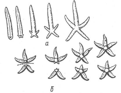
Fig. 223. Asexual reproduction in sea stars:
a — formation of a sea star from a detached arm in Linckia; b — following transverse fission in Nepanthia
Sea stars cause significant economic damage by destroying oysters and other commercially valuable mollusks, including aquaculture species. In recent years, it has been precisely established that sea stars are serious food competitors for many commercial animal species: about 80% of all benthic animals they feed on are also consumed by sea stars.
In the 1960s, a catastrophic population surge of the large (40–50 cm in diameter) multi-rayed sea star Acanthaster planci, commonly known as the crown-of-thorns starfish, was recorded on many coral reefs in the western Pacific Ocean. These animals feed on coral polyps, creating a severe threat of destruction to coral reefs, including
the Great Barrier Reef off the coast of Australia. Here, the culling of crown-of-thorns starfish was carried out by scuba divers who injected formalin directly into the sea stars' bodies. By the 1990s, the population of these sea stars had declined back to normal levels.
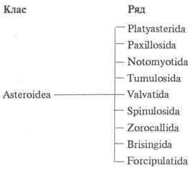
According to recent data, modern Representatives of the Class Asteroidea are divided into 9 orders.
Order Platyasterida. Representatives of this order became extinct mostly in the Paleozoic, with only species of the family Luidiidae (Fig. 224) surviving to the present day, in which the ambulacral tube feet lack suckers. Common at shallow depths in warm seas, relatively large species of the genus Luidia feature long, flexible rays with numerous spines along the edges. Among sea stars, they are unique in having dermal papulae shaped as branching outgrowths. They feed mainly on brittle stars and sea urchins by swallowing them whole.

Fig. 224. Order Platyasterida: Luidia latiradiata
Order Paxillosida. All paxillosids, much like the species of the previous order, lack suckers on their ambulacral tube feet; their aboral (dorsal) surface is typically covered with paxillae, which gives the order its name. A very characteristic feature is the presence of well-defined marginal plates framing the disk and rays. Pedicellariae are always of a simple structure, lacking stalks, and are located directly on the skeletal plates.
One of the largest families in this order is the comb stars (Astropectinidae), in which the marginal plates are usually armed with needle-like spines. These flat, five-rayed stars, frequently colored in orange-red tones, commonly inhabit sandy substrates into which they burrow very rapidly. Polychaete worms of the family Aphroditidae frequently settle within the ambulacral grooves of comb stars. The sea star provides the worms with food, while they, in turn, clean the area around its mouth from food debris.
Representatives of the family Porcellanasteiidae (Fig. 225) are among the most characteristic inhabitants of the ocean depths, where they live on soft, muddy bottoms by burrowing into them. They lack "hepatic" cecal outgrowths and a hindgut with an anus, feeding primarily on detritus.
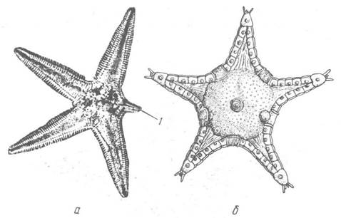
Fig. 225. Order Paxillosida:
a — Astropecten aurantiacus; b — Porcellanaster coerulans; 1 — regenerating ray
Order Notomyotida. This is a small order of deep-sea five-rayed sea stars characterized by paired longitudinal muscle bands along both sides of each arm.
Order Tumulosida. Currently, only four species of the sole extant genus Podosphaeraster are known; they resemble tiny spheres about 1 cm in diameter and externally look more like sea urchins (Fig. 226). However, the five ambulacral grooves in representatives of this order run exclusively along the lower hemisphere, which corresponds to the oral surface.

Fig. 226. Order Tumulosida: Podosphaeraster polyplax (top view): 1 — anus; 2 — ambulacral grooves (dashed lines indicate radii corresponding to the aboral side of the rays)
Order Valvatida. The name of the order derives from the presence of simple, two-valved (bivalved) pedicellariae in valvatids.
This order encompasses numerous structurally diverse sea stars grouped into 12 families; they are particularly abundant in the shallow waters of the tropical zone of the Pacific and Indian Oceans. For instance, this is home to sea stars of the genus Archaster, which exhibit a unique behavior among sea stars—mating during reproduction. Males and females pair up using their rays and then simultaneously release Gametes into the water, resulting in immediate fertilization.
On coral reefs, one frequently encounters a bright blue sea star with five long, cylindrical rays—Linckia laevigata, which, much like other members of the order, is characterized by a specific type of asexual reproduction. Linckias possess The ability to periodically and voluntarily cast off their rays at a certain distance from the disk. The detached portion of the ray crawls away from the parent organism and begins an independent life. At the break site, a star initially forms with a cluster of tiny rays at the tip of the large ray, followed by the gradual development of a normal-looking sea star (see Fig. 223). The parent organism quite rapidly regenerates a new arm. The infamous sea star Acanthaster planci (Fig. 227), mentioned previously, also belongs to this order.

Fig. 227. Order Valvatida: Acanthaster planci
Order Spinulosida. Representatives of this order lack pedicellariae, and most of them have poorly developed marginal skeletal plates; the Number and Length of their rays vary. A striking example is the multi-rayed Solaster papposus, known for its beautiful pink coloration with white-tipped rays.
In the temperate and cold waters of seas and oceans in both hemispheres, representatives of the genus Henricia are widely distributed. These are small five-rayed sea stars with narrow, almost cylindrical rays and a small disk. The skeletal plates of their dorsal side are covered with such dense, minute spines that they create a velvety appearance. Interestingly, this genus includes ovoviviparous species. Their embryos, bound together in a cluster by a single cord, are carried by the mother on the underside of her body beneath the mouth. Meanwhile, in representatives of the family Pterasteridae (Fig. 228), which inhabit primarily great depths, embryonic development takes place within a specialized brooding chamber that has no analogs among other sea stars. This chamber forms on the aboral side of the body between the body wall proper and an overlying integumentary membrane supported by the tips of the dorsal paxillae. The cavity of the chamber communicates with the external environment through a central opening—the osculum, numerous small pores across the entire surface, and lateral openings along the sides of the rays. Driven by rhythmic Movements of the membrane, water circulates constantly within the chamber, supplying oxygen both to the embryos and to the dermal papulae located on the floor of the chamber.

Fig. 228. Order Spinulosida: Pteraster obscurus
Many sea stars host parasitic crustaceans, turbellarians, and other symbionts.
The next three orders — Zorocallida, Brisingida, and Forcipulatida — differ from other sea stars by the characteristic structure of their pedicellariae, which consist of three articulating skeletal elements: a basal piece and two Valves. Typically, pedicellariae are borne on muscular, flexible stalks and may have straight, forceps-like valves or crossed, scissor-like ones. In the order Zorocallida, only straight pedicellariae are present, whereas Brisingida possess exclusively crossed ones. Both orders are species-poor, and we will focus solely on the order Forcipulatida.
Order Forcipulatida. These sea stars typically feature a small disc and long, flexible arms. There is no sharp boundary between the oral and aboral surfaces, as the marginal ossicles are nearly indistinguishable from the aboral ones. Pedicellariae are either straight or crossed. Papulae (skin gills) occur on both the aboral and oral surfaces. Most species in this order belong to the family Asteriidae. A representative example is the genus Asterias (Fig. 229), which is common in shallow waters of the temperate zone in the Northern Hemisphere. One of the most thoroughly studied species is Asterias forbesi, inhabiting the Atlantic coast of the USA. It is a small, five-armed sea star measuring up to 20 cm in diameter. It feeds primarily on mussels and oysters, as well as other mollusks, small crustaceans, worms, and both dead and live fish entangled in nets. In the absence of preferred prey, it is capable of cannibalism. Due to their voracity, these sea stars cause severe damage to oyster fisheries. When moving across horizontal surfaces, they use their tube feet as stilts, whereas on slopes or vertical walls, they pull their bodies forward using the standard stepping action of the ambulacral feet.

Fig. 229. Order Forcipulatida: a — Asterias forbesi; b — Marthasterias glacialis
When feeding on bivalves, A. forbesi first attaches its numerous tube feet to the valves and then begins to pull them apart, exerting a force of up to 4.5 kg. In most cases, they manage to fatigue the bivalve's adductor muscle; as soon as a slight gap opens between the valves, the sea star everts its stomach through its mouth into the opening, digesting the mollusk's soft Tissues right inside its shell.
The family Asteriidae includes one of the largest sea stars, Pycnopodia helianthoides, which inhabits rocky, kelp-covered bottoms along the northeastern Pacific coast. The largest specimens reach up to 80 cm across and weigh as much as 4.5 kg.
Last update: 13/08/2026
Editorial and Educational Adaptation: This material has been compiled based on the primary/original source text. The project team performed an editorial review, corrected technical inaccuracies, structured sections, and adapted the content for an educational format.
What was processed:
- elimination of formatting defects (OCR errors, structural breaks, corrupted characters);
- editorial organization of content;
- standardization of terminology in accordance with academic sources;
- verification of factual statements against the original source text.
All mentions of the author, publication year, and origin of the primary text have been preserved in accordance with the source.