MICROBIOLOGY - M.H. Serhiichuk - 2008
Chapter 9. THE ROLE OF MICROORGANISMS IN THE BIOGEOCHEMICAL CYCLING OF NUTRIENTS IN NATURE
The Nitrogen Cycle
Natural reserves of nitrogen are quite substantial. An area of 1 km2 of the Earth's surface contains over 8 million tons of this essential element. Yet, while plants are literally surrounded by nitrogen, they constantly experience its deficiency—much like a person stranded in the ocean who suffers from thirst.
Nature maintains a continuous nitrogen cycle involving microorganisms, animals, and higher plants. The primary role of microorganisms is to ensure the mineralization of dead organic matter, converting organic nitrogen compounds into mineral forms: ammonium salts, salts of nitric and nitrous acids, ammonia, and molecular nitrogen.
These mineral forms of nitrogen are incorporated into the organic matter of Microbial and Plant Cells.
The Nitrogen Cycle (Fig. 9.1) takes place in several stages (phases). Because it is a closed loop, the starting phase is arbitrary; for convenience, we will begin our Structure/133.html">Discussion with The process of ammonification.
Class="center">
Fig. 9.1. Diagram of the nitrogen cycle
Ammonification is the decomposition of nitrogen-containing Organic compounds resulting in The formation of ammonia (NH3). This process is also referred to as putrefaction or decay, and the microorganisms responsible are called ammonifiers or putrefactive Bacteria.
A wide variety of nitrogen-containing organic compounds undergo ammonification: Proteins and their derivatives (peptones, Peptides, Amino Acids); Nucleic Acids and their derivatives (purine and pyrimidine bases); urea; uric and hippuric acids; humic substances; and Chitin.
Ammonifying ability is exhibited by the majority of microorganisms that synthesize Proteolytic Enzymes, including Proteus vulgaris, Pseudomonas aeruginosa, P. fluorescens, Bacillus subtilis, B. mycoides, B. cereus, Clostridium tetani, C. perfringens, C. putrificum, C. sporogenes, and other bacteria, as well as Fungi.
The Mechanism of protein ammonification involves their Enzymatic Hydrolysis into peptones and peptides, and ultimately into amino acids:

The amino acids produced during protein decomposition are mineralized at varying rates. Some (such as Threonine and Methionine) are quite stable, while others (such as Arginine and Tryptophan) are rapidly degraded by microbial action. The pathways of intracellular or extracellular amino acid transformation vary and may include the following processes:
a - Oxidative Deamination, catalyzed by specific oxidases. This yields NH3 and the corresponding keto acid, which is subsequently transformed into a hydroxy acid or an aldehyde, meaning it does not accumulate:

b - reductive deamination, which proceeds with the Formation of the corresponding organic acid. Such transformations are carried out by anaerobic microorganisms:

c - deamination resulting in the formation of NH3 and unsaturated acids:

d - microorganisms can also carry out decarboxylation:

Anaerobic Decarboxylation of amino Acids can produce toxic products, notably diamines such as cadaverine and putrescine. Cadaverine is formed during The breakdown of Lysine, and putrescine from Ornithine:

Urea also belongs to nitrogen-containing organic compounds. An adult human excretes approximately 30 g of this compound daily. Urea is synthesized by certain fungi (for example, mushrooms contain up to 13% of urea on a dry weight basis). This compound is also formed during the hydrolytic Cleavage of arginine under the action of arginase:

Each year, living organisms on the planet synthesize about 35 million tons of urea, which in soils or Water bodies is rapidly broken down by microorganisms synthesizing urease into ammonium carbonate. Since this is an unstable compound, it quickly decomposes into its constituent parts:

Microorganisms capable of urea degradation are called urolytic. They were first described by L. Pasteur in 1862. They are found in soil, sewage, and the rumen of ruminant animals. These include Representatives of the genera Micrococcus (M. urea), Bacillus (B. pasteurii), and Sporosarcina (S. ureae). These bacteria are capable of growing in an alkaline environment at pH 9.0–10.0, which allows them to hydrolyze significant amounts of urea into ammonia.
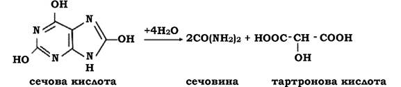
Uric acid is present in urine in small amounts (< 0.04%). It can be degraded by many groups of microorganisms. The Initial Stages of hydrolysis yield tartronic acid and urea:
The hydrolytic cleavage of hippuric acid yields benzoic acid and glycocoll (Glycine), which can be used by microorganisms as a source of carbon and nitrogen Nutrition:

Chitin is a nitrogen-containing compound and a polymer of acetylglucosamine. This compound is degraded by many microorganisms because chitin is constantly present in the soil. It is found in the exoskeletons of invertebrates, the protective shells of insects, and The Cell walls of many fungi.
The ability to degrade chitin is exhibited by microorganisms that synthesize the enzyme chitinase. In the initial stages, this enzyme breaks down chitin into chitobiose and chitotriose, which are subsequently cleaved by chitobiase into acetic acid, glucose, and ammonia.
Ammonia formed in soil or water is relatively quickly oxidized to salts of nitrous acid and then nitric acid. This process is called nitrification.
Before L. Pasteur's work, the accumulation of nitrates in the soil was explained as a purely chemical process—the Oxidation of ammonia by atmospheric oxygen, with the soil acting merely as a chemical catalyst. Pasteur predicted that the formation of nitrite is a microbiological process. The first experimental confirmations of this prediction were obtained by T. Schloesing and A. Müntz in 1879, but nitrifying microorganisms were successfully isolated only in 1890–1892 by S. Winogradsky. To achieve this, he proposed a fundamentally new approach: The Use of selective media. This approach made it possible to discover a new type of METABOLISM in microorganisms known as Chemosynthesis. A characteristic feature of chemosynthetic organisms is their ability to obtain energy for the synthesis of organic matter from atmospheric СО2 through The oxidation of inorganic substances, specifically ammonium salts.
S. Winogradsky also demonstrated that the nitrification process occurs in two stages carried out by different groups of microorganisms. One group oxidizes ammonia to nitrous acid (The first phase of nitrification, NО2-), while the other group oxidizes it to nitric acid (NO3-) (the second phase).
First-phase nitrifiers include representatives of five genera: Nitrosomonas, Nitrosococcus, Nitrosospira, Nitrosolobus, and Nitrosovibrio. The only species studied in more or less sufficient detail is Nitrosomonas europaea. Overall, the first phase can be represented by the following equation:

The second phase is the oxidation of nitrous acid to nitric acid:

Second-phase nitrifiers include representatives of the genera Nitrobacter, Nitrospira, and Nitrococcus. The best-studied among them is Nitrobacter winogradskii.
Nitrifying bacteria develop at pH 6.0–8.6, with an optimum of 7.5–8.0. At pH values below 6.0 and above 9.2, these bacteria do not grow. The optimal Temperature for The Development of nitrifiers is 25–30 oС. However, for some strains, the optimum is around 26 oС or near 40 oС, while others develop normally at 4 oС.
It is believed that conditions favorable for the development of nitrifying bacteria are also favorable for plant growth. This suggests that the presence of an active nitrification regime can serve as one of the indicators of soil fertility.
The ability to oxidize ammonia to nitrites and nitrates has also been established for certain chemoorganoheterotrophic bacteria, such as representatives of the genera Arthrobacter, Xanthomonas, and Pseudomonas. However, the intensity of heterotrophic nitrification is significantly lower than that of autotrophic nitrification.
Nitrates formed in the soil during nitrification undergo further transformations: they can be taken up by plants; leached into water bodies; assimilated into the cells of microorganisms that use them as a nitrogen source; or reduced by microorganisms to molecular nitrogen or ammonia. The latter process is called denitrification. If nitrate reduction is accompanied by the formation of ammonia, the process is called assimilatory denitrification (nitrate reduction). This type of denitrification occurs in both plants and many microorganisms, proceeding in stages with the formation of intermediates (nitrite — NO2-; hyponitrous acid — HNO; hydroxylamine — NH2OH). The first step of the reaction is catalyzed by nitrate reductase B. The Synthesis of the enzyme is triggered when nitrate serves as the sole nitrogen source in the medium. The resulting nitrite is transformed with the participation of nitrite reductase into ammonia; in other words, nitrate reduction during assimilatory denitrification takes place in a stepwise manner:

During dissimilatory denitrification, nitrates act as oxidants for organic matter, providing microorganisms with the necessary energy. Nitrates do not release their oxygen all at once, but rather in a stepwise manner, meaning that nitrate reduction is also a multi-stage process:

The capacity for dissimilatory nitrate reduction is exhibited exclusively by specific facultatively anaerobic bacteria. Soils are predominantly inhabited by denitrifiers of the genera Pseudomonas (P. aeruginosa, P. fluorescens, P. stutzeri) and Paracoccus (P. denitrificans), as well as thermophilic representatives of the genus Bacillus. These and other microorganisms utilize nitrates as hydrogen acceptors in the absence of O2 to oxidize organic compounds into end products such as CO2 and H2O, whereas under aerobic conditions they act as ordinary oxidizers of organic substrates. The survival of denitrifiers under anaerobic conditions can also be supported by nitrites. Such energy-yielding processes are referred to as nitrate Respiration. For example, if Monosaccharides are present in the medium, their oxidation can be represented as follows:

Most denitrifiers are chemoorganotrophs, meaning they require organic compounds in their environment. However, certain chemolithoautotrophs are also capable of reducing nitrates. For instance, the facultatively anaerobic sulfur-oxidizing bacterium Thiobacillus denitrificans can thrive under anaerobic conditions by using nitrates as the terminal hydrogen acceptor. As an energy source, this microorganism utilizes elemental sulfur or thiosulfate, transforming them into sulfates, while the nitrate is reduced to molecular nitrogen:

The schematic processes of assimilatory and dissimilatory denitrification can be represented as follows:

As can be seen from the equations above, microbiological denitrification leads to the depletion of mineral forms of nitrogen in the soil. Annually, approximately 300 million tons of N2 pass from the soil into the atmosphere. However, this process should not be viewed solely as detrimental, since from a global perspective it is of critical importance for sustaining life on the planet. In normally aerated soils, nitrates are the end products; they are highly soluble in water and poorly retained by soil particles, making them readily leached into water bodies. All of this could potentially lead to a decrease in the molecular nitrogen content of the atmosphere. Yet, the N2 released into the atmosphere via denitrification is captured once again by microorganisms and channeled into the synthesis of organic matter, constituting the next stage of the nitrogen cycle—Nitrogen Fixation.
Nitrogen fixation occurs in nature through two pathways: biological and non-biological. The non-biological pathway (e.g., electrical storms) accounts for approximately 0.5% of the global balance, while the total productivity of nitrogen fixation amounts to ~300 million tons per year.
Due to the substantial enrichment of soil with fixed forms of nitrogen, its fixation has attracted the attention of numerous researchers. In 1838, J. Boussingault was the first to notice that clover and other legumes enrich the soil with nitrogen. A similar pattern was observed in 1885 by M. Berthelot, who determined that sterilizing the soil halts the increase in bound nitrogen forms. Subsequently, between 1886 and 1888, H. Hellriegel and H. Wilfarth discovered the relationship between Nitrogen Fixation and the ROOT nodules of legumes. Legumes can grow in the absence of fixed nitrogen forms only when their root system possesses nodules (Fig. 9.2).

Fig. 9.2. Symbiotic nitrogen fixation in the root nodules of legumes:
a – pea root with nodules; b – cross-section of a nodule; c – plant cell filled with bacteria (Rhizobium); d – bacteria inside plant cells acquire unusual shapes (bacteroids, involution forms); e – penetration of bacterial cells through root hairs and growth of the infection thread
In 1888, M. Beijerinck made a significant discovery by isolating in pure culture a symbiotic N2-fixer classified in the genus Rhizobium. In 1893, S. Winogradsky made another breakthrough by isolating an anaerobic free-living N2-fixer from the soil, Clostridium pasteurianum, and later, in 1901, M. Beijerinck described the aerobic free-living N2-fixer Azotobacter chroococcum.
Until 1949, the capacity for MOLECULAR NITROGEN FIXATION was attributed exclusively to representatives of the genera Clostridium and Azotobacter. In 1949, the Nitrogenase system—which drives the reduction of acetylene to Ethylene—was discovered across all diazotrophs, thereby establishing The Link Between N2 fixation and acetylene reduction:

In addition to molecular nitrogen and acetylene, the nitrogenase system also reduces azide, nitrous oxide, cyanide, nitrile, and isonitrile.
The application of the acetylene reduction assay has revealed that the capacity for N2 fixation is also exhibited by representatives of other bacterial genera, such as Azomonas, Aerobacter, Achromobacter, Klebsiella, methanogenic bacteria, certain blue-green Algae, etc.
Representatives of the genus Azotobacter belong to the family Azotobacteraceae. Their cells are oval, 1.5–2.0 µm in diameter, and pleomorphic, ranging from rod-shaped to spherical (Fig. 9.3). They occur singly, in pairs, or form irregular clusters. They are either motile by means of peritrichous flagella or non-motile. They synthesize a capsule that typically encloses two cells. Under adverse conditions, they form cysts (resting forms). The genus Azotobacter includes A. chroococcum, A. agilis, and A. vinelandii. The optimal pH for their growth is 7.0–7.2, with a maximum of 9.0. They are virtually undetectable in soils with a pH of 5.6. The distribution and certain characteristics of Azotobacter species are summarized in Table 9.1.

Fig. 9.3. Azotobacter chroococcum:
a – colonies of A. chroococcum; b – transformation of rod-shaped cells into coccoid forms; c – diplococci surrounded by a capsule
The family Azotobacteraceae also includes the genus Azomonas (A. agilis, A. insignis, A. macrocytogenes). Morphologically, representatives of this genus resemble Azotobacter. During growth, the culture may excrete abundant slime, but it does not form cysts. Colonies on Agar media are colorless, and some strains produce fluorescent pigments. The optimal pH for Nitrogen fixation is close to neutral, although certain strains can fix nitrogen at pH 4.6–4.8. They are found in soils and aquatic environments.
Table 9.1. Species of the genus Azotobacter and their distribution
Species |
Cell size, shape, and arrangement |
Colony characteristics |
Distribution |
A. chroococcum |
2.0 × 3.1 µm; predominantly spherical; in pairs |
Mucoid; darkly pigmented; contain cysts |
Soils |
A. agile |
2.8 × 3.3 µm; spherical, oval; singly and in pairs |
Excrete a yellow pigment with white fluorescence |
Waters of channels and ditches |
A. vinelandii |
1.5 × 3.4 µm; spherical, oval; predominantly in pairs |
Large, mucoid; excrete a yellow pigment with green fluorescence; occasionally contain cysts |
Soils |
Aerobic gram-negative rods and cocci also include representatives of the genus Beijerinckia (B. derxii, B. indica, B. mobilis). The cells are rod-shaped, somewhat curved or pear-shaped, and some tend to branch. Both motile and non-motile cells occur. Motile forms feature peritrichous flagellation. They form capsules (surrounding several cells), and some species form cysts (surrounding a single cell). They grow within a pH range of 3.0 to 9.5–10.0. As the culture ages, colonies may acquire a yellowish-brown, amber-brown, reddish, or pink hue. They are found in soils, particularly frequently in tropical latitudes.
Bacteria isolated from the root nodules of legumes are assigned to the genus Rhizobium (from Greek rhizo — root) of the family Rhizobiaceae. They are most commonly found in soils where legumes grow, but may also occur freely as Saprophytes.
Morphologically, they are pleomorphic rods measuring 0.5–0.9 × 1.2–3.0 µm. They move by means of a single polar (subpolar) or peritrichous flagella (2–6 in number). The optimal pH is 6.0–7.0. On agar media, they form round, convex, translucent, mucous colonies. A characteristic feature of representatives of this genus is their ability to penetrate the root hairs of legumes and stimulate the formation of nodules, inside which the bacteria reside as intracellular symbionts. Within the nodules, the bacteria appear as pleomorphic forms known as bacteroids, which fix N2 by transforming it into a bound form (ammonia) available for use by the host plant. The genus Rhizobium includes: R. leguminosarum (R. leguminosarum biov. viciae, R. leguminosarum biov. trifolii, R. leguminosarum biov. phaseoli), R. loti, and R. meliloti.
In 1989, the genus of root-nodule bacteria Photorhizobium was described, representatives of which exhibit the capacity for symbiotic nitrogen fixation and Photosynthesis. In 1990, another species assigned to the genus Rhizobium was described—R. galegae, which Functions in Symbiosis with goat's rue (Galega).
All symbiotic nitrogen fixers are characterized by such traits as Specificity, virulence, and activity.
Specificity is the ability to form nodules on The Root System of only specific species of legumes. Specificity is not absolute, but nonspecific infection typically results in the formation of inactive or poorly active nodules.
Virulence is the ability of root-nodule bacteria to penetrate through root hairs into the plant root and form nodules. Occasionally, root-nodule bacteria may exhibit low virulence, meaning they fail to infect or poorly infect the legume plant.
The activity of root-nodule bacteria is determined by the intensity of nitrogen fixation, which depends on the content of a red pigment—Hemoglobin, referred to in this case as leghemoglobin; essentially, this is legume hemoglobin located within the vacuoles of plant cells. It facilitates the nitrogen assimilation process by maintaining the redox state at a specific level. Leghemoglobin is responsible for the pink coloration of the nodules. Such pigmented nodules are active. There are cases where a strain exhibits specificity and is virulent, yet inactive. In such instances, nodules are formed, but they are incapable of fixing N2. The isoelectric point of the nodule tissue formed by active cultures lies significantly lower (pH 3.0–4.0) than that of the nodule tissue formed under The Influence of an inactive strain (pH 6.0–6.5).
In reality, only prokaryotes are capable of fixing atmospheric nitrogen and building all nitrogen-containing organic compounds of their cells from it. The reason is that nitrogen is an inert element. The two atoms in its molecule possess a triple bond. Breaking one of the three bonds in its molecule requires expending about 125 kcal per gram-molecule of nitrogen. The other two bonds are broken more easily, with energy expenditures of 63 and 37 kcal, respectively. The pathway of bond cleavage in the nitrogen molecule can be represented as follows:
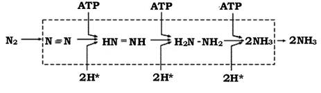
The ATP demand in nitrogen-fixing bacteria is quite high: 12 ATP molecules are consumed for the reduction of a single nitrogen molecule. The functioning of the nitrogenase system is ensured not only by energy supply but also by a continuous flux of electrons for the reduction of N2 to ammonia. The interrelation scheme between the processes underlying the reduction of molecular nitrogen to ammonia is shown in Fig. 9.4.
Ammonia produced during N2 fixation is bound by keto acids, which is accompanied by the synthesis of the corresponding amino acids (Fig. 9.5).

Fig. 9.4. Interrelationship of processes underlying molecular nitrogen fixation:
Fd — ferredoxin; Fl — flavodoxin

Fig. 9.5. Pathways of molecular nitrogen assimilation
Last update: 13/08/2026
Editorial and Educational Adaptation: This material has been compiled based on the primary/original source text. The project team performed an editorial review, corrected technical inaccuracies, structured sections, and adapted the content for an educational format.
What was processed:
- elimination of formatting defects (OCR errors, structural breaks, corrupted characters);
- editorial organization of content;
- standardization of terminology in accordance with academic sources;
- verification of factual statements against the original source text.
All mentions of the author, publication year, and origin of the primary text have been preserved in accordance with the source.