Review of Medical Physiology - William F. Ganong 2002
Endocrine System, Metabolism, and Reproduction
Thyroid Gland
Action of Thyroid Hormones
Other far-reaching effects of THYROID Hormones in the body are secondary and aimed at stimulated oxygen consumption (calorigenic action), although the hormones also influence GROWTH AND DEVELOPMENT in mammals, help regulate Lipid METABOLISM, and increase Carbohydrate Absorption from the intestine (Table 18-3). They also enhance oxygen dissociation from Hemoglobin by increasing the concentration of 2,3-diphosphoglycerate (DPG) in erythrocytes (see Chapter 35).
Thyroid hormones enter The Cell, and T3 binds to thyroid Hormone Receptors (TRs) in The Nucleus; T4 can also bind, albeit with lower affinity. Subsequently, the hormone-receptor complex binds to DNA via zinc fingers and increases (or in some cases decreases) the expression of various genes encoding Proteins that regulate cellular Functions (see Chapter 1). Thus, nuclear thyroid hormone receptors belong to the superfamily of hormone-responsive nuclear Transcription factors.
Two human TR genes are known: the $\alpha$-receptor Gene on chromosome 17 and the $\beta$-receptor gene on chromosome 3. Alternative Splicing of each transcript yields at least two distinct mRNAs and, consequently, two different receptor proteins. The TR$\beta_2$ gene is expressed almost exclusively in the Brain, whereas TR$\alpha_1$, TR$\alpha_2$, and TR$\beta_1$ are widely distributed. The TR$\alpha_2$ gene differs from other forms in that it does not bind T3, and its function remains incompletely understood. Thyroid receptors bind to DNA as monomers, homodimers, and heterodimers with other nuclear receptors, notably the retinoid X receptor (RXR). Although this heterodimer does not bind 9-cis-retinoic acid—the usual RXR Ligand—TR binding to DNA is significantly enhanced. There are also coactivator and corepressor proteins that modulate thyroid receptor activity. This complexity likely enables thyroid hormones to exert their diverse physiological effects throughout the body, though the overall physiological significance of this intricate machinery remains largely unknown.
Class="center">Table 18-3. Physiological Actions of Thyroid Hormones1
Target Tissue |
Action |
Mechanism |
Chronotropic |
Increased number and affinity of $\beta$-adrenergic receptors |
|
Inotropic |
Enhanced responses to circulating catecholamines; increased expression of $\alpha$-Myosin heavy chains (with higher ATPase activity) |
|
Adipose tissue |
Catabolic |
Stimulation of lipolysis |
Catabolic |
Increased protein degradation |
|
Bone |
Developmental |
Stimulation of normal skeletal growth and development |
Developmental |
Stimulation of normal mental development |
|
Intestine |
Metabolic |
Increased rate of carbohydrate absorption |
Metabolic |
Stimulation of LDL receptor formation |
|
Other |
Calorigenic |
Stimulation of oxygen consumption in metabolically active Tissues (exceptions: Testes, Uterus, Lymph Nodes, Spleen, anterior pituitary); increased metabolic rate |
1 Modified and reprinted with permission from McPhee SJ et al: Pathophysiology of Disease. 3rd ed. McGraw-Hill, 2000.
For most of these effects, T3 acts more rapidly and potently (three to five times more strongly than T4) (Fig. 18-11). This is attributed to the fact that T3 is bound less tightly to Plasma Proteins and binds more avidly to thyroid hormone receptors; rT3 is inert.
Calorigenic Action
T4 and T3 increase oxygen consumption in nearly all metabolically active tissues. Notable exceptions include the brain, testes, uterus, lymph nodes, spleen, and anterior pituitary. In fact, T4 suppresses $\text{O}_2$ consumption in the anterior pituitary, possibly by inhibiting TSH secretion. The increase in metabolic rate induced by a dose of T4 is measurable after a latent period of several hours and can persist for six days or longer.
Some of the calorigenic effects of thyroid hormones result from the metabolism of the Fatty acids they mobilize. Additionally, thyroid hormones increase The activity of membrane-bound $\text{Na}^+$-$\text{K}^+$-ATPase in many tissues.
Secondary Calorigenic Effects
When the metabolic rate in adults is increased by T3 or T4, nitrogen excretion rises correspondingly. If caloric intake does not increase, endogenous proteins and fat stores are catabolized, leading to weight loss. In hypothyroid children, small doses of thyroid hormones induce a positive nitrogen balance by stimulating growth, whereas large doses promote Protein Catabolism, much as they do in adults. Potassium released during protein catabolism appears in the urine, alongside elevated urinary hexosamine levels and increased uric acid excretion. Large doses of thyroid hormones generate sufficient excess heat to cause a slight rise in body Temperature (see Chapter 14), which in turn activates heat dissipation mechanisms. Peripheral resistance decreases due to cutaneous vasodilation; however, Cardiac Output increases owing to the combined action of thyroid hormones and catecholamines on The Heart. Consequently, pulse pressure and heart rate increase, while Circulation time decreases.
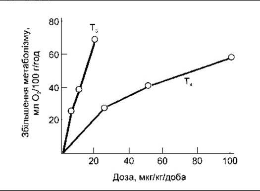
Fig. 18-11. Changes in thermogenesis in thyroidectomized rats following subcutaneous administration of T4 and T3 (reproduced with permission from Barker SB: Peripheral actions of thyroid hormones. Fed Proc 1962;21:635).
During elevated metabolic rates, vitamin deficiency syndromes may become more pronounced due to the body's increased demand for Vitamins. Thyroid hormones are also required for the hepatic conversion of carotene into vitamin A, and the accumulation of carotene in the Blood (carotenemia) in cases of hypothyroidism causes a yellowish Skin discoloration (carotenoderma). This form of jaundice differs from others in that the sclerae do not turn yellow.
Typically, the skin contains abundant proteins bound to Polysaccharides, hyaluronic acid, and chondroitin sulfate. In hypothyroidism, these complexes accumulate, promoting Water retention and The Development of the characteristic skin thickening known as Myxedema. Following the administration of thyroid hormones, proteins are catabolized, diuresis increases, and the symptoms of myxedema disappear.
Milk secretion is reduced in hypothyroidism and stimulated by the administration of thyroid hormones, a fact occasionally utilized in the dairy industry. Thyroid hormones do not stimulate metabolism in the uterus, yet they are essential for normal menstrual cycles and fertility.
Effects on The Nervous System
In hypothyroidism, mental processes are slowed, and protein levels in the CEREBROSPINAL FLUID are elevated. Administration of thyroid hormones normalizes these changes. Large doses induce accelerated mental processes, irritability, and restlessness. Overall cerebral blood flow and the cerebral consumption of glucose and oxygen remain normal in both hypothyroid and hyperthyroid adults. Nevertheless, thyroid hormones do enter the adult brain and have been detected in the Gray matter across various regions. Furthermore, the brain converts T4 into T3. Following thyroidectomy, the activity of brain 5'-deiodinase increases sharply, returning to normal within 4 hours after an intravenous dose of T3. Some actions of thyroid hormones on the brain are secondary, linked to an enhanced response to catecholamines and a concomitant increase in the activation of the reticular activating system (see Chapter 11). These hormones also exert a profound effect on brain development, most notably impacting CNS structures such as the Cerebral Cortex AND Basal Ganglia, as well as the cochlea. A deficiency of thyroid hormones during development results in mental retardation, motor rigidity, and deaf-mutism.
Thyroid hormones affect all Reflexes. The reaction time for protective reflexes (see Chapter 6) decreases in hyperthyroidism and increases in hypothyroidism. Measurement of the Achilles reflex relaxation time has gained attention as a clinical test for assessing thyroid function, although it is also influenced by other pathologies.
Relationship with Catecholamines
The actions of thyroid hormones and catecholamines (norepinephrine and epinephrine) are closely intertwined. Epinephrine increases metabolic rate, stimulates the nervous system, and produces cardiovascular effects similar to those caused by thyroid hormones, albeit of shorter duration. Norepinephrine generally exhibits comparable effects. The toxicity of catecholamines is markedly increased in rats treated with T4. Although plasma catecholamine levels are normal in hyperthyroidism, the cardiovascular effects, tremor, and increased sweating induced by thyroid hormones can be diminished or entirely eliminated by sympathectomy or by drugs such as propranolol, which blocks ß-adrenergic receptors. Indeed, propranolol and other ß-blockers are extensively used in the Treatment of thyrotoxicosis and severe exacerbations of hyperthyroidism, known as thyroid storms. However, although ß-blockers are weak Inhibitors of the interconversion of T4 to T3 in peripheral tissues and may slightly lower plasma T3 levels, they have little effect on the other actions of thyroid hormones.
Effects on the Heart
Thyroid hormones exert multiple effects on the heart. Some of these are mediated by the direct action of T3 on myocytes, but the interplay among thyroid hormones, catecholamines, and the sympathetic nervous system also significantly influences cardiac function. For example, a generalized upregulation of metabolism can trigger hemodynamic changes and increase cardiac output.
Thyroid hormones increase both the number and affinity of ß-adrenergic receptors in the heart, thereby enhancing its sensitivity to the inotropic and chronotropic effects of catecholamines. They also influence the type of myosin present in The cardiac muscle, partly through the direct activation of myocytes. The heart contains two myosin heavy chain (MHC) isoforms: a- and ß-MHC. These are encoded by two highly homologous genes located in tandem on the short arm of human chromosome 17. Each myosin molecule consists of two heavy chains and two pairs of light chains (see Chapter 3). Myosin containing ß-MHC exhibits lower ATPase activity than myosin containing a-MHC. In the adult human atrium, a-MHC predominates, and its levels increase under The Influence of thyroid hormones, which leads to an elevated heart rate. Conversely, when a-MHC Gene Expression is suppressed and ß-MHC gene expression is stimulated, hypothyroidism is observed.
Effects on Muscles
In cases of hyperthyroidism, most patients develop muscle weakness (thyrotoxic myopathy); if the hyperthyroidism is severe and prolonged, the myopathy can become profound. Muscle weakness is partly a consequence of accelerated protein catabolism. Thyroid hormones influence MHC gene expression not only in the cardiac muscle but also in skeletal muscles (see Chapter 3). However, they exert complex effects, and their exact relationship to myopathy remains incompletely understood. Hypothyroidism is likewise associated with muscle weakness, cramps, and rigidity.
Effects on Carbohydrate Metabolism
Thyroid hormones increase The rate of carbohydrate absorption from the gastrointestinal tract; this action is independent of their calorigenic effect. Consequently, in hyperthyroidism, plasma glucose levels rise rapidly following a carbohydrate-rich meal, occasionally exceeding the renal threshold, though they subsequently drop just as quickly.
Effects on Cholesterol Metabolism
Furthermore, thyroid hormones reduce circulating plasma cholesterol levels prior to any rise in metabolic rate, indicating that this action is independent of stimulated O2 consumption. As noted in Chapter 17, the reduction in plasma cholesterol concentration results from an increased synthesis of LDL receptors in the Liver, which leads to enhanced hepatic clearance of cholesterol from the circulation. Despite considerable efforts, it has not yet been possible to develop a clinically viable thyroid hormone analogue that lowers cholesterol without simultaneously increasing the metabolic rate.
Effects on Growth
Thyroid hormones are essential for normal growth and skeletal development (see Chapter 22). In hypothyroid children, bone growth is retarded, and the closure of epiphyseal growth plates is delayed. In the absence of thyroid hormones, Growth Hormone secretion is also suppressed; thus, thyroid hormones act to potentiate the effects of growth hormone in tissues.
Another clear illustration of The Role of thyroid hormones in supporting normal growth and maturation is their effect on amphibian metamorphosis. Tadpoles treated with T4 and T3 undergo premature metamorphosis into miniature frogs, whereas hypothyroid tadpoles never transform into frogs at all.
Last update: 10/08/2026
Editorial and Educational Adaptation: This material has been compiled based on the primary/original source text. The project team performed an editorial review, corrected technical inaccuracies, structured sections, and adapted the content for an educational format.
What was processed:
- elimination of formatting defects (OCR errors, structural breaks, corrupted characters);
- editorial organization of content;
- standardization of terminology in accordance with academic sources;
- verification of factual statements against the original source text.
All mentions of the author, publication year, and origin of the primary text have been preserved in accordance with the source.