NEONATAL SURGERY - 1976
2. SPECIAL SECTIONS
8. Malformations of Individual Organs and Systems
Hirschsprung's Disease.
In most cases, the clinical manifestations of Hirschsprung's disease in newborns are characterized by a complete large bowel obstruction (acute, decompensated form of Hirschsprung's disease). Cure is possible only through surgery. The choice of surgical method is determined by the infant's condition, The Nature of clinical manifestations, the availability of appropriate conditions for the Procedure, and the surgeon's experience with this specific neonatal operation. In critical conditions, lack of necessary facilities, or insufficient surgical experience, The First stage involves a palliative procedure aimed at fecal diversion from the intestine—typically creating one of the variants of a colostomy. The choice of colostomy method depends on the form of the disease, specifically The Nature and extent of the aganglionic zone.
In recent years, we have followed the recommendations of Rickham (1971). For a short, low spastic zone, we construct an artificial anus on the ascending colon. For an extensive zone, we exteriorize the end of the dilated colon. Such a tactic allows for a subsequent radical operation in patients with a short, low aganglionic zone under the Protection of the artificial anus. Conversely, in cases of a long spastic zone, the absence of an artificial anus on the ascending colon makes it possible to mobilize and pull down the bowel to the required length.
Surgical technique. Patient position: supine. Under general anesthesia, a small incision is made along the Skin crease, 2 – 4 cm above the right iliac crest, to open the Abdominal cavity. The ascending colon is delivered into the wound. An opening is created in the mesentery of the bowel, through which a Glass rod is passed. Two Lembert (seromuscular) sutures are placed on the proximal and distal loops directly beneath the glass rod to form a double-barreled colostomy. Both ends of the double-barreled loop are brought out into the wound and secured with sutures first to the Peritoneum, and then to the aponeurosis and Muscles. The glass rod prevents the bowel from slipping back into the abdominal cavity; it is left in place for 8 – 10 days. If the patient's condition is critical and emergency intestinal decompression is required, the bowel is opened immediately, and its edges are sutured to the skin. In the majority of patients, the bowel is packed with gauze soaked in vaseline oil, and its lumen is opened with a electrosurgical unit on the following day, once adhesion between the wound edges and the bowel has occurred. When opening, no more than 3/4 of the bowel circumference is incised, leaving the mesenteric border intact. Once The Need for the stoma has passed, intestinal patency is restored extraperitoneally by dissecting the bowel edges and closing the defect in the intestinal wall.
In cases where prolonged functioning of the colostomy is required or particularly meticulous perineal asepsis is necessary, the standard technique does not yield the desired effect. A narrow tract (1/4 or 1/3 of the bowel diameter), through its peristaltic action, carries along fecal particles that ultimately pass into the distal bowel segment. Therefore, in certain cases, it is advisable to completely transect the bowel, close its distal segment blindly, and bring the proximal stump out to the skin as a rosette, having previously excised an oval patch of skin to prevent stenosis of the artificial anus.
In patients with an extensive aganglionic zone on the left, the abdominal cavity is opened in the iliac region. The sigmoid colon is brought out into the wound, and the transition point from the narrowed segment to the dilated one is identified. The bowel is transected at the transition zone between the spastic and dilated parts. The stump of the distal segment is closed with a double-row suture and returned to the abdominal cavity. The end of the proximal segment is brought out into the wound and sutured first to the peritoneum, and then to the edges of the skin wound.
Following appropriate preparation and stabilization of the infant's condition, a radical operation is performed. This involves the resection of the non-functioning, aganglionic zone and the restoration of intestinal patency.
Currently, the most widely adopted Procedures are the Swenson, Soave, and Duhamel operations (S. Ya. Doletsky, 1956; Yu. F. Isakov, 1965; G. A. Bairov, 1968; Yu. F. Isakov, A. I. Lenyushkin, S. Ya. Doletsky, 1972). In newborns, the Duhamel method offers fundamental advantages as it is simpler and less traumatic. In each specific case, when choosing a radical surgical method, the surgeon weighs a whole group of factors, among which the surgeon's personal experience plays a significant role.
Technique of the Duhamel radical operation. Patient position: supine. An indwelling catheter is placed in the Urethra. The operation is performed by two surgical teams (4 people). The first stage involves opening the abdominal cavity via a midline incision. The sigmoid colon is brought into the wound, and the narrowed segment along with another 4 – 5 cm of the dilated zone is resected. The bowel mesentery is mobilized accordingly. Then, following a preliminary injection of novocaine, the peritoneum is incised posteriorly to the rectum at the level of its reflection fold, and a tunnel is created between the rectum and the anterior surface of the sacrum down to the external sphincter. Meticulous hemostasis is performed. The rectum is transected directly above the peritoneal reflection fold. The distal stump is closed tightly with two rows of sutures or a mechanical stapler, and a rubber cap (finger from a surgical glove) is placed over the proximal end.
The Second Stage involves manual dilation of the anal sphincter, and the rectal mucosa is treated with a mercuric chloride solution and alcohol. Along the lower semi-circumference of the anus, 0.5 cm from the mucocutaneous junction, the rectal wall is incised through all layers, taking great care to avoid any potential damage to the sphincter. This incision is connected to the lumen of the tunnel between the posterior rectal wall and the sacrum. Using ring forceps through the wound, the end of the mobilized sigmoid colon is pulled down. The correct position of the bowel mesentery and the degree of its tension are monitored by the assistant from the abdominal side. The segment of the bowel to be removed is resected at the level of the anus. The posterior semi-circumference of the pulled-down sigmoid colon is sutured through all layers with nylon stitches along the wound edge. The anterior surface is joined to the posterior wall of the rectum through all layers using interrupted sutures. The resulting spur is crushed using a Bairov clamp or clamped at its edges with powerful forceps whose tips must Touch.
Ravich (1970) anastomoses the bowel walls with a mechanical stapler and excises the excess intestinal wall.
The extrarectal space is drained through a separate stab incision in the skin between the anus and the coccyx. Simultaneously, the surgeons operating from the abdominal side secure the rectal stump to the wall of the pulled-down sigmoid colon with a few sutures. The parietal peritoneum is fixed to the wall of the pulled-down bowel to recreate a semblance of a peritoneal reflection fold. The abdominal cavity is closed layer by layer.
The clamps or the crushing clamp slough off independently 7 – 9 days after the operation. By this time, the bowel walls have fused, forming a new rectal ampulla that is somewhat larger than normal. Complications of the operation are most commonly caused by improper surgical technique. For instance, if the bowel is brought out outside the anal sphincter, fecal incontinence will subsequently develop.
Incorrect placement of clamps leads to The formation of a "sail" and impaired defecation. This complication is corrected surgically.
Technique of the Soave radical operation. Preoperative preparation, patient positioning, and the surgical team are the same as in the Duhamel operation. Laparotomy is performed. The sigmoid colon is brought into the wound, and its mesentery is infiltrated with novocaine. The resection site is marked, and the bowel is mobilized accordingly, taking into account the Topography of the vascular arcades. It is advisable to follow the advice of A. I. Lenyushkin (1972) and preserve the superior rectal artery, since nerve trunks of the inferior hypogastric and superior hemorrhoidal plexuses run parallel to it. After preparation and mobilization of the bowel, 5 – 6 cm away from the peritoneal reflection fold, the seromuscular layer of the bowel is carefully incised transversely along the avascular zone. It is important to correctly enter the submucosal layer without compromising the integrity of the mucous membrane. The characteristic dark-cherry color of the submucosal layer facilitates orientation.
Next, careful Separation of the seromuscular layer from the mucous membrane is initiated. In some cases, the dissection of the mucosal cylinder is facilitated by the injection of novocaine. As the mucosa is being separated, stay sutures are placed on the edges of the seromuscular cylinder to facilitate subsequent maneuvers. Thus, using dissecting Sponges, the mucous membrane is successively separated from the seromuscular cylinder along its entire circumference. Demucosalization is completed 1.5 – 2 cm from the anal sphincter (at the PROJECTION OF THE internal sphincter). After mobilization is complete, the perineal phase of the operation begins.
The patient is placed in the lithotomy position. The perineal skin is prepared according general rules, and the rectal mucosa is treated with mercuric chloride solution (1:1000). The anal sphincter is dilated. Ring forceps are inserted through the anus into the bowel lumen to grasp the wall of the mobilized bowel. With the assistance of the surgeon working from the abdominal side, the segment of the bowel to be resected is everted and pulled down through the anus. The everted mucous membrane is treated with mercuric chloride solution. The outer cylinder of the pulled-down bowel is transected 3 cm away from the mucocutaneous junction. The pulled-down sigmoid colon is drawn down to the intended level, carefully monitoring THE POSITION OF the mesenteric border and the degree of mesenteric tension. The severed edge of the everted rectal mucosa is sutured to the serosa of the pulled-down sigmoid colon with several catgut stitches. A thick rubber tube is inserted into the lumen of the pulled-down bowel, over which the bowel is tied with a gauze strip, ensuring reliable hemostasis. The tube is left in the bowel lumen for 2 – 3 days.
Concurrently, the assistants working in the abdominal cavity suture the edge of the seromuscular cylinder to the wall of the pulled-down bowel. If the integrity of the mucous membrane was compromised during dissection and the surrounding Tissues became infected, the internal cavity of the seromuscular cylinder must be irrigated with antibiotic solutions before closure. The abdominal wall is closed layer by layer.
After 15 – 20 days, once the cylinders have fused, the second stage of the operation is performed—the amputation of the bowel. Using an electrosurgical unit, the bowel is excised stepwise along the edge of the mucosal cylinder. Interrupted catgut sutures are placed on the edges of the rectal and pulled-down sigmoid mucosa (A. I. Lenyushkin, 1972).
The Soave operation allows for the resection of the aganglionic bowel zone with minimal disruption of pelvic anatomical relationships and yields good functional outcomes. Relative disadvantages of the operation include the technical difficulties of the demucosalization phase, which is time-consuming (mucosal injuries and channel infection are not uncommon), and the potential for cicatricial stenosis to form at the site where the rectal and sigmoid mucosae fuse.
Technique of the Swenson radical operation. The abdominal cavity is opened via a midline laparotomy incision. The sigmoid colon is brought into the wound, the level of resection is determined, and the bowel mesentery is mobilized. Novocaine is then infiltrated around the pelvic segment of the bowel to facilitate subsequent mobilization. The peritoneal reflection fold is incised around the bowel, and the bowel is mobilized. Dissection is carried out as close to the intestinal wall as possible. During mobilization of the rectum posteriorly, its wall is dissected down to the level of the internal sphincter; anteriorly, down to the level of the Ureters and vas deferens—i.e., the rectum is mobilized in an antero-posterior-oblique direction (Yu. F. Isakov), which helps avoid potential trauma to the vas deferens and Seminal Vesicles, and prevents disruption of bladder innervation. Following mobilization, hemostasis is performed, and the adequacy of mobilization is checked. Control sutures are placed at the site of the intended resection. This concludes the first, intra-abdominal, stage of the operation.
The second, perineal, stage begins with dilation of the anal sphincter. Long ring forceps are inserted into the bowel lumen, the wall of the resected segment is grasped, and the bowel is gradually everted and pulled out through the anus. The everted mucous membrane is treated with a mercuric chloride solution, and the perineal skin is re-prepared. The anterior wall of the everted rectum is incised longitudinally through all layers 1 – 1.5 cm from the anus. At this level, the bowel is transected transversely and shaped into 3 flaps. The degree of mesenteric tension and the correct positioning of the mesenteric border are verified. Then, the first row of the anastomosis is placed—the seromuscular layer of the pulled-down sigmoid segment is sutured to the muscular layer of the rectum using interrupted nylon stitches. After placing the first row of sutures, 0.5 cm away from them, the bowel walls are transected stepwise and a second row of sutures is placed through all layers. The anastomosis thus created is reduced into the pelvic cavity. Simultaneously, the assistant concludes the abdominal portion of the operation by suturing the peritoneum to the pulled-down bowel and closing the anterior abdominal wall wound layer by layer.
A prerequisite for performing the "classic" Swenson procedure is that the diameters of the sigmoid and straight rectums match.
In recent years, we (S. Ya. Doletsky, 1972) have divided the perineal stage of the operation into two steps. First, the everted rectum is transected 1 – 1.5 cm from the mucocutaneous junction, and its edge is sutured with interrupted catgut stitches to the wall of the pulled-through bowel. A thick tube is inserted into the lumen of the pulled-through bowel, around which the bowel is tied with a gauze strip. About 8 – 10 cm of the pulled-through bowel is left, similar to the Soave procedure. After 10 – 15 days, once bowel wall fusion has occurred, the excess pulled-through bowel is excised, and the mucosal edges are sutured together with interrupted stitches. This Modification of the operation creates optimal conditions for anastomotic healing.
When the aganglionic zone extends to the Small Intestine, the prognosis is unfavorable. Prevot et al. (1972), utilizing the side-to-side ileocoloanal anastomosis proposed by Marten (1968), achieved good results in 5 children.
Postoperative period. The management of patients following surgery for congenital intestinal obstruction follows general guidelines. Following surgery for Hirschsprung's disease, with the pulled-through bowel in place, the infant is positioned with elevated and abducted legs. Striving for the quickest possible transition to enteral feeding is well-justified. The gastric tube is left in place until the passage of intestinal contents is established. At least 3 times a day, the gastric contents are gently aspirated with a syringe, and The Stomach is rinsed with a warm isotonic sodium chloride solution. Oral feeding begins after the discharge of stagnant contents through the tube ceases. Following intestinal surgery without bowel anastomoses, this typically occurs on days 2 – 3. If the operation involved bowel resection and creation of an anastomosis, oral feeding can rarely be initiated before days 3 – 4.
Initially, fractional feeding is prescribed. After gastric lavage, the patient is given 5 ml of isotonic solution, cooled tea, or a 10% glucose solution. If this does not cause vomiting, 5 ml of fluid is given every hour; after 3 – 4 hours, an attempt is made to offer 5 ml of expressed breast milk during one of the feedings. In the absence of vomiting, The amount of milk and fluid is increased to 10 ml per feeding on the following day. Then, a two-hour interval is adopted, gradually increasing the volume of milk. Standard recommendations on this matter cannot be provided. If vomiting recurs, a 2 – 3-hour break is taken, after which feeding is resumed. If the infant nurses well and there is no vomiting, oral feeding is continued, and the amount of milk is progressively increased.
Prior to initiating enteral feeding, the necessary amounts of fluids, electrolytes, and Proteins must be administered intravenously.
Sutures are removed on days 7 – 9, depending on the child's condition and the degree of wound healing.
In Conclusion, it should be noted that in this chapter we examine only the modern principles of Diagnosis and Treatment of congenital intestinal obstruction in newborns, which are most widely accepted in pediatric surgical clinics both in our country and abroad. The syndromic approach to presentation has helped avoid numerous repetitions.
Anorectal malformations
Anorectal malformations account for a significant proportion of all congenital anomalies. Many infants born with these defects perish in the first days of life from intestinal obstruction or other complications. The surviving children often remain disabled regardless of the treatment received. Therefore, the search for rational Methods of correcting anorectal malformations has continued for many decades.
As early as antiquity, the Greeks, Arabs, and Italians were aware of such pathology as the absence of the anal opening. The first attempt to treat an infant with anal atresia was performed in the 7th century. The late 18th century marked the beginning of an intensive search for Surgical methods to correct these severe malformations. In 1789, Mantell operated on 2 children by making a perineal incision to locate and divide a fistula. Amussat (1835) first proposed an operation consisting of perineal dissection, thorough mobilization of the rectum, and suturing it to the perineal skin. In subsequent years, surgeons proposed various modifications to these known operations. In 1885, Kraske, and in 1886, Robson, reported on the resection of the coccyx and sacrum for high atresia with a urethral fistula.
In 1950, Romualdi and Rehbein independently applied intrarectal abdominoperineal proctoplasty, the technique of which was published by Romualdi in 1955.
Currently, perineal proctoplasty, combined abdominoperineal proctoplasty, and Romualdi's operation are used for atresia of the anus and rectum.
Embryogenesis and various variants of anorectal malformations are schematically depicted in Figs. 87 and 88.
Class="center">Fig. 87. Embryogenesis and potential anorectal malformations in females (after Stephens, with modifications and additions).
In the Cytology/cytology/16.html">Early stages of embryonic development, shortly after the Formation of the allantois—an outgrowth of the hindgut—the latter begins to expand, forming the cloaca, a blind pouch into which the mesonephric duct (the primary Kidney duct) opens (a). The cloaca is covered by the cloacal membrane from the side of the future Perineum. By the 4th week, the cloaca begins to divide into two parts: a dorsal part, which forms the rectum, and a ventral part, called the urogenital sinus. This division (c) occurs through the formation and caudal growth of a mesodermal fold (Tourneux's fold) and The Development of mesenchymal outgrowths from the lateral walls of the cloaca (Rathke's folds). Upon fusing, these structures form the urorectal septum. It consists of two endodermal plates (urogenital and rectal), between which mesoblastic tissue containing primordial Germ Cells is embedded.
By the 8th week, perforation (d) of the urogenital sinus membrane and the rectum takes place. The urorectal septum, having merged with the central part of the cloacal membrane, projects outward as the perineal spur—the primordium of the future perineum. However, the appearance of the anal opening depends not only on the rupture of the anal membrane. Even earlier, starting from the 5th week, the ectoderm approaches the outer surface of the anal membrane, forming an anal pit that deepens toward the gut. The rupture of the membrane connects the anal pit of ectodermal origin (marked in black in Fig. d) with the rectum, which develops from the entoderm.
Simultaneously with these processes, the genitalia and perineum are formed. In female embryos, the internal genitalia develop from the Müllerian ducts, which initially run alongside the mesonephric ducts and elongate downward into the mesenchyme of the urorectal septum (Fig. b – MD). Their middle and lower ends approach and fuse (d), forming the Uterus and Vagina. The unfused sections form the fallopian tubes (z). The Wolffian ducts (WD) regress.
In female embryos, following the division of the cloaca, the posterior halves of the genital folds (Fig. e – GF) fuse and, together with the perineal spur of the urorectal septum, form the perineum and the vaginal vestibule cleft (i, l). The genital swellings do not fuse (Fig. e – GS) and develop into the Labia Majora. The anterior halves of the genital folds likewise do not fuse and become the Labia minora.
By the 4th month of embryonic development, the perineum begins to grow intensively in the anteroposterior direction (m), causing the anal opening to shift to its normal anatomical position (n, o).
The occurrence of anorectal malformations depends on the stage at which normal embryogenesis was disrupted. Disorders in the formation and division of the internal cloaca lead to the following types of defects: p – persistent cloaca; r – rectovesical fistula (in the absence of fusion of the middle portions of the Müllerian ducts, this defect is combined with the formation of a bicornuate uterus; in the absence of fusion of the lower portions, with vaginal duplication); s – rectovaginal fistula; t – rectovestibular fistula; u – rectovestibular fistula with a normally functioning anal opening; f – anal atresia without a fistula; x – imperforate anus.
An arrest in development at the next stage results in the birth of an infant with an imperforate anal membrane (ts).
When the perineal spur of the urorectal septum is insufficient, the genital folds serve as the primary source for perineal formation. Becoming hypertrophied, they may fuse over a normally positioned anal pit of a patent anal canal, leading to a malformation known as a "covered anus" (ch).
Anal ectopia (sh) is a consequence of perineal underdevelopment and the absence of normal migration of the anal opening.

Fig. 88. Embryogenesis and potential developmental defects of the anorectal region in boys (adapted from Stephens with modifications and additions).
The processes of cloacal division occurring during the asexual stage of development are identical in male and female embryos (a – d, see caption to Fig. 87). Significant differences emerge from the moment the internal and external genitalia, as well as the perineum, begin to form.
Concurrently with the Development of the Testes and the transformation of the mesonephric ducts into the vas deferens (d, g, h), the Müllerian ducts undergo regression. During the formation of the perineum in boys, the genital folds approach each other along the midline (e, i), covering the opening of the urogenital sinus. Gradually fusing, the genital folds transform the urethral groove into the external portion of the urethra (l, n, o). The Scrotum is formed from the labioscrotal swellings that fuse along the sagittal line. As in female embryos, the most intensive growth of the perineum in the anteroposterior direction and the migration of the anus are observed during the 4th month of development (k, m).
The genesis of anorectal malformations in boys and girls is fundamentally identical; existing differences are determined by Anatomical Features.
A persistent communication between the urogenital and anorectal sinuses resulting from impaired cloacal division manifests clinically in boys as a cloaca or, more commonly, a rectourethral or rectovesical fistula. The fistula may be localized in the area of the Laimer's triangle (p) (rectovesical fistula) or in the region of the prostatic urethra (s) (rectourethral fistula). Upon obliteration of the fistula, the defect manifests as atresia of the rectum and anus (p).
The consequences of developmental disruption in subsequent stages of embryogenesis include imperforate anus (t), imperforate anal membrane (u), and anal membrane stenosis (f). Underdevelopment of the perineum leads to anal ectopia (x) and the formation of a covered anus with a perineal fistula (c). The dashed line delineates malformations restricted to the anal opening region.

The first attempt to classify anorectal anomalies belongs to Amussat (1835).
Among the classifications proposed in subsequent years, the one by Ladd and Gross (1934) gained the widest acceptance. However, no Classification, including that of Ladd and Gross, accounted for the full spectrum of anorectal pathology.
International Classification of Anorectal Anomalies (1970)

In 1970, a new international classification of anorectal anomalies was approved by the Congress of Pediatric Surgeons in Melbourne, providing the most comprehensive coverage of the diverse forms of this pathology. The classification is based On the Relationship between the underdeveloped end of the bowel and the puborectal part of the levator ani Muscle.
Clinical and radiological presentation. Each of the numerous Anomalies of the anorectal region has a corresponding clinical picture.
To determine the management strategy during the neonatal period, the following are identified: 1) the height of the atresia relative to the levator muscles; 2) the competence of the sphincters and their neural apparatus; 3) the presence of associated anomalies (Stephens, Smith, 1971).
The diagnosis relies on the following parameters: clinical examination findings; invertograms; the presence of gas in other Organs; fistula contrast imaging; contrast studies of the Urinary Tract and bowel; and endoscopy data.
The predominant symptoms are alterations in the formation of the perineal organs: absence of the anal opening, abnormal positioning, presence of a perineal fistula, etc. Fistulous forms are characterized by pathological Changes in the urine, passage of gas through the urethra, etc. In almost all forms of anorectal defects, the child develops symptoms of intestinal obstruction or secondary megacolon.
A plain abdominal radiograph, especially in non-fistulous forms of atresia, reveals signs of intestinal obstruction in the form of distended bowel loops and fluid levels. Special diagnostic techniques are of particular importance.
The invertogram (according to Wangesteen and Rice, 1930) makes it possible to determine the height of rectal or anal atresia. The procedure is performed with the infant in a HEAD-down position. In this position, the gas residing in the lower Large Intestine rises, displaces the meconium masses, and accumulates in the dome of the atretic bowel. The anal fold is marked. The height of the atresia is determined by the distance from the radiopaque marker to the edge of the gas bubble (Fig. 89). Errors are possible, however, since gas may be absent in the lower large intestine During the first hours after birth. If the examination is performed at a later postnatal stage, thick, viscous meconium filling the lower bowel prevents the gas from reaching the dome. Consequently, radiographs show the gas located significantly higher than the actual dome of the atretic bowel. The clinical practice has introduced an improvement that largely avoids the described error.
Fig. 89. Neonatal invertogram. Diagnosis: atresia of the anus and rectum. The diastase between the gas bubble outlining the blind rectal pouch and the radiopaque marker is 1 cm. The indicated surgery: perineal proctoplasty.

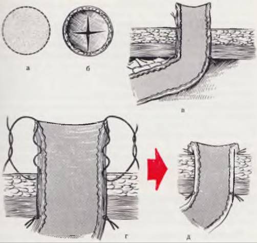
Fig. 90. Specific features of colostomy in a newborn.
An oval skin window 1 cm in diameter is excised (a). The aponeurosis is incised in a cruciate manner (b). The bowel is sutured to the parietal peritoneum (c). A spout-like stoma is created by suturing the end of the bowel to the skin using a purse-string (gather) suture (d, e).
Following gastric aspiration with a tube, the newborn is placed in the Trendelenburg position for 15 – 20 minutes, as also recommended by Swenson and Donnellan (1967). Then, turning the infant head-down, their legs, flexed at the hip joints, are brought toward the abdominal wall. This increases intra-abdominal pressure and improves gas progression into the blind pouch of the atretic bowel. Anteroposterior and lateral radiographs are obtained after straightening the infant's legs. Instead of a conventional radiopaque marker, it is recommended to use a barium paste. Applied in the area of the intergluteal cleft, it clearly outlines the posterior and lower BOUNDARIES OF THE pelvic soft tissues and facilitates orientation when determining the localization of the atretic bowel's blind pouch.
When interpreting an invertogram, it is necessary to rely not only on the distance between the gas and the contrast medium, but also on the relationship between the blind pouch and bony landmarks indicating the position of the levators.
Urethrocystography is a valuable diagnostic method for fistulous forms of atresia. Both ascending and descending urethrography can be performed, which reveal direct signs of a fistula (its opacification) and indirect signs (contrast medium entering the large intestine). Applying this method in newborns is challenging, and accurate data cannot always be obtained. Therefore, only a positive result—the presence of direct or indirect signs of a fistula—is taken into account.
For fistulous forms of atresia, fistulography is used, which helps determine the direction of the fistula, its length, and the level of atresia.
A barium enema examination in newborns with anorectal malformations is rarely used. The prolonged transit time of the contrast medium through the small intestine makes the procedure lengthy.
Puncture of the blind pouch of the atretic bowel is performed when low, non-fistulous forms of atresia are suspected. The depth of needle insertion upon obtaining meconium can serve as an indicator of the height of the atresia. Contrast medium injected after aspirating the meconium reveals the shape and position of the bowel. Probing the fistulous tract provides valuable additional information regarding the direction, length, width, and other CHARACTERISTICS OF THE fistula.
High anomalies. Anorectal agenesis without a fistula in boys and girls is a developmental defect in which the rectum ends blindly at some level above the levator ani muscles. Often, a fibrous cord extends toward the urethra in boys, or toward the vagina or perineum in girls. The internal sphincter is absent, and the external sphincter is rudimentary.
The diagnosis is established based on: 1) the absence of an anus; 2) the absence of gas and meconium in the urine; 3) the Location of gas on the invertogram at or above the pubococcygeal line; 4) the absence of a fistula, urethral deformities, or bends on the urethrocystogram that would indirectly indicate a fistula, with all of the contrast medium remaining in the Urinary Bladder.
Fistulous forms of anorectal agenesis present in boys as agenesis of the anorectal segment with a fistula into the urinary bladder and urethra, and in girls as a fistula into the urinary bladder, a common cloaca for the pelvic organs, and the vagina.
In rectovesical fistula in boys, the rectum opens directly into the urinary bladder, most frequently near its base. All sphincters are rudimentary. The fistula is frequently associated with other malformations.
The diagnosis is established based on the following findings: 1) the absence of an anus or its diagnostic markers; 2) the presence of gas and meconium in all portions of urine; 3) the location of gas on the invertogram above the pubococcygeal line; 4) the presence of gas in the urinary bladder; 5) visualization of the fistula by contrast medium on the fistulogram; 6) the passage of contrast medium through the fistula into the rectum.
In rectourethral fistula, the most common form in boys, the fistulous tract is narrow and in most cases opens into the posterior urethra adjacent to the vas deferens, although it may open above or below this urethral segment. The puborectal sling of the levator ani is shortened, closely embracing the urethra below or at the level of the fistula. The internal sphincter is underdeveloped, and the external sphincter is variable.
The diagnosis is established based on: 1) the absence of an anus; 2) the presence of gas and meconium in the urine; 3) the absence of urinary changes within the bladder; 4) the location of gas on the invertogram at or below the pubococcygeal line; 5) the possible presence of gas in the urinary bladder; 6) visualization of the fistula on the urethrogram.
In rectovesical fistula in girls, the rectum opens into the bladder between two separate vaginas that empty into the bladder via a common cloaca.
The diagnosis is established based on the following findings: 1) a single cloacal opening on the perineum; 2) the presence of gas and meconium in all portions of urine; 3) the location of gas on the invertogram above the pubococcygeal line; 4) the presence of gas in the urinary bladder; 5) visualization of a fistula on the cystogram; 6) the passage of contrast medium from the bladder into the rectum.
Both horns of the vagina may also be opacified with contrast medium.
In rectocloacal fistula in girls, unlike the previous form, the rectum, vagina, and urethra open into a common cloaca. The vagina may be short and single, or divided by a septum. The puborectal sling closely surrounds the cloaca below the fistula. The internal sphincter is underdeveloped, and the external sphincter is variable.
The diagnosis is established based on the following findings: 1) the presence of a single cloacal opening in the Vestibule of the vagina; 2) the presence of gas and meconium in the urine; 3) the inability to determine the height of the gas level on the invertogram; 4) the possible detection of the fistula using vaginoscopy and vaginography; 5) in cases of a wide fistula, the possibility of detecting it via a barium enema examination.
In high rectovaginal fistula in girls, the vagina is usually normal, and the fistula opens posteriorly along the midline with a large orifice. Both the internal and external sphincters are underdeveloped.
The diagnosis is established based on the following findings: 1) the presence of two openings in the vaginal vestibule and the absence of an anal orifice or its diagnostic markers; 2) the discharge of meconium from the vagina; 3) the inability to determine the height of the gas level on the invertogram; 4) the possibility of detecting the fistula via vaginoscopy and vaginography; 5) in cases of a wide fistula, potential detection through contrast enema examination of the large intestine.
Rectal atresia in both boys and girls is a malformation in which the terminal end of the atretic rectum is located high above the anal opening. A fibrous cord extends from it toward the sacrum or the anal canal. All sphincters are sufficiently well developed and maintain a normal spatial relationship with the formed anal opening and anal canal.
The diagnosis is established based on the following findings: 1) the presence of a normally formed anus; 2) the detection of an obstruction during digital rectal examination; 3) the location of gas on the invertogram at or below the pubococcygeal line; 4) upon administration of a contrast medium, the distal canal is visualized, allowing the extent of the atresia to be determined on the radiograph.
Intermediate anomalies. Anal agenesis without a fistula in boys and girls presents a similar clinical picture. The rectum ends blindly at the level of the bulbospongiosus muscles in boys and near the lower end of the vagina in girls. The puborectal sling surrounds the distal end of the rectum. The internal sphincter is underdeveloped, and the external sphincter is rudimentary.
The diagnosis is established based on the following findings: 1) the absence of an anal opening on the perineum; 2) the absence of gas and meconium in the urine; 3) the location of gas on the invertogram below the pubococcygeal line; 4) the absence of a fistula and urethral deformity on the urethrogram.
Anal agenesis with a rectobulbar fistula is observed in boys, whereas low rectovaginal and rectovestibular fistulas occur in girls.
In rectobulbar fistula in boys, the terminal end of the rectum opens via a wide or narrow fistula into the bulbous urethra or a more distal portion of the urethra. The rectum and the fistula lie within the puborectal sling, and a significant caudal portion of the fistula penetrates the tissues. The internal sphincter is absent, and the external sphincter is variable.
The diagnosis is established based on the following findings: 1) the absence of an anal opening; a narrow perineum, hypospadias, or a scrotal cleft may be present; 2) the presence of gas and meconium in the urine; 3) the free passage of a probe inserted via the urethra into the rectum; 4) the location of gas on the invertogram below the pubococcygeal line; 5) the possibility of detecting the fistula on the urethrogram; 6) The entry of contrast medium into the rectum during urethrography.
In low rectovaginal fistula in girls, the puborectal sling loops around the end of the bowel.
The diagnosis is established based on the following signs: 1) absence of the anal opening on the perineum and presence of two openings in the vaginal vestibule; 2) discharge of meconium from the vagina; 3) inability to determine the height of the gas bubble on the invertogram; 4) possibility of detecting the fistula using vaginoscopy and vaginography.
In rectovestibular fistula in girls, the terminal end of the rectum is located on the pubococcygeal line or slightly lower. A thin fistula, 1 – 2 cm long, opens in the vestibule near the posterior vaginal wall. Thus, the terminal segment of the bowel is located above the levators, and the fistula passes through them.
The diagnosis is established based on the following signs: 1) the anal opening is absent. There are three openings in the vaginal vestibule: the urethra, vagina, and a thin fistula, with hyperemia of the mucous membrane around the openings; 2) a probe passes only upward through the fistula; 3) the length of the fistula can be measured with a catheter; 4) fistulography makes it possible to determine the length, width, and direction of the fistula.
Anorectal stenosis in boys and girls manifests as stenosis of the anal canal with a normally formed anus. The stenosis extends to the lower end of the rectum. All sphincters are developed normally.
The diagnosis is established based on the following signs: 1) a normally formed anal opening is present; 2) the obstruction is detected by Palpation (rather than with a probe!); 3) the length of the stricture can be determined using a fistulogram.
Low malformations. The anal opening is in its usual location. In this condition, both boys and girls exhibit complete closure of the anal opening by an excessively developed posterior genital fold, which has the appearance of a thick seam. The puborectal sling surrounds a normal anorectal canal. The external sphincter is variable.
The diagnosis is established based on the following signs: 1) presence of a thick fold on the perineum; 2) presence of gas below the pubococcygeal line on the invertogram; 3) possibility of determining the thickness of the fold by percutaneous puncture.
Anal stenosis in boys and girls is a malformation similar to the previous one, but the anal opening represents a stenotic canal or a fistula of the median fold.
The diagnosis is established based on the following signs: 1) a hypertrophic fold with a fistula is present on the perineum; 2) anal stenosis is visible on the fistulogram.
Anal opening on the perineum. Anterior perineal anus in boys and girls is a malformation in which the normal anal opening is located anterior to its usual site, between it and the Base of the scrotum in boys and the frenulum in girls.
The diagnosis is established based on the following signs: 1) a normal anus is present, located more anteriorly; 2) X-ray Examination with a contrast medium reveals an unchanged anorectal canal.
Cutaneous-anal fistula is more common in boys. There is excessive growth of the genital fold in the form of a flap covering the anus. The fistula opening is displaced anteriorly. It may open anywhere behind the Penis, but most often at the base of the scrotum, and in girls, between the posterior vaginal frenulum and the normal LOCATION OF THE anus. The anal canal is fully formed up to the flap, and the fistula is covered with a thin membrane. The internal sphincter is absent, and the external one is variable.
The diagnosis is established based on the following signs: 1) a distinct fistula opening or traces of meconium are present on the perineum; 2) the fistula is filled with meconium. The anal pit or hypertrophic fold is pronounced; 2) the probe passes backward along the fistula into the anal canal; 3) the fistulogram confirms the presence and extent of the fistula; 4) a direct bowel puncture confirms the diagnosis.
3. Anal opening in the vulvar region. Vulvar anus is a variant of anterior anus located in the vaginal vestibule. The anal opening is normally developed, but somewhat narrowed, usually surrounded by moist epithelium. All sphincters are developed normally.
The diagnosis is established based on the presence of three openings in the region of the vaginal vestibule.
Anovulvar fistula is another variant of a closed anal opening with a fistula in the posterior part of the vulva.
The diagnosis is established based on the following signs: 1) three openings are present in the vulvar region; 2) the fistula opening is located at the edge of the vaginal vestibule with moist epithelium anteriorly and skin posteriorly; 3) a rudimentary tail is present; 4) a probe in the fistula passes subcutaneously backward and cranially; 5) fistulography confirms the diagnosis.
Fig. 91. Skin incisions in the anal area during proctoplasty.
A longitudinal incision tends to undergo circular scarring and should not be used (a). A cruciate incision (b), excision of an oval-shaped skin area (c), and a Z-plasty incision (d) are not accompanied by cicatricial stenosis.

Anovestibular fistula is embryologically identical to rectovestibular fistula, but the anatomical resemblance is very slight. The rectum lies close to the vagina. The puborectal sling normally encircles the rectum. The internal sphincter is rudimentary, and the external one is variable.
The diagnosis is established based on the following indicators: 1) three openings are present in the vaginal vestibule; 2) the fistula opens in the navicular fossa and is surrounded by a moist mucous membrane; 3) a probe in the fistula passes immediately cranially and posteriorly; 4) the fistulogram confirms the diagnosis.
Mixed malformations. Imperforate anal membrane. In this developmental anomaly, a membrane bulging with meconium is present in the area of a normally formed anal opening.
Anal membrane stenosis is a similar anomaly, but with partial perforation of the membrane.
Mixed malformations also include vesicointestinal fistula; duplication of the anus, rectum, or urogenital organs;
Perineal groove. The latter is observed in girls. In this anomaly, a moist groove extends from a normally formed vaginal vestibule to the anus. The groove tends to undergo epithelialization. The perineal canal usually connects the normally formed anal canal with the fossa navicularis of the vestibule.
A special group, not included in the above classification, consists of rectovestibular fistulas with a normally formed anus.
The diagnosis is established based on the following signs: 1) the presence of a fistula in the vaginal vestibule with a normal anus; 2) the discharge of liquid stool and gas through the fistula; 3) the free passage of a probe inserted into the fistula into the anorectal canal.
Anorectal malformations are frequently combined with developmental defects of other organs. According to our clinic's data (T. I. Kontselidze, 1970), associated malformations and anomalies occur in 28% of children with anorectal malformations. The most common are developmental defects of the Urogenital System, Musculoskeletal System, Digestive System, etc.
Treatment. Developmental defects of the anus and rectum that hinder meconium discharge and lead to symptoms of intestinal obstruction require surgical correction in the first days of the patient's life. These include all forms of complete atresia and the majority of fistulous forms. A mislocated normally formed anal opening or atresia with wide fistulas into the Reproductive System in girls should be corrected at a later stage.
In cases of developmental defects of the anus and rectum requiring early correction, surgery is performed on the first day after birth, before the onset of symptoms of intestinal obstruction. Depending on the form of
atresia, colostomy (Fig. 90), perineal proctoplasty, and combined types of abdominoperineal proctoplasties can be used.
Perineal proctoplasty is used in newborns with low and intermediate deformities. The surgery consists in mobilizing the blind end of the rectum. In non-fistulous forms of atresia, the intestine is mobilized by 1.5 — 2 cm, after which its mucous membrane is sutured to the perineal skin (Fig. 91). The intestine is preliminarily cleared of meconium.
In perineal fistulas in boys and girls, and in low fistulas into the reproductive system in girls, fistulectomy is performed.
Abdominoperineal proctoplasty is used for high deformities and can also be used for certain forms of intermediate deformities, in particular anal agenesis with a rectourethral (bulbar) fistula and anorectal stenoses.
In newborns, Gross abdominoperineal proctoplasty is used, which is indicated for very high forms of atresia, such as anorectal agenesis with a rectovesical fistula in boys and girls (Fig. 92). For other forms, the intrarectal abdominoperineal proctoplasty according to Romualdi can be applied.
Fig. 92. Passing the intestine through the puborectal sling.
Surgical treatment of high rectal atresia provides the opportunity to ensure fecal continence.
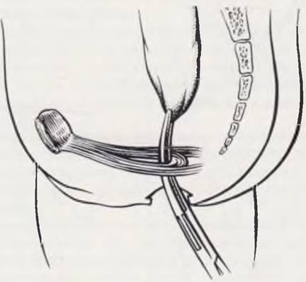
Technique of the Gross abdominoperineal proctoplasty. A midline laparotomy is performed. The sigmoid and rectum are mobilized after incising the parietal peritoneum. Mobilization of the rectum is carried out down to the fistula. The fistula is carefully dissected, its vesicular end is ligated and severed. On the perineal skin, at the site where the anus should be, a circular incision is made, the Muscles of the external sphincter are carefully separated, and behind the urethra (into which a catheter has been inserted), an instrument is gently advanced into the anorectal canal. The severed end of the sigmoid or rectum is grasped with an instrument and pulled through the anorectal canal onto the perineum. The integrity of the damaged peritoneum is restored from the abdominal cavity. On the perineum, the wall of the exteriorized intestine is sutured to the subcutaneous tissue, the intestine is opened, and its mucosa is sutured to the skin. If a significant part of the intestine has been successfully brought down to the perineum, it can be left intact for several days. In such a case, a catheter is inserted into the intestine before closing the laparotomy wound and secured to the exteriorized bowel. The resection of the excess bowel and suturing of the mucosa to the skin are performed after 18 — 20 days.
Technique of the Romualdi intrarectal abdominoperineal proctoplasty as modified by A. I. Lenyushkin. A midline laparotomy is performed. The sigmoid and rectum are mobilized. The sigmoid colon is transected 5 cm above the transition fold. Its distal end is cleared of meconium, and the mucosa is separated from the serromuscular layer. The isolated cylinder of mucosa, which transitions into the fistula, is ligated and resected. Then, a tunnel is created to pull
the intestine down to the perineum. To do this, an oval, cruciate, or Z-shaped skin incision is made from the perineum, and the external sphincter muscles are gently separated with an instrument. A finger of the left hand is inserted into the resulting canal, while the index finger of the right hand is simultaneously inserted into the rectal canal from the abdominal cavity. Guided by the metal catheter placed in the urethra, both fingers are brought as close to it as possible. Under the control of the right index finger, a clamp is introduced into the perineal wound and used to perforate the blind end of the demucosalized section of the rectum. Through the created tunnel, the end of the mobilized intestine is brought out to the perineum and fixed with interrupted sutures to the subcutaneous tissue and external sphincter. The exteriorized segment, 4 — 5 cm long, is left hanging freely with a rubber tube secured inside it. Resection of the redundant bowel is performed after 20 — 25 days.
Last update: 10/08/2026
Editorial and Educational Adaptation: This material has been compiled based on the primary/original source text. The project team performed an editorial review, corrected technical inaccuracies, structured sections, and adapted the content for an educational format.
What was processed:
- elimination of formatting defects (OCR errors, structural breaks, corrupted characters);
- editorial organization of content;
- standardization of terminology in accordance with academic sources;
- verification of factual statements against the original source text.
All mentions of the author, publication year, and origin of the primary text have been preserved in accordance with the source.