NEONATAL SURGERY - 1976
2. SPECIAL CHAPTERS
8. Malformations of Individual Organs and Systems
Malformations of the Bile Ducts and Gallbladder
Malformations of the bile ducts and gallbladder are relatively rare, accounting for no more than 6% of all congenital anomalies. In the neonatal period, these anomalies primarily manifest as disorders of bile secretion associated with malformations of the gallbladder and bile ducts. The latter conditions (atresia and Aplasia of the biliary tract) occur on average once every 20,000 to 30,000 births. The occurrence of biliary atresia is attributed to Specific features of embryonic development.
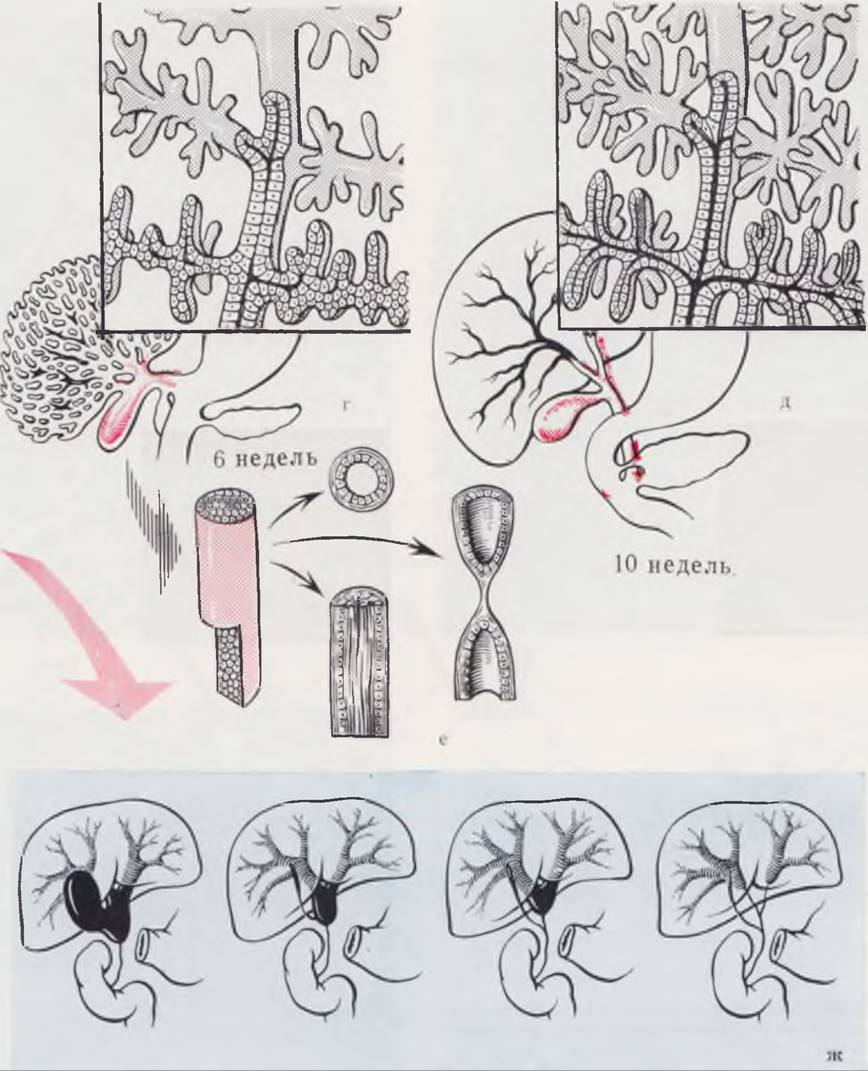
The Embryogenesis OF THE biliary duct System and Its main developmental anomalies are schematically illustrated in Fig. 93.
Class="center">Fig. 93. Stages of Formation of the biliary duct system. Malformations of the bile ducts.
The hepatic diverticulum, from which the secretory PARTS OF THE Liver develop together with the duct system and gallbladder, begins to form during the 4th week of embryonic development. Initially, it appears as a thickened area near the anterior intestinal portal (a) and consists of proliferating entodermal Cells. From the primary diverticulum bud, a labyrinth of branching and anastomosing Cell cords grows in ventral and cranial directions (b, c). The secretory parts (hepatic cords) arise from the distal segments of these cords, while the hepatic ducts arise from the proximal segments. The gallbladder originates from an outgrowth of the hepatic diverticulum (v, g), and as the gallbladder develops and elongates, numerous hepatic ducts—representing the proximal sections of the biliary system—open into its duct. The distal parts of the biliary system develop in parallel with the secretory units. Initially, the growing cell cords lack excretory ducts. The latter appear and anastomose as active secretory hepatic cords are formed (see diagrams in the top right corner of Fig. g, d). Subsequently, As a result of the fusion of the distal and proximal sections of the biliary ducts (d)—which develop independently until connection—a unified biliary system is formed.
The Development of extrahepatic bile duct atresia is attributed to impaired normal embryogenesis during the stage of hepatic diverticulum formation (a, b) or to aberrant Development of the distal segments of the biliary system (v, g, d). Variants of this type of atresia are shown in Fig. z.
The development of extrahepatic bile duct atresia (zh) is explained by a disruption in The formation of the proximal biliary tract segments (v) or by a disturbance in the resolution of the solid cord stage (e), which this segment of the biliary tract undergoes alongside other sections of the intestinal tube between the 20th and 45th days of embryonic development. Congenital giant cell hepatitis is considered to play a significant role in the Pathogenesis of atresias.

Clinical presentation. The clinical manifestations of biliary atresia are typical in most cases. The infant is born with jaundice of the Skin and sclera (in intrahepatic atresia), or jaundice develops within the first days after birth. Stools are acholic (clay-colored). Individual portions of stool may be slightly pigmented due to the diffusion of bile pigments through the intestinal wall. The urine resembles the color of dark beer. A gradual progression of jaundice is characteristic. The liver (showing signs of bile stasis and biliary cirrhosis) and Spleen enlarge. Notable features include abdominal distension (intestinal paresis) and dilation of the anterior abdominal wall Veins. Blood tests reveal a significant increase in direct (conjugated) bilirubin levels. In later Stages of the disease, ascites develops, accompanied by hemorrhagic manifestations. Without Treatment, infants die by 6 to 9 months of age from Biliary Cirrhosis of the liver.
Fig. 94. Congenital Diseases of the Liver and biliary tract. Macro- and microscopic manifestations.
a — congenital hepatitis, photolaparoscopy: the Inferior surface of the right lobe of the liver is visible; the gallbladder is filled with clear fluid. Histological specimen of a liver biopsy: a multinucleated symplast formed by the fusion of neighboring hepatocytes; hematoxylin-eosin staining; ×250; b — total form of biliary tract malformation, photolaparoscopy: the inferior surface of the liver is visible; the gallbladder rudiment located in the cystic notch. Histological specimen of a liver biopsy: bile ducts presented as epithelial cords; proliferation of cholangioles; biliary cirrhosis; hematoxylin-eosin staining, ×250; c — malformation of the Extrahepatic bile ducts, photolaparoscopy: the inferior surface of the liver is visible; gallbladder rudiment. Histological specimen of a liver biopsy: cholestasis in normally formed interlobular bile ducts; hematoxylin-eosin staining, ×250.
Special attention is drawn to Structure/19.html">The Importance of instrumental Palpation during endoscopic examination of the liver surface. Clear visibility of the gallbladder rudiment, located in the depression of the cystic notch and shown in Fig. b, was achieved using a probe-palpator (visible on the right side of the photolaparogram, also in Fig. c).

Timely Diagnosis of congenital biliary atresia presents considerable difficulties due to the similarity of clinical manifestations among various diseases accompanied by prolonged jaundice syndrome.
Differential diagnosis is performed with prolonged physiological jaundice of the newborn, hemolytic disease of the newborn due to blood group incompatibility, intrauterine hepatic parenchymal damage (neonatal giant cell hepatitis), Viral Hepatitis, obstruction of the bile ducts by mucus and bile plugs, generalized forms of cytomegalovirus infection, Toxoplasmosis, Syphilis, and septic jaundice.
Physiological jaundice is the most frequent manifestation of the Relative immaturity of The Liver and enzyme systems in a newborn infant. In typical cases, its diagnosis is straightforward. However, errors may occur in cases of prolonged jaundice. Unlike biliary atresia, prolonged physiological jaundice of newborns presents with overly pigmented stools due to The excretion of large amounts of thick bile. The urine is dark. Blood tests show a moderately elevated bilirubin level, predominantly at the expense of the indirect (unconjugated) fraction (whereas in biliary atresia, the direct, conjugated fraction prevails). In physiological jaundice, clinical symptoms tend to regress, whereas in biliary atresia, they steadily progress.
In most cases of hemolytic jaundice, there is an incompatibility involving the Rh factor or ABO blood group system. As in physiological jaundice of the newborn, indirect (unconjugated) bilirubin predominates in the blood. Pronounced, rapidly progressive jaundice and a severe General condition of the sick infant are characteristic. If timely exchange transfusion is not performed, symptoms of kernicterus rapidly develop.
The diagnosis of congenital syphilis, toxoplasmosis, or cytomegalovirus infection is either ruled out or confirmed using serological tests, special assays, and the identification of disease-specific cells or intracellular inclusions.
The greatest difficulties are encountered in the Cytology/practical/136.html">DIFFERENTIAL DIAGNOSIS OF biliary atresia, obstruction of the bile ducts by mucus or bile plugs, and congenital giant cell hepatitis. The clinical manifestations of these conditions and The Nature of bilirubin METABOLISM disorders in them are largely identical, which precludes a precise diagnosis. As a result of diagnostic errors, infants are admitted to specialized departments in advanced stages with pronounced symptoms of biliary cirrhosis, which significantly worsens treatment outcomes.
Differential diagnosis of bile duct obstruction by mucus plugs, giant cell hepatitis, and biliary atresia begins by attempting to identify patients whose prolonged jaundice is caused by mucus or bile plugs blocking the ducts. For this purpose, all patients undergo the magnesium sulfate test (Gross, 1958). Five milliliters of a 25% magnesium sulfate solution are administered via a gastric tube twice daily for 3 days. Magnesium solution is a potent choleretic agent. Its administration into The Stomach induces contraction of the biliary tract, which in some cases leads to the dislodgement of plugs and the recovery of the infant.
At our current level of knowledge, differential diagnosis between biliary atresia, congenital giant cell hepatitis, and preoperative Determination of the type of atresia are only possible through The Use of specialized Instrumental Diagnostic Methods, such as laparoscopy and percutaneous needle liver biopsy (A.B. Okulov, 1965) (Fig. 94). Attempts to opacify the bile ducts via cholecystocholangiography are unsuccessful in most cases, and even when successful, do not provide sufficient information for a definitive diagnosis (M.A. Filippkin, 1968). In cases of suspected biliary atresia or congenital dilatation of the bile ducts, ultrasonic echography may be useful (Suruga et al., 1966).
Laparoscopy and percutaneous liver biopsy in such patients are performed under general anesthesia following the guidelines described above. In children with extrahepatic biliary atresia or total biliary atresia, the liver exhibits the characteristic appearance of biliary cirrhosis (the liver surface is dark green and finely granular or nodular). The extrahepatic bile ducts are completely absent, or a rudimentary gallbladder filled with clear fluid is visible. In patients with congenital giant cell hepatitis, the general appearance of the liver is very similar to that seen in biliary atresia. The gallbladder is formed and flaccid.
The Nature and severity of liver parenchymal damage, as well as the state of the intrahepatic bile ducts, are clarified through histological examination of liver core biopsy specimens.
Patients with extrahepatic biliary atresia show marked dilation of the intrahepatic bile ducts. In intrahepatic bile duct aplasia, the ducts are either absent in the histological section or appear as epithelial cords lacking a lumen. The severity of liver parenchymal damage depends on the time elapsed since the child's birth.
Characteristic Changes in the liver parenchyma are detected in biopsy specimens from patients with giant cell hepatitis. The architecture of the hepatic lobules is generally preserved. There is a disruption of the trabecular structure with dissociation of hepatic cells. A key feature is the presence of giant cells, which may replace a large portion of the hepatic parenchyma. These multinucleated giant cells are large symplastic structures formed by the fusion of hepatocytes whose cell membranes have undergone destruction. This histological picture is considered typical for giant cell hepatitis.
It is not uncommon to observe a combination of congenital giant cell hepatitis and a form of biliary atresia (E. N. Ter-Grigorova, V. S. Ter-Grigorov, 1967; Perry, 1949; Peterman, 1957; Silverberg, 1960). We have conducted studies in 18 patients. In all cases, performing concurrent laparoscopy and percutaneous liver biopsy enabled the differential diagnosis between biliary atresia and giant cell hepatitis, and allowed the specific form of atresia to be determined prior to surgery. As a result, giant cell hepatitis was diagnosed in 6 patients, sparing them from unnecessary surgery.
Treatment. Biliary atresia requires surgical intervention (Fig. 95). During preoperative preparation, special attention is paid to normalizing Blood Coagulation parameters, as patients with biliary atresia are prone to bleeding.
Surgical technique. The Abdominal cavity is opened via an incision parallel to the right costal margin, positioned 1.5–2 cm below it. An exploration of the liver and extrahepatic bile ducts is performed to ascertain the specific form of atresia.
In cases of partial extrahepatic biliary atresia, the remnants of the bile ducts are anastomosed to the duodenum. If extrahepatic ducts are absent while intrahepatic ducts are present, an attempt is made to divert bile into the gastrointestinal tract by creating a double hepatogastric and hepatoduodenal anastomosis according to G. L. Bairov, or surgeons attempt to establish an external biliary fistula with its subsequent transplantation into the intestine.
In total biliary atresia, to partially divert bile into the digestive tract and prolong the child's life, Thoracic duct-to-Esophagus anastomosis is performed, or liver transplantation is attempted (Starzl, 1968).
Kasai (1959, 1968, 1974) successfully applied the method of hepatoportoenterostomy in treating previously uncorrectable forms of biliary atresia.
The rationale behind this method was based on histological studies demonstrating the presence of microscopic bile ducts, varying in size and number, which are capable of satisfactorily performing bile excretion when proper drainage into the lumen of the newborn's Small Intestine is established.
Performing hepaticojejunostomy in 57 patients with uncorrectable forms of biliary atresia using the author's technique resulted in active bile excretion in 19 patients, with 8 of them remaining jaundice-free for 3 years. The longest follow-up period with a favorable outcome was 17 years.
The results of Surgical treatment directly depend on the type of atresia and the timing of the surgical intervention. The best outcomes are achieved within the first 2 months of life in infants with operable extrahepatic forms of atresia.
Fig. 95. Types of surgical Procedures for biliary atresia.
a — cholecystoduodenostomy; b — choledochoduodenostomy; c — Roux-en-Y choledochojejunostomy; d — Bairov gastroduodenohepatostomy; e — creation of an external biliary fistula; f — Roux-en-Y hepaticojejunostomy according to Morio Kasai; g — thoracic duct-to-esophagus anastomosis according to Suruga.

Last update: 10/08/2026
Editorial and Educational Adaptation: This material has been compiled based on the primary/original source text. The project team performed an editorial review, corrected technical inaccuracies, structured sections, and adapted the content for an educational format.
What was processed:
- elimination of formatting defects (OCR errors, structural breaks, corrupted characters);
- editorial organization of content;
- standardization of terminology in accordance with academic sources;
- verification of factual statements against the original source text.
All mentions of the author, publication year, and origin of the primary text have been preserved in accordance with the source.