Human Anatomy - H. I. Koliadenko 2009
The Doctrine of Vessels (Angiology)
General Information
Significance of the Nervous system. The Human Body stands at the highest stage of evolutionary development in the animal kingdom; therefore, its Functions can be controlled only by a highly organized mechanism, which is The Nervous System. Its primary functions are to ensure the integration of all Organs and systems of the body, making it possible to perceive the Organism as a single whole, and to maintain a close relationship between the body and the external environment.
The activity of the nervous system is reflex-based. A reflex is the body's response to a stimulus mediated by the Central Nervous System (CNS). Reflex connections between organs and with the central nervous system are made possible by such specific properties as excitability, impulse propagation over a certain distance, and response to a stimulus.
Structure of Nervous Tissue. Nervous tissue consists of Neurons (Nerve Cells) and intercellular substance (neuroglia). Neuroglia is subdivided into macroglia, which performs supportive and trophic functions, and microglia, represented by mobile cells that perform the function of phagocytosis.
The structural unit of the nervous system is the nerve Cell—the neuron, or neurocyte (Fig. 140). A neuron has Two Types of processes: an axon and dendrites. There is only one axon, or neurite, which conducts nerve impulses away from the neuron's cell body. A neuron may have several dendrites, which conduct nerve impulses from The Cell body toward the effector organ or other neural structures. Depending on the number of processes, neurons are classified as unipolar (with one process), bipolar (with two processes), and multipolar (with many processes). Depending on the direction of excitation, neurons are divided into afferent and efferent. Afferent, or sensory, neurons perceive external and internal stimuli and transmit them to the central nervous system. Efferent, or motor, neurons transmit neural excitation from the central nervous system to the effector organ.
Class="center">
Fig. 140. Diagram of a neuron:
1 — cell body with nucleus; 2 — axon; 3 — nerve fiber; 4 — dendrite
On the outside, nerve processes are covered by one or two sheaths enclosing an axial cylinder. These processes are called nerve fibers. Depending on the number of sheaths, fibers are divided into myelinated and unmyelinated. Myelinated fibers have two sheaths: an outer neurolemma (Schwann sheath) and an inner myelin sheath, which consists of a major neurolemma and Lipoproteins. Unmyelinated fibers are covered by only a single layer of glial Schwann cells. The somatic nervous system is built of myelinated fibers, whereas the majority of fibers in the Autonomic (vegetative) nervous system are unmyelinated. Functionally, these two types of fibers also differ: myelinated fibers conduct nerve impulses at a speed of 50–120 m/s, while unmyelinated fibers conduct them at 3–7 m/s. The former are also referred to as thick white fibers, and the latter as thin gray fibers.
Together, nerve cells form the Gray matter of the Brain, while nerve processes, by connecting with each other, form the White matter OF the brain and nerve fibers capable of excitation and Nerve Impulse Conduction. A sensory nerve fiber, branching out, forms numerous nerve endings located on the Skin surface, in the hypodermis, and in Internal Organs, which are called receptors. Receptors are sensory nerve endings that perceive stimuli. They are subdivided into exteroceptors (external), interoceptors (internal), and effectors (terminal endings of axons in effector organs).
Synapses are specialized structures of the nervous system. Although synapses vary in structure, all of them conduct nerve impulses in only one direction. Under an Electron microscope, one can observe The structure of a synapse (Fig. 141), which consists of the expanded, funnel- or ring-shaped ending of a nerve fiber containing oval or round vesicles. Its thickened surface features the presynaptic membrane, while the postsynaptic membrane is located On the surface of another cell. Between these membranes lies the synaptic cleft. The walls of the vesicles release a substance known as a neurotransmitter. The postsynaptic membrane of the synapse has numerous folds containing receptors sensitive to the neurotransmitter. Depending on the functional orientation of the nerve fiber, this substance can be excitatory or inhibitory. Consequently, synapses are divided into excitatory and inhibitory. Neurotransmitters transmit excitation from one nerve cell to another.

Fig. 141. Diagram of synapse structure:
1 — neurite; 2 — mitochondrion; 3 — synaptic vesicles; 4 — synaptic cleft; 5 — dendrite; 6 — pre- and postsynaptic membranes
Reflex Nature of the nervous system's activity. For a reflex to occur, a special pathway along which neural excitation propagates is necessary. A chain of consecutively connected neurons forms a reflex arc, which serves as the material substrate of a reflex.
The simplest reflex arc consists of two neurons: an afferent (sensory) and an efferent (motor) neuron. An example of a two-neuron reflex arc is the tendon-Muscle knee-jerk reflex.
The reflex arc is the Structural and functional unit of the nervous system (Fig. 142). It begins with a receptor that perceives and transforms a stimulus into a nerve impulse, which is then transmitted to the centripetal, or afferent (sensory), nerve fiber, and from there to an associative (intercalated) neuron. This interneuron transmits the impulse to the centrifugal, or efferent (motor), nerve fiber, which is directly connected to the effector, or working organ. Thus, the roles of neurons in a reflex arc are unequal; each of them is responsible for a separate link of reflex activity. There may be several interneurons. The more interneurons there are, the more complex the reflex arc. Centripetally, the impulse travels through a chain of interneurons to the Cerebral Cortex, and centrifugally reaches the effector from there. Such a reflex arc is complex, and it mediates a multitude of intricate, coordinated human movements.
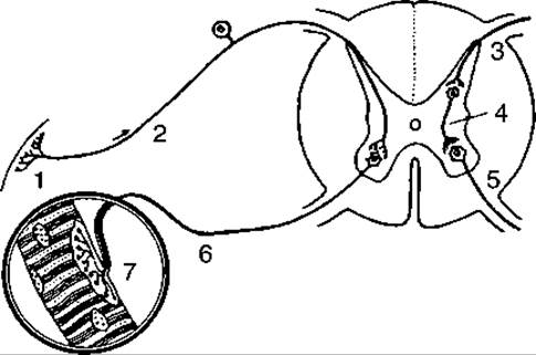
Fig. 142. Diagram of a reflex arc — two-neuron (left) and three-neuron (right):
1 — receptor; 2, 3 — afferent (sensory) neuron; 4 — interneuron; 5, 6 — efferent neuron; 7 — nerve ending in striated muscle
S.P. Botkin discovered and proved The phenomenon of nervism, which I.P. Pavlov understood as "a physiological trend that attempts to extend the Influence of the nervous system to as large a scope of the organism's activity as possible."
METABOLISM/2.html">THE CONCEPT OF analyzers. The definition of analyzers as organs of sense was first introduced into physiology by the great Russian scientist I.M. Sechenov. Later, I.P. Pavlov experimentally substantiated and developed this concept. According to Pavlov, an analyzer consists of a peripheral part (receptor) that perceives Changes in the external environment; a conducting pathway represented by a sensory centripetal nerve and a chain of interneurons; and a central part located in the cerebral cortex. According to this doctrine, the receptors of Sense Organs constitute the perceptive part. Excitation is transmitted to the brain, where it undergoes complex analysis and synthesis. These intricate processes take place in the cerebral cortex, enabling a person to adapt as effectively as possible to environmental influences and interact closely with the environment.
According to I.P. Pavlov's teaching, an analyzer is only part of a reflex arc. It is a region of the cerebral cortex connected to a motor or secretory neuron via descending interneurons. I.P. Pavlov deeply and consistently studied and proved the reflex nature of the nervous system, elucidating the leading role of the higher Divisions of the brain in regulating the functioning of internal organs.
The work of P.K. Anokhin proved that the reflex arc is closed through feedback afferentation; that is, the effector organ does not passively receive impulses from the central nervous system, but sends back feedback impulses that introduce necessary corrections into The regulation of reflex influences on the organ. In this regard, the reflex arc is referred to as a reflex ring.
Divisions of the nervous system. The nervous system consists of two parts: central and peripheral. The central nervous system includes the BRAIN AND SPINAL cord, while the Peripheral Nervous System includes nerves, nerve plexuses, ganglia, and nerve trunks. The brain and Spinal Cord are formed by the combination of nerve cells, which together make up the gray matter of the brain, whereas the white matter is formed by the combination of associative, commissural, and projection fibers that perform a conducting function. Gray matter is localized on The surface of the cerebral hemispheres, and in the spinal cord, it is located in the center. The white matter of the Brainstem and spinal cord lies on the periphery, covering the gray matter from above.
NERVES OF THE peripheral nervous system consist of bundles of nerve fibers connected by loose Connective Tissue through which Blood Vessels supplying the nerve pass. Externally, the nerve is covered by a Cytology/practical/45.html">Dense connective tissue sheath called the epineurium. Nerve plexuses, nodes, or ganglia are also built of nerve cells located outside the brain and spinal cord. These include spinal ganglia, cranial nerve ganglia, and numerous ganglia of the autonomic (vegetative) nervous system.
The central and peripheral nervous systems are further subdivided into the somatic and autonomic nervous systems.
The somatic nervous system innervates the soma—that is, the body proper—which encompasses skeletal Muscles, the Integumentary System (external body coverings), sensory receptors (analyzers), and the mucous membranes of the nasal and oral cavities.
The autonomic, or vegetative, nervous system innervates internal organs, glands, and blood vessels. Blood vessels are an integral component of muscles, which receive dual innervation from both the somatic and autonomic nervous systems.
Embryogenesis OF THE nervous system. In the early Selection/3.html">Stages of development, in a 2.5-week-old embryo, the neural plate forms from ectodermal cells on the DORSAL SIDE OF the body. Driven by Cell Division, the plate grows and invaginates to form the neural groove, flanked by neural folds. The edges of the groove gradually fuse to form the neural tube. The folds merge with one another to give rise to the ganglionic plate. The neural tube consists of three layers: the inner layer (ependyma), which lines the interior of the neural tube; the middle layer, which develops into the gray matter of the brain; and the outer layer, which gives rise to the white matter. The neural tube progressively grows, increases in size, and differentiates into an anterior (expanded) and a posterior (narrowed) region. The brain develops from the anterior part, while the spinal cord develops from the posterior part.
The walls of the neural tube are composed of two types of cells: neuroblasts and spongioblasts. Neuroblasts develop into neurons, whereas spongioblasts differentiate into macroglial cells.
Along the lateral surfaces of the neural tube, a longitudinal furrow forms on each side, dividing the tube into an alar (upper) plate and a basal (lower) plate. Subsequently, interneurons develop from the alar plate, while motor neurons originate from the basal plate.
The lateral sulci serve as the sites where the ganglia of the autonomic (vegetative) nervous system begin to develop. By the third week of development, the rostral (cephalic) end of the neural tube expands, forming three successive primary brain vesicles: the Forebrain (prosencephalon), Midbrain (mesencephalon), and Hindbrain (rhombencephalon). By the end of the fourth week of embryonic development, the forebrain vesicle divides into two: the Telencephalon, which gives rise to the cerebral hemispheres, and the Diencephalon, which forms the diencephalon (interbrain). The midbrain (mesencephalon) does not divide and serves as the foundation for The Development of the midbrain of the same name.
During the fifth week of development, the third brain vesicle divides into two vesicles (metencephalon)—giving rise to the Pons and Cerebellum (the metencephalon or hindbrain)—while the myelencephalon develops from the lower part of the rhombencephalon to form the Medulla Oblongata. The brain vesicles grow at uneven rates, resulting in The formation of three flexures: the midbrain (cephalic) and cervical flexures, which are directed ventrally, and the pontine flexure, which is directed dorsally.
The brain contains four cavities known as ventricles, which develop from the embryonic brain vesicles. Two Lateral ventricles—right and left—form within the region of the cerebral hemispheres. The Third ventricle is located in the diencephalon, the cerebral aqueduct (aqueduct of Sylvius) runs through the midbrain, and the Fourth ventricle is situated within the hindbrain and medulla oblongata.
The spinal and Cranial Nerves develop from the glial plate.
Last update: 08/08/2026
Editorial and Educational Adaptation: This material has been compiled based on the primary/original source text. The project team performed an editorial review, corrected technical inaccuracies, structured sections, and adapted the content for an educational format.
What was processed:
- elimination of formatting defects (OCR errors, structural breaks, corrupted characters);
- editorial organization of content;
- standardization of terminology in accordance with academic sources;
- verification of factual statements against the original source text.
All mentions of the author, publication year, and origin of the primary text have been preserved in accordance with the source.