Human Anatomy - H. I. Koliadenko 2009
Endocrine (Ductless) Glands
Characteristics of Endocrine Glands
The Thyroid Gland (glandula thyreoidea) is located in the anterior neck region, inferior to the Hyoid bone, covering the lateral surface of the Larynx and the anterolateral surface of the Trachea. The gland consists of two lobes and an isthmus, weighing between 30 and 60 g. Externally, it is enveloped in a fibrous capsule, which is smooth and transparent in young children. The glandular tissue is composed of individual lobules, each made up of microscopic vesicles known as follicles. These follicles contain a viscous colloid substance harboring the Hormones thyroxine and triiodothyronine, which are released directly into the bloodstream. They regulate METABOLISM, thermogenesis, Nervous system excitability, and tissue GROWTH AND DEVELOPMENT.
The thyroid gland undergoes changes throughout a person's lifespan. In newborns, for instance, it weighs only a few grams. During Puberty, it grows rapidly; by ages 11–15, its mass reaches 15 g, and by ages 16–20, it increases to 25–70 g. After 40–50 years of age, the gland decreases in size, and adipose tissue accumulates beneath its capsule. By ages 60–65, a portion of the follicles is replaced by Connective Tissue, arterial and Blood vessel counts decline, and a partial Atrophy of the gland takes place.
The Parathyroid glands (glandula parathyreoideae) are four small, oval or round bodies, each weighing between 0.01 and 0.05 g. They lie adjacent to the posterior surface of the right and left lobes of the thyroid gland—one pair positioned superiorly and the other inferiorly.
The parathyroid glands produce parathyroid hormone (parathormone), which facilitates calcium Absorption in the body and maintains its blood levels. The removal of these glands leads to impaired bone growth, heightened Central Nervous System excitability, Muscle spasms, and death from tetany.
The Thymus (thymus; Fig. 139) is situated in the upper portion of the anterior Mediastinum, positioned between the right and left mediastinal pleurae. It comprises asymmetrical right and left lobes connected by loose connective tissue. Externally, the gland is covered by a delicate connective tissue sheath, from which septa extend deep into the organ, dividing it into individual lobules. Each lobule consists of an outer cortex and an inner medulla. The gland is built of Epithelial Tissue whose Cells, connected via cellular processes, form a reticulum with interstices filled with lymphocytes. The thymic mass in a newborn is 10–15 g, reaching its peak of 25–35 g during puberty at ages 13–15. Following sexual maturation, the gland undergoes involution (regression).
Class="center">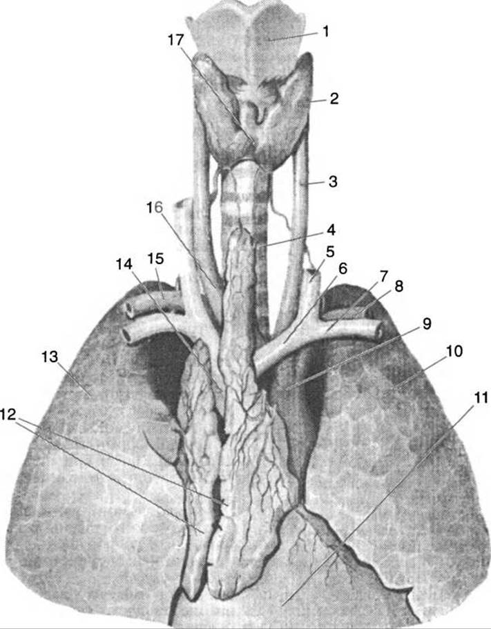
Fig. 139. The thymus and thyroid gland:
1 — thyroid Cartilage; 2 — thyroid gland; 3 — common carotid artery; 4 — trachea; 5 — Internal jugular vein; 6 — brachiocephalic vein; 7 — subclavian vein; 8, 15 — Subclavian Artery; 9 — aortic arch; 10 — left lung; 11 — Pericardium; 12 — thymus; 13 — right lung; 14 — SUPERIOR VENA CAVA; 16 — brachiocephalic artery; 17 — thyroid isthmus
Recent studies have established that the thymus controls the development and distribution of T-lymphocytes, which are responsible for cellular Immunity (resistance to numerous diseases). The function of the thymus includes The regulation of growth processes (calcium metabolism control and Carbohydrate Metabolism stimulation), as well as the suppression of the Gonads prior to sexual maturity. Isolating the thymic hormone has not yet been successful.
The Pancreas is a mixed-secretion gland. As an exocrine gland, it possesses a duct that opens onto the duodenal papilla, where its secretions are discharged. Its endocrine function is carried out by clusters of cells located in the body and tail of the gland, known as pancreatic islets (islets of Langerhans). These islets secrete the hormones Insulin and Glucagon. Insulin regulates Carbohydrate Metabolism in the body and increases Glycogen storage in the Liver. A drop in blood insulin levels raises blood glucose, leading to Diabetes Mellitus. The hormone glucagon promotes The breakdown of liver glycogen into glucose (it does not affect muscle glycogen), thereby increasing blood glucose levels. When glucose is present in excess in the blood, it cannot be utilized by the body and is excreted in the urine; the individual experiences weight loss and intoxication from incomplete carbohydrate breakdown products, blood pressure drops, fainting occurs, and death from diabetic coma may ensue.
The Adrenal Glands (glandula suprarenales) are located directly on the superior poles of the Kidneys. A gland weighs between 7 and 20 g and is about 5 cm long. It consists of two layers: an outer cortex and an inner medulla. The dark brown cortical substance secretes vital hormones: glucocorticoids (hydrocortisone, cortisone, and corticosterone), mineralocorticoids, and Sex Hormones. Glucocorticoids influence carbohydrate, protein, and Lipid Metabolism, affect Hematopoietic Organs, and exhibit anti-inflammatory effects. Mineralocorticoids participate in the regulation of mineral balance. Sex hormones promote The Development of sex organs during childhood as well as secondary sexual characteristics.
The adrenal medulla is dark brown, slightly darker than the cortex, and is formed by chromaffin tissue. Its medullary hormones, adrenaline (epinephrine) and noradrenaline (norepinephrine), accelerate Heart rate, raise blood pressure, and constrict small Arteries, with the exception of the Blood Vessels supplying The Heart, Lungs, and Brain. Adrenaline relaxes the smooth Muscles of the digestive tract, dilates the pupils, stimulates lacrimal and salivary gland secretion, and boosts metabolism. It enters the bloodstream during excitation of the Sympathetic division of the Autonomic nervous system.
Paraganglia are small clusters of chromaffin tissue situated along The pathway of the aorta and within sympathetic ganglia On the surface or inside organs. They are also found along the carotid arteries, near the heart (supercardiac), in the lumbar-aortic region, and elsewhere. Functionally, they are similar to the adrenal medulla, releasing the hormone adrenaline upon excitation.
The gonads. The gonads include the Testes in males and the Ovaries in females. These glands perform two primary Functions: the first involves hormone secretion, while the second involves The production of Germ Cells (Gametes).
The testes secrete a group of hormones known as androgens. Among the androgens, or Male Sex Hormones, testosterone is the most prominent; male sexual activity depends directly on its concentration in the blood. Furthermore, testosterone, acting alongside Other Hormones, promotes enhanced Protein Synthesis, which in turn positively influences skeletal Muscle Development.
In the ovaries, ova (eggs) mature and hormones known as estrogens are produced. Because one of the primary hormones is secreted by the follicular walls, it was named folliculin (estrone). Folliculin drives the development of secondary sexual characteristics. Another equally important hormone is progesterone, or lutein, secreted by the corpus luteum; it prepares the uterine lining for egg implantation, influences placental development, and inhibits ovulation during Pregnancy.
The Pineal Gland, or epiphysis cerebri (corpus pineale), is an unpaired Structure of oval shape that develops from the Diencephalon. In children, the pineal body is relatively larger than in adults, with an average mass of 0.2 g. The gland reaches its maximum development by age 7, after which it undergoes involution. Calcium phosphate and calcium carbonate salts—known as brain sand (corpora arenacea)—are deposited in place of the glandular tissue. The pineal hormone, melatonin, suppresses gonadal development and prevents precocious puberty.
It is believed that melatonin is secreted primarily at night, while light suppresses its effects; hence, the epiphysis is often referred to the body's biological clock.
The hypophysis, or Pituitary Gland (hypophysis), is a small oval gland weighing up to 0.5–0.7 g, situated in the hypophyseal fossa (sella turcica) and enclosed by a Cytology/practical/45.html">Dense connective tissue capsule, with its superior surface covered by the diaphragma sellae. The pituitary consists of three distinct lobes: the anterior lobe (adenohypophysis), the posterior lobe (neurohypophysis), and the intermediate lobe. The hypophysis releases over 20 hormones into the bloodstream.
The anterior lobe produces somatotropin (Growth Hormone). If its secretion is deficient or absent altogether—a condition known as hyposecretion of the anterior lobe—childhood growth is severely stunted, leading to Pituitary dwarfism. Pituitary dwarfs display proportionally developed body parts, normal cognitive abilities, and mental development, although secondary sexual characteristics are absent.
Hypersecretion of the anterior pituitary lobe during childhood causes gigantism, whereas in adults it results in the enlargement of certain skeletal elements (jaws, hands, feet, ears, Nose), a condition known as acromegaly. This can also be triggered by a pituitary tumor.
The anterior lobe of the hypophysis also produces corticotropin (adrenocorticotropic hormone, ACTH), which acts upon the adrenal glands; these, in turn, secrete Glycoproteins—substances that enhance the body's resistance to infection.
In addition to growth hormones, the anterior pituitary produces prolactin (lactogenic hormone), which stimulates milk secretion. This hormone exerts a positive effect on follicle and egg development in females, promotes sperm maturation in males, and stimulates the production of sex hormones in both men and women.
The posterior pituitary lobe produces vasopressin (antidiuretic hormone), which triggers smooth Muscle contraction, thereby raising blood pressure and reducing urine output. The hormone oxytocin stimulates the contraction of the smooth muscles of the Uterus. During Lactation, oxytocin also influences milk production.
The intermediate lobe is poorly developed and produces melanocyte-stimulating hormone (intermedin), which stimulates pigment formation to regulate Skin coloration.
Last update: 08/08/2026
Editorial and Educational Adaptation: This material has been compiled based on the primary/original source text. The project team performed an editorial review, corrected technical inaccuracies, structured sections, and adapted the content for an educational format.
What was processed:
- elimination of formatting defects (OCR errors, structural breaks, corrupted characters);
- editorial organization of content;
- standardization of terminology in accordance with academic sources;
- verification of factual statements against the original source text.
All mentions of the author, publication year, and origin of the primary text have been preserved in accordance with the source.