MEDICAL BIOLOGY, ANATOMY, PHYSIOLOGY AND HUMAN PATHOLOGY - Ya.I. Fedoniuk 2010
BIOLOGY
CHAPTER 2. BIOCOENOTIC LEVEL OF ORGANIZATION OF LIFE AND HUMAN'S PLACE IN IT
2.5. MEDICAL ARACHNOENTOMOLOGY
Phylum Arthropoda
Class Insecta (Insects)
The largest Class of the phylum Arthropoda, uniting all tracheate Arthropods with three pairs of limbs. Hence the alternative name of the class is Hexapoda. The science of insects is called entomology.
The body of insects is divided into three regions: the HEAD, Thorax, and abdomen. The head bears a pair of segmented antennae, mouthparts, a pair of compound (facet) eyes, and often simple ocelli. The thorax consists of three segments, each bearing a pair of legs. In flying insects, wings are attached to the second and third thoracic segments or exclusively to the second one. The body integument is represented by a chitinized cuticle, hypodermis, and basement membrane. The musculature is highly differentiated and formed by striated Muscles. The body cavity is a mixocoel filled with hemolymph. The Digestive System consists of the Oral Cavity with Salivary Glands, Pharynx, Esophagus, crop, gizzard, midgut (often with pyloric appendages), and hindgut with an anus. The excretory Organs are Malpighian tubules (emptying into the hindgut) and the fat body (which accumulates toxic substances). The respiratory organs are tracheae (a system of branching tubules of varying diameters) opening via respiratory apertures (stigmas). The terminal tubules (tracheoles) penetrate even into individual Cells. Thus, the Respiratory system performs the function of the Circulatory system in transporting oxygen to the Tissues. Due to the peculiarities of the respiratory organs, the circulatory system is relatively weakly developed. It is open. The Heart and aorta are located on the dorsal side. Hemolymph circulates through the circulatory system. The Nervous system consists of supraesophageal and subesophageal ganglia connected by circumesophageal commissures, which together form the circumesophageal ring, and the ventral nerve cord. The supraesophageal ganglion, often referred to as the Brain, achieves special development. Sense Organs—Touch, smell, Vision, taste, and Hearing—reach a high level of development. Insects exhibit complex behaviors based on instincts. They reproduce exclusively sexually, are dioecious, and display well-marked Sexual Dimorphism (differences between females and males). Postembryonic development is either direct (in lower insects) or involves metamorphosis: incomplete metamorphosis (egg–larva–imago) and complete metamorphosis (egg–larva–pupa–imago).
Medical significance. Among insects, there are: 1) vectors of disease pathogens, including mass-scale ones (epidemics); 2) ectoparasites; 3) venomous insects; 4) disease causative agents. The orders of greatest medical significance are: Anoplura (Siphonaptera/lice - wait, Anoplura is lice), Siphonaptera (fleas), Hemiptera (bugs), Blattodea (cockroaches), and Diptera (flies).
Order Anoplura (Sucking Lice). Wingless, Blood-sucking insects. Obligate parasites of mammals. Each species parasitizes a specific host and does not move to individuals of other species. The human head louse, body louse, and pubic louse parasitize The Human Body (Fig. 2.51).
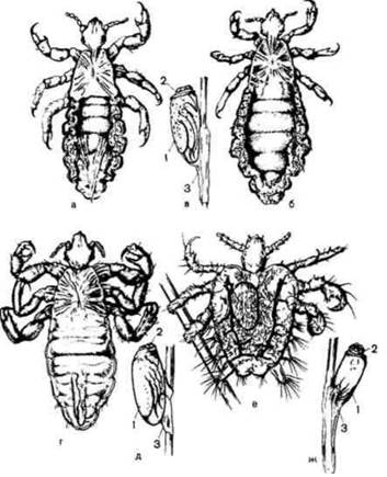
Fig. 2.51. Human lice:
a — head louse, male; b - female; c - head louse nit (egg); d — body louse, male; e - body louse nit; f - pubic louse; g - pubic louse nit; 1 - egg; 2 - operculum; 3 - adhesive substance
Head louse (Pediculus humanus capitis).
Localization - hairy part of the head (scalp).
Morphophysiological characteristics. Grayish insects. The length of the male is 2–3 mm, and of the female, 3–4 mm. The body is dorsoventrally flattened, consisting of the head, thorax, and abdomen. The head bears a pair of short, thick antennae, a pair of simple eyes (sometimes absent), and a piercing-sucking mouthpart, which in a resting state is retracted into the head and not visible externally. The thoracic segments are fused and bear three pairs of legs. The tarsus has a movable claw used by the lice to cling to Hair. Wings are absent. There are deep indentations along the sides of the abdomen. The last abdominal segment is forked in the female and rounded in the male. The copulatory apparatus is visible at the end of the abdomen in males.
Life cycle. Development involves incomplete metamorphosis (egg–larva–imago). Eggs (nits) are glued to the hair by the secretion of adhesive glands.
The entire development takes place on the human body. A larva hatches from the egg and transforms into an imago after three molts. Both the larva and the imago feed on blood. The lifespan of an adult louse is 27–38 days. During this time, the female lays up to 150 eggs. Development from the egg to the sexually mature stage takes 2–3 weeks.
Medical significance. The head louse is a permanent human ectoparasite and the CAUSATIVE AGENT OF pediculosis (infestation with lice). Louse bites are accompanied by itching, scratch marks, and Skin irritation. Heavy infestation can lead to matted hair (plica polonica). However, the primary medical importance of the head louse lies in its being a specific vector for the causative agents of epidemic (louse-borne) relapsing fever and (less commonly) typhus. The causative agents of endemic relapsing and typhus fevers are transmitted by ticks.
Prophylaxis. Personal: to prevent pediculosis, it is necessary to wash the body regularly while changing underwear, and to keep clothing and housing clean. Public: detection and Treatment of pediculosis, Sanitary supervision of places where large numbers of people gather, such as railway stations, hotels, trains, steamships, bathhouses, and hair salons. Insecticidal soaps are used to destroy head lice, and special remedies are used to destroy nits. Mechanical Methods include haircutting and shaving of hair, etc.
Body louse (Pediculus humanus humanus).
Localization - undergarments, clothing; moves onto the body when feeding on blood.
Morphophysiological characteristics and life cycle. In appearance and development, it is very similar to P. humanus capitis. Currently, they are classified as different subspecies of the same species, P. humanus. It is larger in size: the male is 2.1–3.75 mm long, and the female is 2.2–4.75 mm long. The body is light gray or whitish. Distinctive external features include less deep indentations along the margin of the abdomen, less pronounced pigmentation of the lateral abdominal segments, and thinner and longer antennae on the head. Development proceeds with incomplete metamorphosis (egg–larva–imago). Eggs are glued to The surface of clothing. All Selection/3.html">Stages of development occur on humans. The lifespan is up to 48 days. During this time, the female lays 300 eggs—twice as many as the head louse. The developmental cycle lasts up to 16 days.
Medical significance. The body louse is a permanent human ectoparasite, the causative agent of pediculosis, and a specific vector for the pathogens of epidemic (louse-borne) typhus and relapsing fever. The Role of the body louse as a specific vector of the typhus pathogen was established in 1909 by the French bacteriologist and parasitologist Charles Nicolle (Nobel Prize, 1928).
Epidemic relapsing and typhus fevers are obligate transmissible diseases, anthroponoses, which can assume epidemic proportions, affecting large populations. In 1874, G.M. Minkh, and in 1881, I.I. Mechnikov, proved through self-experimentation that the blood of patients with epidemic relapsing fever is infectious. In 1876, O.O. Mochutkovsky established through self-experimentation that the blood of patients with epidemic typhus is also infectious.
The causative agents of relapsing fever (Obermeier's spirochete) and epidemic typhus (*Rickettsia prowazekii*) are transmitted from infected individuals by lice, which, having engorged on blood, remain capable of infecting humans throughout their lives. The Mechanism of human infection has certain specific features related to the biology of the pathogens. Spirochetes from the louse's gut penetrate its hemolymph, where they multiply and accumulate. Rickettsias invade the epithelial Cells of the louse's gut, undergoing intensive Replication. As these epithelial cells disintegrate, rickettsias enter the lumen of the gut and are subsequently excreted with the feces. Spirochetes and rickettsias do not migrate to the salivary Glands of the louse. Consequently, the bite of an infected louse is not infectious in itself, though it causes itching and scratching. Infection with relapsing fever occurs when an infected louse is crushed during scratching, rubbing its hemolymph containing spirochetes into the bite wounds or skin excoriations. This mode of transmission is known as contamination. Similarly, infection with epidemic typhus occurs by rubbing the feces of an infected louse containing rickettsias into bite wounds or scratches (contamination).
Prophylaxis: same as for head lice.
Pubic louse, or crab louse (Phthirus pubis).
Localization: pubic hair, occasionally eyebrows, eyelashes, and axillary hair.
Morphophysiological characteristics and life cycle. The pubic louse is smaller than the head or body louse: males are about 1 mm in length, and females are 1.5 mm. The body is short and wide, with the thorax and abdomen being virtually indistinguishable. The lifespan of an adult pubic louse is 17-26 days, during which the female lays about 50 eggs. Development proceeds with incomplete metamorphosis.
Medical importance: a permanent ectoparasite and the causative agent of phthiriasis (pediculosis pubis). It does not transmit disease pathogens.
Prophylaxis: same as for other species of lice.
Order Fleas (Aphaniptera). Fleas are wingless, blood-sucking insects and ectoparasites of humans and animals. Typical representatives include the human flea and the rat flea.
Human flea (Pulex irritans). The body is laterally compressed. The head bears a pair of simple eyes, short antennae, and a piercing-sucking mouthparts. Wings are absent. The hind pair of legs is longer than the others and adapted for jumping. The abdomen consists of 10 segments; in males, the tip of the abdomen is curved upward. The body surface features hairs, bristles, Teeth, and spines of systematic importance (Fig. 2.52).
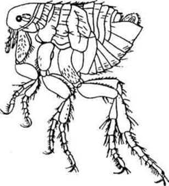
Fig. 2.52. Human flea (Pulex irritans).
Development involves complete metamorphosis (egg-larva-pupa-adult). Eggs are laid in indoor habitats (floor cracks and crevices, dry debris) or, in natural conditions, in rodent burrows. A legless, worm-like white larva hatches from the egg, feeding on decaying organic matter and flea feces. The larva spins a cocoon and transforms into a pupa (Fig. 2.53).

Fig. 2.53. Flea development:
1 - egg; 2 - larva; 3 - pupa; 4 - cocoon cleaned of foreign particles; 5 - normal appearance of the cocoon.
Adult fleas live up to 1.5 years and feed on blood.
Depending on ambient Temperature and humidity, the flea development cycle lasts from 19 to 270 days.
Medical importance. Fleas bites are painful, irritating nerve endings in the skin and causing itching accompanied by scratching, secondary infections, and suppuration. However, their primary significance lies in being vectors of plague pathogens. Plague (syn.: black death) is an acute, highly dangerous, naturally focal, facultatively vector-borne bacterial disease. Natural reservoirs of plague include various rodents: rats, marmots, susliks, hamsters, gerbils, and voles. The most dangerous vectors are the oriental rat flea (Xenopsylla cheopis), the marmot flea (Oropsylla silantievi), and others. The human flea also transmits the plague pathogen. Fleas acquire the infection while feeding on the blood of infected animals. The plague bacterium multiplies in the flea's Stomach, forming a plug that blocks its lumen (the "plague block"). When the flea attempts to feed again, the block obstructs blood flow, causing the flea to regurgitate the Bacteria-laden mass into the bite wound, thereby introducing A large number of bacteria into the host's body. Infection can also occur when a person rubs the feces of infected fleas containing plague bacteria into bite wounds or skin scratches. Other routes of transmission also exist, which is why plague is classified as a facultatively vector-borne disease. In addition to plague, fleas can transmit the causative agents of endemic typhus fevers and tularemia.
Prophylaxis: Maintaining indoor cleanliness, wet cleaning, and eliminating flea breeding sites such as floor cracks and crevices. Insecticides are used to eradicate fleas in buildings or on clothing. In natural environments, rodents (and consequently their fleas) are exterminated in their burrows using rodenticides.
Order True Bugs (Hemiptera sin. Heteroptera). The forewings are hardened at the base and membranous toward the free ends. Most species feed on plant sap. Some bugs have transitioned to a parasitic lifestyle. Those of medical importance include the bed bug and the kissing bug.
Bed bug (Cimex lectularius). Geographic
distribution: ubiquitous in human dwellings.
Morphophysiological characteristics. The body is reddish-brown and dorsoventrally flattened. Females are 6 mm long, and males are 5 mm. The head features long segmented antennae, a pair of compound eyes, and piercing-sucking mouthparts. The thorax consists of three segments, each bearing a pair of legs. The upper surface of the second segment bears a pair of rudimentary wing pads, behind which lie scent glands producing a specific odor. The segmented abdomen is rounded in females, whereas in males it is narrower, with an asymmetric posterior end featuring a notch that houses the sickle-shaped copulatory organ (Fig. 2.54).
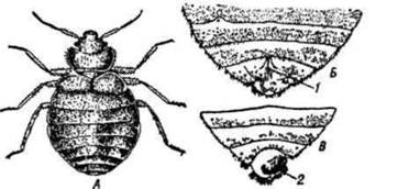
Fig. 2.54. Bed bug (Cimex lectularius):
a — female; b — posterior end of the female body; c — posterior end of the male body; 1 — opening of the copulatory pouch; 2 — copulatory organ.
Life cycle. Development occurs via incomplete metamorphosis. The female lays eggs in the same places where she lives—in cracks in beds, furniture, and walls, mattress seams, under wallpaper, or behind picture frames.
They reproduce very rapidly. A single female can lay up to 12 eggs per day, and up to 500 over her lifetime.
Bed bug nymphs go through 5 stages; the final, fifth stage is called a nymph. A newly hatched larva closely resembles an adult bug.
Both nymphs and adults feed on blood. They predominantly attack humans at night. They are capable of surviving long periods of starvation (several months). They are highly resistant to low temperatures; at -17°C, they remain viable for about a day. They spread by being transported as adults, nymphs, or eggs via clothing, furniture, carpets, and other items. They frequently parasitize laboratory animals under unsanitary maintenance conditions.
Medical significance. Temporary ectoparasites of humans. Bug saliva contains a toxic secretion, which makes their bites painful. Redness and Swelling develop at the bite site. Their bites disrupt normal human Sleep, which negatively impacts health. Transmission of vector-borne disease agents by bed bugs has not been established.
Prevention. Bug eggs are destroyed mechanically, while nymphs and adults are eliminated using insecticides.
The kissing bug (Triatoma infestans) is a vector of Trypanosoma cruzi, the causative agent of American trypanosomiasis (Chagas disease). It belongs to the family Reduviidae, genus Triatoma. They are large in size, ranging from 1.5 to 3.5 cm (Fig. 2.55). They have a nocturnal lifestyle. They inhabit wild animal burrows and bird nests. Some species are closely associated with humans, found in adobe houses and thatched roofs. They are temporary ectoparasites of animals and humans. They feed on blood. When attacking humans, the bugs bite near the eyes or on the Lips, where the skin transitions into the mucous membrane (hence the common name "kissing bug"). After engorging on blood, the bug turns 180° and deposits a drop of feces containing trypanosomes at the bite site. These pathogens penetrate either through the bite wound or via skin scratch marks.
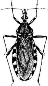
Fig. 2.55. Kissing bug (Triatoma infestans).
The infection rate among bugs in focus areas of trypanosomiasis reaches up to 50%.
The primary preventive measure is the improvement of socio-living conditions (construction of modern-type housing). Eradicating bugs is complicated by the fact that they parasitize various animals.
Order Blattodea (Cockroaches). Over 300 species are known, with
about 20 species found in Ukraine.
Of medical interest are synanthropic species—the oriental cockroach (Blatta orientalis) and the German cockroach, or Prussian cockroach (Blatta germanica).
The size of the former is 20–26 mm, while the latter is 8–11 mm. The body is flattened dorsoventrally. The head is directed downwards with mouthparts and is almost completely covered from above by a large shield-like pronotum. The antennae are setaceous and multi-segmented. The legs are adapted for running. There are two pairs of wings.
The forewings are leathery, while the hindwings are membranous (concealed beneath the tegmina at rest). In male oriental cockroaches, the forewings are well-developed, whereas in females they are reduced (Fig. 2.56).
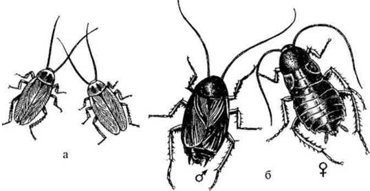
Fig. 2.56. Synanthropic cockroaches:
a — German cockroach (Blatta germanica); b — oriental cockroach (Blatta orientalis).
In both males and females of the German cockroach, the wings are well-developed. Postembryonic development occurs with incomplete metamorphosis. They inhabit human dwellings, particularly kitchens and garbage chutes. They feed on foodstuffs consumed by humans and various types of organic waste. They are frequently found in dining facilities. During the day, they hide in cracks and crevices, emerging at night to forage for food.
Medical significance.
Cockroaches act as mechanical vectors of disease pathogens, primarily gastrointestinal ones. They can transmit agents of typhoid fever, dysentery, tularemia, diphtheria, helminth eggs, and protozoan cysts.
Order Diptera (True Flies). Diptera are characterized by the presence of only a single (anterior) pair of wings. The posterior pair is modified into small appendages called halteres, which function as balancing organs.
Family Culicidae (Mosquitoes). In Eurasia, three genera of mosquitoes are most commonly encountered:
Anopheles (malaria mosquito), Culex, and Aedes (non-malaria mosquitoes). Only females are blood-sucking. Anopheles mosquitoes transmit malaria pathogens to humans. Some Aedes species transmit the pathogens of tularemia, Japanese encephalitis, lymphocytic choriomeningitis, yellow fever, Dengue fever, and anthrax; certain Culex species transmit Japanese encephalitis and tularemia pathogens. There are about 10 species of the genus Anopheles in Eurasia, the most common being the common malaria mosquito (A. maculipennis). Mosquito biology was most thoroughly studied by V.M. Beklemishev (1890–1962) and his disciples.
Morphophysiological characteristics. At all stages of development, malaria and non-malaria mosquitoes differ from each other (Fig. 2.57). Eggs of Anopheles mosquitoes differ from those of Culex not only in shape but also in the method of deposition.

Fig. 2.57. Main differences between malaria and non-malaria mosquitoes:
1 - egg floats of Anopheles; 2 - larval spiracles; 3 - pupal respiratory trumpets; 4 - antennae; 5 - maxillary palps; 6 - proboscis; 7 - eyes; 8 - thorax; 9 - abdomen of an adult mosquito.p>
Anopheles females lay eggs scattered and singly on the Water surface. Each egg is bordered by a concave belt and has a float chamber. Culex eggs lack a belt and chambers; they are laid on the water surface in rafts resembling a small boat. Aedes eggs are laid on damp soil near drying water bodies and, less frequently, on the water surface, either in clusters or scattered.
Culex and Aedes larvae possess a respiratory siphon shaped like a tube on the penultimate segment of the abdomen. Therefore, they position themselves in the water at an angle, attaching the siphon to the water surface. Anopheles larvae lack a respiratory siphon and lie horizontally on the water surface.
Pupae are comma-shaped and differ in The Structure of their respiratory tubes (siphonal horns). In Anopheles, the respiratory tubes are conical, while in Culex, they are cylindrical.
At the imaginal stage, there are differences in the structure of head appendages, wing coloration, and resting posture. In Anopheles females, the maxillary palps are approximately equal in length to the proboscis, whereas in non-malaria mosquitoes, they are short, making up 1/3–1/4 of the proboscis length. In Anopheles males, the maxillary palps are as long as the proboscis, with club-shaped thickenings at the end; in non-malaria mosquitoes, they are usually longer than the proboscis, without club-shaped thickenings. Antennae are located on the sides of the mosquitoes' mouthparts. In males, they are covered with long hairs, while in females, they are short (Fig. 2.58). Wing Veins are covered with scales that may form a pattern of spots. A. maculipennis has 4 dark spots in the middle part of the wing, whereas Culex mosquitoes lack such spots. When resting, Anopheles mosquitoes hold their abdomen elevated and positioned at an angle to the surface. In other mosquitoes, the body is curved during rest, with the abdomen inclined toward the substrate or parallel to it.

Fig. 2.58. STRUCTURE OF THE head of a malaria (Anopheles) and non-malaria (Culex) mosquito:
A - Culex; B - Anopheles; 1 - female; 2 - male; a - antennae; b - proboscis; c - palps.
Life cycle and biological features. Development involves complete metamorphosis (egg - larva - pupa - imago). Eggs, larvae, and pupae develop in water. A newly emerged winged Anopheles mosquito initially stays near water bodies in coastal vegetation. During this time, mosquitoes (both females and males) feed exclusively on plant juices. After a few days, at dusk, males form swarms. A female flies into the swarm and leaves it with one of the males. Following Fertilization, the female searches for a host and sucks the blood of humans or animals. Blood is essential for egg development.
For blood feeding, females have piercing-sucking mouthparts. In males, the mouthparts are adapted for feeding on plant juices. Anopheles females feed primarily indoors, which is why they fly from water bodies to human settlements. The distribution range of mosquitoes around anopheles-producing water bodies reaches approximately 3 km. Having engorged on blood, the female flies to a shelter and remains there until blood Digestion and the simultaneous maturation of fertilized eggs are completed. Then the female flies to a water body and lays her eggs. This described cycle of a female's life from THE START OF blood feeding to egg deposition is called the gonotrophic cycle (from Greek *gonos* - seed, germ Cell, *trophe* - Nutrition). After laying eggs, the female again searches for a host and feeds on blood. Such cycles may occur 2–3 to 6 times during the summer. The lifespan of a female in the summer period is about 1 month. Males die shortly after mating; their lifespan is several days.
For egg laying, Anopheles use water bodies with standing or slowly flowing water. The number of eggs in a single batch ranges from 60 to 350. Larvae hatch from the eggs and breathe atmospheric air. Anopheles larvae live exclusively in clean or nearly clean water bodies. Water bodies with a significant amount of organic matter and suspended particles, as well as shaded ones, are unsuitable for them. Therefore, in combating A. maculipennis mosquitoes, a good effect is achieved by planting trees with large and spreading crowns along the banks of water bodies. The duration of larval development depends on water temperature. Development begins at temperatures not lower than +10° C, with the optimum temperature being +25° C. The minimum larval development period is 15 days. Larvae feed on bacteria and plant residues by filtering water and swallowing any particles that can pass through the oral opening. This biological feature of mosquito larvae is used to destroy them by spraying powdered toxic substances onto the water surface. Larvae transform into pupae, and pupae into imagoes. The most widespread mosquito in our country, A. maculipennis, was previously considered a single species. Today, it is known to comprise 6 sibling species that differ in A number of biological features, particularly their overwintering behavior. In all A. maculipennis sibling species, fertilized females overwinter. Overwintering sites for some of them are cellars, where they enter diapause, while for others, they are sheds, where they feed on animal blood all winter. Males die in the autumn.
Mosquitoes of the genus Aedes differ from Anopheles in their biological features. The breeding sites for most Aedes species are temporary water bodies: puddles, ditches, and marshy areas. Larvae of certain species can develop in small containers, including buckets, barrels, tin cans, etc. Decaying organic matter is not harmful to them. A characteristic feature of Aedes mosquitoes is the asynchronous hatching of larvae from a single egg batch, which stretches over weeks or even months. This is an adaptation to life in periodically drying water bodies. If a water body dries up before larval development is complete and the larvae die, upon a new flooding, larvae hatch from the remaining eggs. This ensures the survival of the species. Some Aedes species, termed monocyclic, develop a single generation over the summer, while others, polycyclic, develop several generations. Adult mosquitoes are most active in the evening, but can also attack hosts during the day, especially in cloudy weather. During the day, they hide in grass, bushes, and pits near water bodies.
Prophylaxis and control measures. Personal protection involves defense against mosquito bites. Public measures include the eradication of mosquitoes at all stages of development. Mosquito control encompasses a range of measures: 1) elimination of small water reservoirs unused by humans; 2) spraying pesticides in water bodies that serve as breeding sites; 3) oiling of water bodies to prevent oxygen access; 4) clearing water bodies of vegetation; 5) land drainage (melioration); 6) biological control methods: Introduction of mosquitofish (*Gambusia*), which feed on mosquito larvae (though gambusia can only survive at water temperatures no lower than +5° C); use of natural predators (e.g., attracting bats), and pathogens of fungal, bacterial, and viral diseases of mosquitoes; Genetic Methods (release of sterile males into the wild); 7) zooprophylaxis — placing livestock farms between potential mosquito breeding sites and residential buildings during settlement planning, since mosquitoes readily feed on animal blood; 8) destruction of adult forms in overwintering sites (cellars, attics, sheds) using insecticides. All insecticides must be applied in a way that avoids harming wildlife.
Family Psychodidae (moth flies). Sandflies of the genus *Phlebotomus* are temporary ectoparasites and specific vectors of leishmaniasis and pappataci fever pathogens. Morphophysiological characteristics. Small insects with long legs, bright yellow, gray, or brownish in color, with a body length of 1.3–3.5 mm. The body and wings are densely covered with hairs (Fig. 2.59). They attack at night and during twilight. They are found both near human dwellings and in the wild, where they inhabit caves, rodent burrows, and other animal shelters.
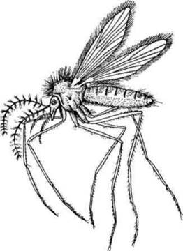
Fig. 2.59. Sandfly
Life cycle. Development involves complete metamorphosis.
Larvae develop in decaying refuse. They feed on organic matter. Pupae do not feed. Males feed on plant sap, while females suck the blood of humans and animals.
At optimal temperatures, 46 days elapse from egg laying to imago development. Adult sandflies stay close to their breeding sites.
Sandfly control measures include cleaning areas of refuse, applying contact insecticides in residential premises, and eradicating rodents in their burrows.
Family Muscidae (house flies). Of this family, synanthropic flies are of medical importance as mechanical vectors of disease pathogens (house fly, stable fly) and agents of myiasis.
The house fly (Musca domestica) is a mechanical vector of pathogens causing infectious and parasitic diseases.
(Phlebotomus pappatasii). The body length of females is 5.8-7.5 mm.
Males are slightly smaller. The mouthparts are sponging-sucking. By secreting abundant saliva
containing Enzymes, the fly dissolves solid food and ingests it. The tarsi feature claws and adhesive pads densely covered with hairs, allowing flies to walk on ceilings and vertical surfaces. The legs are also covered with hairs. Through these hairs, the fly can transmit various infection pathogens, helminth eggs, and protozoan cysts. The presence of flies is particularly dangerous in environments housing patients with tuberculosis, typhoid fever, or dysentery. Up to 6 million bacteria have been found on a fly's body, and up to 28 million in its gut. Outbreaks of intestinal disease epidemics typically occur during the summer months when fly populations reach their peak.
Development proceeds with complete metamorphosis. The female lays eggs wherever decaying organic matter is present (manure, human feces, garbage containers, landfills). A single clutch contains 100-150 eggs, and a female may produce 5-6 such clutches. Larvae hatch from the eggs within 12-36 hours; they are worm-like, white, legless, with a reduced head. Larvae pupate in the soil, burrowing to depths exceeding 30 cm. The pupae are immobile. The developmental cycle from egg to imago takes 10-26 days. The average lifespan of a fly is 30-35 days. Control measures against flies in populated areas include: protecting food products from flies; urban sanitation and improvement; implementing sanitary measures to ensure garbage and waste collection, along with timely cleaning of areas; composting manure; destroying larvae in breeding sites using insecticides; and eliminating adult flies through mechanical and chemical means.
The stable fly, or biting house fly (Stomoxys calcitrans), is a mechanical vector of anthrax and Sepsis pathogens. It is distributed globally. In terms of biology and Morphology, it closely resembles the house fly. The body color is gray with dark stripes on the thorax and spots on the abdomen. The proboscis is highly elongated and features chitinous "teeth" on its tip. By rasping its proboscis against the skin, the fly scrapes off the epidermis and feeds on blood, simultaneously injecting toxic saliva and causing severe irritation. Both females and males primarily attack animals, but occasionally humans as well. Control measures are the same as for the house fly.
The Wohlfahrt fly (Wohlfartia magnifica). It is viviparous. The larvae cause myiasis. This fly is found in central and southern Europe. Adult insects inhabit fields and feed on flower nectar. During flight, the female deposits 120-150 larvae into open cavities such as the eyes, Nose, and ears, as well as onto wounds and ulcerated surfaces of mammals and occasionally humans, especially children sleeping outdoors. The larvae are highly mobile. Using specialized adaptations, they rapidly penetrate tissues, erode them down to the bone, create passages, and destroy Blood Vessels (Fig. 2.60).

Fig. 2.60. Scalp lesions caused by Wohlfahrt fly larvae.
Clinical manifestations of myiasis include suppuration, hemorrhages, gangrenous processes, and severe pain.
Eye involvement can lead to blindness. Fatal cases have been documented. The duration of larval parasitism in animals lasts up to 3-5 days. They pupate in the soil, burrowing to depths of up to 32 cm. Control measures against fly larvae are complicated by their deep penetration into tissues.
They are removed surgically with forceps, followed by appropriate wound treatment.
The tsetse fly (Glossina palpalis) is the vector of the causative agent of African trypanosomiasis (sleeping sickness), Trypanosoma brucei gambiense. It is viviparous. Distributed in Western Africa. Size: 10-13.5 mm. Lifespan: 3-6 months. During this period, the female deposits a single living larva onto the soil surface 6-12 times. In the soil, the larvae transform into pupae, from which adult flies emerge after 3-4 weeks. They predominantly inhabit bush thickets along the banks of rivers and lakes, near human dwellings.
They feed mainly on human blood, and less frequently on the blood of domestic and wild animals. To control these flies, bushes and trees along water bodies, near human settlements, and along roads are cleared. Insecticides are also used.
Biting midges and associated blood-sucking pests (gnats). The term "gnat" encompasses all blood-sucking dipteran insects that massively attack humans and animals (mosquitoes, sandflies, biting midges, black flies, horseflies, and certain species of blood-sucking flies) (Fig. 2.61).

Fig. 2.61. Main Components of the gnat complex:
1 - horsefly (Tabanus bromis); 2 - deer fly (Chrysops flavipes); 3 - notch-horned clegs (Haematopota pluvialis); 4 - mosquito (Aedes communis); 5 - black fly (Simulium sp.); 6 - biting midge (Culicoides nubeculosus); 7 - sandfly (Phlebotomus pappatasii).
Breeding sites of blood-sucking gnats are primarily river floodplains and deltas, artificial irrigation areas, rice-growing regions, and stagnant water bodies. The species composition of gnats depends on the landscape and geographical zone. In the Siberian taiga, tundra, and other regions, they appear in
vast numbers, attacking animals and humans in clouds and causing severe harm. In some cases, sensitization (hypersensitivity) to subsequent bites develops, which can lead to severe conditions, even anaphylactic Shock (p. 242). People are deprived of normal rest, and work productivity declines. Individual protection measures include Pavlovsky nets, protective clothing made of special mesh fabric (preventing the blood-sucker's proboscis from reaching the skin surface), and repellents (such as dimethyl phthalate).
Family Ceratopogonidae (Biting Midges). The bulk of blood-sucking biting midges belong to the genus Culicoides. These are the smallest of the flying blood-sucking insects (Fig. 2.61, 6). Body length is 1-2.5 mm. Females attack humans and animals during morning and evening hours. Larvae and pupae develop in moist soil, forest litter, tree hollows, and small stagnant water bodies. Biting midges are mechanical vectors of the tularemia pathogen, and in the tropics, they act as specific vectors and intermediate hosts of nematodes (filariae).
Family Simuliidae (Black Flies). In appearance, they resemble tiny flies (Fig. 2.61, 5). Body length is 2.5-4.5 mm. Females of most species suck blood. They attack exclusively outdoors during daylight hours. Development takes place in rivers and streams. Black flies are mechanical vectors of the tularemia pathogen, while in the tropics, black flies of the genus Simulium serve as specific vectors of filariae (Onchocerca volvulus, O. coecutiens).
Family Tabanidae (Horseflies). In appearance and size, they resemble a large fly (Fig. 2.61, 1-3). The large head features brightly colored eyes. Development proceeds with complete metamorphosis. Males feed on plant sap. Females also feed on plant sap, but following fertilization, they require a blood meal for egg development. They attack during hot weather and buzz during flight. A horsefly bite is painful and particularly unpleasant because the insect may deliver 8-10 punctures with its proboscis. Horsefly saliva is toxic. Blood flows from the wound for a long time due to the injection of hemolysins. Most species lay eggs on the leaves of littoral vegetation. Horseflies are mechanical vectors of tularemia and anthrax pathogens, whereas horseflies of the genus Chrysops transmit filariae (Loa loa) in Africa.
Last update: 08/08/2026
Editorial and Educational Adaptation: This material has been compiled based on the primary/original source text. The project team performed an editorial review, corrected technical inaccuracies, structured sections, and adapted the content for an educational format.
What was processed:
- elimination of formatting defects (OCR errors, structural breaks, corrupted characters);
- editorial organization of content;
- standardization of terminology in accordance with academic sources;
- verification of factual statements against the original source text.
All mentions of the author, publication year, and origin of the primary text have been preserved in accordance with the source.