Nephrology for the Family Physician - O.I. Bakaliuk 2003
Certain Urological Conditions in Therapeutic Practice
Nephrolithiasis
Crystal formation, much like calculus formation, is a complex physicochemical process driven by a multitude of factors.
There are three distinct theories explaining The Mechanism of Kidney stone formation: crystallization, colloid, and inhibitor theories.
According to the crystallization theory, this process obeys the physical laws of solution crystallization, whereas The Role of the micelle (the stone's core) is considered negligible.
The colloid (matrix) theory, formulated in 1968 by C.W. Vermeulen et al., attributes primary importance to the micelle itself and its biological properties. Salt precipitation is normally prevented by protective colloids—mucopolysaccharides containing a protein component known as an aminosugar. When these are deficient against a Background of excessive salt concentration and fluctuations in urine pH, individual crystals tend to aggregate around the core of the future stone (the micelle), which may consist of fibrin, a Blood clot, desquamated epithelium, pus debris, or a foreign body.
To be fair, an elevated salt concentration in the urine is still a prerequisite for the onset of stone formation. Subsequently, stone growth may continue even at normal (!) urinary concentrations of that salt. Thus, the initiation of lithogenesis may be triggered by a single, transient abnormal state of the urine, which can occur, for instance, during a brief but drastic fluid restriction, the consumption of certain foods, and so forth.
The inhibitor theory attributes The process of crystal and stone formation to an imbalance between crystallization-promoting and anti-crystallization factors in the urine (E.A. Yuryeva et al., 1985). Promoting factors include high concentrations of poorly soluble compounds, Calcium Ions, urinary acidity, and low diuresis, whereas anti-crystallization factors encompass magnesium, zinc, citrate, pyrophosphate, and polyphosphate ions (such as ATP and ADP), glycosaminoglycans, acidic Glycoproteins, urinary fibrinolytic activity, unpolarized Tamm-Horsfall protein, uronic acid, and various Enzymes (such as urease and alkaline phosphatase).
O.V. Lyulko et al. (1998) and Y.M. Postolov (1998), based on a review of literature and their own research, emphasize the crucial role of renal papillae status in stone Pathogenesis—specifically, the impairment of their hemodynamics (necrosis with The Development of small infarctions) against the backdrop of elevated oxalate and calcium levels within the papillae.
The development of renal papillary necrosis is promoted by the anatomical and PHYSIOLOGICAL CHARACTERISTICS OF the medullary Blood supply and, crucially, by the presence of microorganisms. The urease activity of Bacteria (Proteus, staphylococci, Klebsiella, Ureoplasma)—which easily find an ecologically "advantageous" niche for persistence in the renal medulla (slow blood flow, relatively long capillaries, high osmotic pressure)—along with their endotoxins, leads to the impairment of tubular enzyme systems and a reduction in the Kidneys' anti-crystallization capacity (N.V. Merkusheva et al., 1997; R.A. Older et al., 2000). The consequence of this process is enhanced crystal formation and increased levels of oxalates and calcium within the papillae.
This process is directly correlated with the activity level of the inflammatory process. This pattern is explained by the "capillary pathology" theory, which posits the existence of so-called renal parenchymal microliths—the deposition of calcium salts beneath the urothelium damaged by Bacterial toxins. In cases of highly virulent infection, such changes lead to dystrophic and necrotic alterations of the urothelium, most frequently at the apex of the papilla, which then serves as a peculiar matrix for stone formation (Randall's plaque) (S. Leslie, 2000). During subsequent inflammatory flare-ups, this plaque increases in size, the overlying epithelium breaks down, and its surface becomes a focal point for crystal deposition. Notably, such microliths may also form in the region of the Loop of Henle and the interstitium (Y.M. Postolov, 1998).
Urolithiasis (kidney stone disease) is a genetically determined multifactorial disease that manifests under specific human living conditions (climatic and geochemical factors, national traditions, dietary habits, ambient Temperature, soil moisture, drinking Water composition and its saturation with mineral salts, heavy metal salts, etc.) (O.I. Tischenko et al., 1998; I.M. Trakhtenberg et al., 1998). In terms of prevalence, it ranks second after Pyelonephritis and occurs across all age groups and in every country worldwide, with a frequency ranging from 1.3% to 4%.
Endemic foci of the disease exist, including the North Caucasus, the republics of Central Asia, Iran, India, Brazil, and within Ukraine—the Lviv, Odesa, Zakarpattia, Donetsk, and Dnipropetrovsk regions.
Determinacy in urolithiasis refers to "fatal errors of METABOLISM"—the presence of congenital enzymopathies (Tubulopathies) resulting from the deficiency or complete absence of a specific enzyme. Enzymopathies that promote stone formation most commonly include congenital or acquired oxaluria, urosuria/uraturia, cystinuria, glycinuria, Aminoaciduria, galactosemia, and fructosuria (R. Scott, 1995).
The most prevalent enzymopathies in adults are oxaluria (50% of patients), followed by uraturia (25%), phosphaturia (10–20%), and galactosemia and fructosuria (12–13%). Cystine stones are relatively rare; their formation is associated with impaired cystine reabsorption in the tubules. Galactosemia is rooted in a defect in converting galactose to glucose, leading to subsequent hypergalactosemia and galactosuria. In fructosuria, a deficiency of fructose-1-phosphate aldolase results in pronounced fructosuria.
Important roles in the pathogenesis of urolithiasis are also attributed to such risk factors as renal infection, particularly involving the interstitial zone (pyelonephritis), hormonal imbalances (hyperparathyroidism), general metabolic shifts (Diabetes Mellitus, obesity), Pregnancy, and functional or organic urinary outflow disorders.
The Clinical presentation of urolithiasis is well-defined: pain, gross Hematuria, and less frequently, obstructive anuria. The pain varies in intensity, duration, and radiation pattern, depending on the size and Location OF THE stone.
The most reliable symptom of urolithiasis is a Renal Colic attack. The pathogenesis and Clinical Features of renal colic are described above.
The likelihood of spontaneous stone passage is likewise limited by its location and size, as well as the tone of the renal pelvis and Ureters. In some patients, gross hematuria is observed following stone passage. Prolonged retention of a calculus within the Urinary Tract lumen threatens the development of Hydronephrosis and secondary infection.
Diagnosing urolithiasis is straightforward when the clinical picture is typical. Difficulties arise in cases of atypical stone location and pain radiation, lowered blood pressure, or reflex tension of the anterior abdominal wall Muscles. A positive costovertebral angle tenderness (pounding) sign is observed, along with localized tenderness upon Palpation over the stone projection area. Indirect signs of urolithiasis include positive Abrahams' sign (R. Abrahams: tenderness elicited by pressing the midpoint of the line connecting the umbilicus to the ninth left rib Cartilage),
and Bittorf's sign (A.Bittorf: onset of lumbar pain upon pressure applied to the testicles or scrotal Skin). Blood tests reveal a left shift in the leukocyte formula and an elevated ERYTHROCYTE SEDIMENTATION RATE (ESR), whereas urinalysis shows moderate proteinuria and microhematuria.
The results of chromocystoscopy (total renal blockage) and isotope renography (obstructive pattern) determine the urgency and direction of subsequent medical management.
The Use of ultrasound Diagnostics (Fig. 85) has significantly expanded The ability to detect radiopaque and radiolucent stones even at the preclinical stage.
Plain radiography, excretory urography, and retrograde pyelography allow for the precise determination of calculus Location and Structure, as well as renal Morphology (Figs. 86, 87, 88).
Class="center">
Fig. 85. Kidney stone (ultrasound).
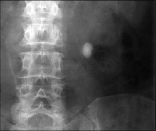
Fig. 86. Kidney stone (plain abdominal radiograph).
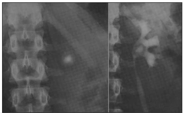
Fig. 87. Kidney stone (plain abdominal radiograph and excretory urogram).
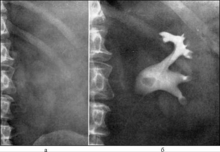
Fig. 88. Kidney stone: a — plain abdominal radiograph (no abnormalities detected); b — left retrograde pyelogram (radiolucent calculus in the left renal pelvis).
Cytology/practical/136.html">Differential Diagnosis OF urolithiasis is most frequently required to rule out acute appendicitis, the gastralgic variant of myocardial infarction, perforated gastric ulcer, mesenteric or renal vessel thrombosis, acute cholecystitis, acute pancreatitis, paralytic ileus, Ectopic Pregnancy, and ovarian Hemorrhage.
The management of urolithiasis is a challenging and critical task.
Conservative therapy for this condition is indicated in all cases where the calculus does not impair urine outflow or provoke frequent flare-ups of urinary tract infection. Furthermore, it serves as an integral component of other Treatment modalities.
The cornerstone of treatment is medical Nutrition therapy (dietotherapy) combined with pharmacotherapy and mineral water consumption.
Dietary management for uric acid calculi is described in the section «Renal Involvement in Gout», and for calcium oxalate calculi in «Kidneys and Pregnancy». It should be noted that urinary pH should be alkalinized gradually to prevent phosphate precipitation.
In phosphaturia, to modify urinary reaction (acidification), the following agents are employed: ammonium chloride (1–3 g/day), Methionine (0.5 g 3–4 times/day), sodium phosphate (1 g 4 times/day), madder extract (marelin, 2–3 tabl/day), succinamide (2–3 tabl/day), benzoic acid (0.05 g 2 times/day), and mineral waters (Narzan, Arzni, Naftussya); in oxaluria — magnesium oxide (0.2–0.4 g 3 times/day), magnesium carbonate (0.5 g 4 times/day), and mineral waters (Yessentuki No. 20, Luzhanska, Naftussya).
According to Ye.M. Neiko (2000), the administration of «Naftussya» in urolithiasis provides, In addition to a diuretic effect, enhanced peristalsis of the urinary tract, as well as analgesic, anti-inflammatory, and detoxifying properties. A distinctive feature of «Naftussya» is the universality of its action, regardless of The chemical composition of the calculi, the type of urinary diathesis, or urinary pH values. «Polyana Kvasova» possesses similar properties (V.H. Kudyk et al., 1998).
In phosphate urolithiasis, Almagel is also utilized to actively bind phosphates within the intestine.
Agents possessing antispasmodic and bacteriostatic effects have gained widespread use: cystenal, urolesan (5–8 drops 3 times/day on a sugar cube under the Tongue), enatin (1 g/day), avisan (0.05 3–4 times/day), and phytolysin (1 cm of paste dissolved in 1/2 cup of warm water twice a day).
Recently, Canephron (Bionorica, Germany) has been used for the Prevention of recurrent uric acid Nephrolithiasis following lithotripsy, as well as in the combination treatment of immune-inflammatory nephropathies. It contains herbal extracts of lovage, rosemary, centaury, and rose hip, which exhibit anti-inflammatory, antiseptic, diuretic, hypotensive, antispasmodic, and alkalinizing effects (Yu.A. Pytel et al., 1999; V.O. Moiseienko et al., 2001; T.D. Nykula, 2001). Canephron is prescribed at 30–40–50 drops or 1–2 dragees 3–4 times a day in courses lasting 10–14 days.
In the presence of concomitant urinary tract infection, semi-synthetic Penicillins and nitrofurans are the drugs of choice.
Physical therapy modalities used for urolithiasis include iontophoresis (Electrophoresis) with antispasmodics, inductothermy, phonophoresis, diadynamic therapy, amplipulse therapy, microwave therapy, paraffin, ozokerite, and peloid therapy, as well as fresh or mineral water baths (general or sitz) at a temperature of 36–37 °C for 8–15 minutes every other day, and sinusoidal modulated currents combined with preliminary inductothermy of the ureteral region.
Citing Ye.M. Neiko (2000), we present herbal tea mixtures whose components are readily available and whose therapeutic efficacy is well-documented.
For uric acid stones and acidic urine reaction:
1. Common bean pods (15.0), bilberry leaves (15.0), yarrow herb (15.0), blackthorn flowers (15.0), field horsetail herb (20.0), St. John's wort herb (20.0) — steep one full tablespoon of the mixture in a Glass of cold water for 6–8 hours, then boil for 15 minutes, take three times a day, 30 minutes before meals.
2. Corn silk (30.0), greater celandine herb (30.0), goldenrod herb (30.0), agrimony herb (20.0), barberry herb (20.0) — steep 1 tablespoon of the mixture in a glass of water, take three times a day, before meals.
3. Ash ROOT (10.0), cherry stems (5.0), nettle leaves (20.0), tricolor violet flowers (10.0), mountain arnica flowers (15.0) — pour 500 ml of water over 3 tablespoons of the mixture, boil down to 150 ml, take 30 drops three times a day.
4. Couch grass rhizomes (30.0), agrimony herb (20.0), wild carrot seeds (40.0), rupturedwort herb (10.0) — pour a glass of boiling water over 1 tablespoon of the mixture, infuse in an oven for 8 hours, strain, and take 2 tablespoons three times a day before meals, while warm.
For phosphate stones and alkaline urine reaction:
1. Rupturedwort herb (20.0), parsley fruit (20.0), bearberry leaves (60.0) — steep one full tablespoon of the mixture in a glass of cold water for 6–8 hours, then boil for 15 minutes, take three times a day, 30 minutes before meals.
2. Lovage root (10.0), parsley fruit (10.0), birch leaves (25.0), juniper fruit (25.0) — preparation method is the same as in No. 1.
3. Linden flowers (20.0), oak bark (20.0), bearberry leaves (20.0) — pour 2 cups of boiling water over 1 tablespoon of the mixture and infuse. Take 1 glass of the warm infusion before dinner.
4. Burnet-saxifrage root (10.0), field horsetail herb (10.0), bearberry leaves (20.0), birch leaves (20.0), juniper fruit (20.0) — preparation method is the same as in No. 1.
For oxalate stones, regardless of urine reaction:
1. Knotweed herb (120.0), celandine herb (20.0), yarrow flowers (20.0) — pour 3 cups of warm water over 3 tablespoons of the mixture, leave for 6–8 hours, boil, infuse for 10 minutes, strain, and take by tablespoons throughout the day.
2. Knotweed herb (150.0), peppermint leaves (20.0), heather flowers (20.0) — preparation method is the same as in No. 1.
3. Sweet violet root (10.0), cornflower flowers (5.0), field horsetail herb (15.0), thyme herb (20.0), St. John's wort herb (30.0) — preparation method is the same as in No. 1.
For mixed-composition stones, the following herbal mixtures are recommended:
1. Stinging nettle leaves (5.0), prickly chaff-flower root (5.0), sweet flag rhizome (5.0), peppermint leaves (5.0), field horsetail herb (15.0), black elderberry flowers (15.0), linden blossom (15.0), juniper fruit (15.0), rose hip fruit (15.0) — pour a glass of boiling water over 1 tablespoon of the mixture, infuse, strain, and take 1 glass twice a day 30 minutes before meals.
2. Wild strawberry leaves (10.0), rosemary leaves (10.0), field horsetail herb (10.0), licorice root (10.0), caraway fruit (10.0), juniper fruit (10.0) — preparation method is the same as in No. 1.
3. Oregano herb (5.0), coltsfoot leaves (5.0), marshmallow root (20.0) — pour a glass of boiling water over 2 teaspoons of the mixture, infuse, strain, and take 1/2 glass three times a day before meals.
4. Rupturewort herb (50.0), lovage root (10.0), juniper fruit (10.0), spiny restharrow root (10.0) — preparation method is the same as in No. 1.
5. Asarabacca herb with root (15.0), birch leaves (50.0), lingonberry leaves (25.0), licorice root (25.0) — preparation method is the same as in No. 1.
Herbal decoctions and infusions are usually taken for 2–3 weeks; a repeated course of treatment is recommended after a 1–2 week break.
Below are also recipes for treating urolithiasis and salt diathesis using juices and raw vegetables (indicated amounts of vegetables or juices per 1 serving) (S.M. Drohovoz, 1995) and therapeutic baths (Ye.S. Tovstukha, 1994).
Urolithiasis: 1–2 lemons; juice and soft pulp of ordinary pumpkin (2–2.5 kg/day); carrots (308.0) + beets (84.0) + celery (140.0); carrots (308.0) + beets (84.0) + coconut (56.0); carrots (308.0) + beets (84.0) + cucumbers (84.0); carrots (336.0) + parsley (112.0); carrots (280.0) + spinach (168.0).
Salt diathesis (acidic): 1–2 oranges, carrots (336.0) + parsley (112.0); carrots (252.0) + parsley (56.0) + celery (140.0).
Uric acid diathesis: carrots (196.0) + parsley (56.0) + celery (112.0) + spinach (84.0); carrots (308.0) + beets (84.0) + coconut (56.0); carrots (308.0) + beets (84.0) + cucumbers (84.0); carrots (336.0) + parsley (112.0); carrots (280.0) + spinach (168.0); carrots (252.0) + parsley (56.0) + celery (140.0).
For therapeutic baths in nephrolithiasis and associated pyelonephritis, the following plants are used (indicated amount of plant material per 1 bath): a) common heather herb (100 g) + common knotweed herb (50 g) + peppermint herb (50 g) + lemon balm herb (50 g) + silver birch leaves (25 g) + hop cones (25 g); b) ground ivy herb (100 g) + common oregano herb (100 g) + silver birch leaves (50 g) + common hop cones (50 g); c) lady's bedstraw herb (100 g) + common lungwort herb (100 g) + common oregano herb (50 g) + common yarrow herb (50 g).
Method of bath preparation: pour 3 liters of water over 300–400 g of the mixture, boil for 5 minutes, and infuse for 4 hours. Strain through 4 layers of gauze and add to bath water with a temperature of 38–40 °C. Baths are started 4–6 months after the beginning of herbal therapy and are administered for 2–3 months, 1–2 times a week.
Surgical treatment does not protect the patient from further stone formation; therefore, it should be minimally invasive.
Indications for Surgical Treatment of urolithiasis include frequently recurring pyelonephritis and the threat of hydronephrotic transformation of the kidney; open surgical interventions are performed in approximately 2% of cases (J.W. Segura, 1990). The rate of surgical interventions for urolithiasis in Ukraine is 10.7% (L.P. Pavlova et al., 1998).
Recently, Methods of remote stone fragmentation (lithotripsy) have been introduced into clinical practice. Technical aspects of the Procedure, especially in patients with comorbid conditions, as well as the efficacy of extracorporeal Shock wave lithotripsy depending on stone size and location, have been studied by O.F. Vozianov et al. (1998) and O.V. Lyulko et al. (1998). A non-explosive method of urinary calculus destruction (D.M. Haydamachenko, 1998), contact pneumatic ureterolithotripsy (O.F. Vozianov et al., 1998), etc., have also been proposed.
Emergency therapy for renal colic is described below.
Last update: 08/08/2026
Editorial and Educational Adaptation: This material has been compiled based on the primary/original source text. The project team performed an editorial review, corrected technical inaccuracies, structured sections, and adapted the content for an educational format.
What was processed:
- elimination of formatting defects (OCR errors, structural breaks, corrupted characters);
- editorial organization of content;
- standardization of terminology in accordance with academic sources;
- verification of factual statements against the original source text.
All mentions of the author, publication year, and origin of the primary text have been preserved in accordance with the source.