Nephrology for General Practitioners - O.I. Bakaliuk 2003
Selected Urological Conditions in Therapeutic Practice
Renal Tuberculosis
According to WHO data (1996), 22 million people suffer from tuberculosis, of whom about 3 million are doomed to death. This is the price of complacency paid by the world, which assumed that tuberculosis was a disease of the past.
The dramatic global situation is further compounded by the lack of a law on compulsory Treatment for tuberculosis patients, as well as its fatal combination with AIDS. The latter virus, by disrupting the body's Immune Response, dramatically accelerates the transition of tuberculosis from an inactive infection to a life-threatening condition, and the probability of activating a dormant tuberculosis infection in this situation increases by 10% per year.
Among patients with newly diagnosed active disease, Urogenital Tuberculosis accounts for 2.2% (M.M. Savula et al., 1999), and the overall incidence of Renal tuberculosis ranges from 2.8 to 14.0 per 100,000 population (J. Neuman et al., 1992). Among the urogenital Organs, the Kidneys, Epididymis, Prostate Gland, Fallopian tubes, and Uterus are most frequently affected. In men, simultaneous involvement of the kidneys and genital organs occurs more frequently than in women.
Tuberculosis of the kidneys ranks first among extrapulmonary localizations (30–40%) and develops after The activity of Primary tuberculosis subsides (10–15 years later). Renal changes accompany Pulmonary Tuberculosis in 60–90% of cases, and Bone and joint Tuberculosis in 30–52% of cases (A.L. Shabad, 1978). Women fall ill more often than men, and frequently the first manifestations of renal tuberculosis (with predominant involvement of the right Kidney) occur during Pregnancy under the guise of gestational renal failure.
The main pathway of infection entering the kidney is hematogenic. It has been proven that initial tuberculous changes develop in the cortical layer. However, after renal infection, most patients experience healing of the tuberculous foci, and subsequently, they remain asymptomatic, becoming active under METABOLISM/18.html">The Influence of exo- or endogenous adverse factors. The period from the moment of infection to The Development of the disease in individual cases reaches 20 years. Four factors have been described upon The ratio of which the form and course of experimental renal tuberculosis depend (H. Ishioda et al., 1998): deposition conditions, virulence of the pathogen, state of the body's defense mechanisms, and size of the infecting dose.
Under certain conditions, a lymphogenic pathway of infection is also admitted. In confirmation of this, data are cited that in 68–85% of cases, renal tuberculosis is unilateral, specifically on the same side where the pleuropulmonary focus was located. All PARTS OF THE Urinary System are drawn into the pathological process: the renal parenchyma, renal pelvis, Ureters, and Urinary Bladder.
Foci of a specific process (tubercles) with a tendency to fusion and breakdown are formed in the renal medulla. Caseous masses in the center of such a focus undergo liquefaction, forming a cavity. Subsequent fibrosis and thickening of the cavity walls lead to partial Isolation of the focus from the healthy parenchyma. Sometimes the tuberculous process is localized in the renal papillae (papillitis) with involvement of the neck of the renal pelvis, leading to the development of its stenosis and obliteration.
Taking into account the localization of the process, the following forms of renal tuberculosis are distinguished: parenchymal tuberculosis, specific papillitis, cavernous tuberculosis, Fibro-cavernous tuberculosis, and tuberculomas (caseomas). In cases where stenosis and obliteration of the pelvicalyceal segment occur, the process ends with the complete dissolution of the renal parenchyma, which is termed tuberculous Pyonephrosis. The appearance of tubercles in the submucosal layer of the pelvis and ureters with ulceration, followed by their scarring along with paresis of nerve endings, leads to impaired urine passage and the occurrence of refluxes, which facilitates the further spread of the process.
An important diagnostic method for renal tuberculosis is excretory urography, which reveals narrowing and elongation, or conversely, widening and shortening of a calyx or calyces, their "detachment," "moth-eaten" appearance of the renal papilla surface, presence of cavities, strictures, and ureteral dilations (Fig. 89).
In some cases, the process ends in complete renal calcinosis (Fig. 90).
Taking into account the degree of renal involvement, A.L. Shabad (1973) proposed the following clinical and radiological Classification of renal tuberculosis:
- non-destructive (infiltrative) renal tuberculosis;
- initial destruction of renal structures (papillitis or small cavities up to 1 cm in diameter);
- limited destruction of renal structures (large cavities or polycavernous tuberculosis in one of the renal segments);
- total or subtotal destruction of renal structures (polycavernous tuberculosis of two segments, tuberculous pyonephrosis, renal calcinosis).
Class="center">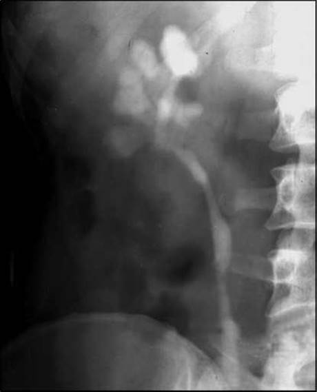
Fig. 89. Renal tuberculosis (excretory urogram).
Changes in the urinary bladder appear relatively late, yet they are quite characteristic and are detected during cystoscopy. It is worth noting The Significance of Lezhnev's symptom—bullous edema of the ureteral orifice.
Diagnosis of renal tuberculosis is extremely difficult. While early forms of pulmonary tuberculosis are diagnosed in 80–85% of cases, this percentage for renal tuberculosis is only 25–50% (R.F. Ozersky et al., 1990), and in 48–70% of patients, renal tuberculosis is diagnosed only at the stage of formed cavities, with every third nephrectomy performed for this disease.
In the Cytology/cytology/16.html">Early stages of the disease, the endogenous Intoxication syndrome dominates (weakness, asthenia, low-grade fever), which is more frequently encountered in tuberculous involvement of both kidneys or a solitary kidney. Body Temperature (in 20–30% of patients) is usually low-grade, and fever occurs rarely. Arterial Hypertension is detected in 5–18% of patients and is more often observed in bilateral involvement at the polycavernous stage. Pain in the lumbar region (in 50–65% of patients) typically feels like a sensation of heaviness, and in some cases resembles Renal Colic (resulting from obstruction of the ureteral lumen by a Blood clot, caseous masses, etc.). Somewhat later and less frequently, painless dysuria occurs, which is attributed to The Effect of bacterial toxin on the neuroreceptor apparatus of the kidney or predominant involvement of the urinary bladder (I.S. Kamyshan et al., 1995).
Certain Specific features of the clinical course of renal tuberculosis in elderly patients should be noted: minimal expression of general symptoms and predominance of dysuric disorders, "masking" of specific involvement signs by symptoms of other diseases (renal failure, urolithiasis, prostate adenoma), and faster development of nephroangiosclerosis and Chronic Kidney Disease.
Urinary sediment is characterized by moderate (up to 1 g/day) and acylindric proteinuria (85–95%), microhematuria (70–75%), and leukocyturia (the latter being the earliest sign of renal tuberculous involvement, present in 96–99% of patients). Monosymptomatic macrohematuria is observed in 10–12% of cases. The urine reaction is persistently acidic (!). Despite leukocyturia, Microbial growth on standard nutrient media is not observed, and nonspecific bacteriuria is detected in 20–35% of patients, albeit already at the stage of cavernous renal involvement.

Fig. 90. Metatuberculous calcinosis of the right kidney (plain radiograph).
A persistently acidic urine reaction combined with abacteriuric pyuria should always alert the physician to the possibility of tuberculous renal involvement. Occasionally, sudden (usually once or twice) macrohematuria is observed as a manifestation of a microcavity breaking through into the tubular system.
A definitive diagnosis of renal tuberculosis can be reliably established using only two Methods: bacteriological, when growth of Mycobacterium tuberculosis is successfully obtained on special media (Lowenstein-Jensen egg-potato medium, or deep inoculation on Preis-Shkolnikova blood medium, yielding positive results in 50–60% of cases), and morphological, characterized by specific tissue changes in the kidneys or urinary bladder such as Pirogov-Langhans giant Cells, or nonspecific changes including diffuse or focal mucosal hyperemia predominantly in the trigone area, cystic or follicular cystitis, and granulations. It should be noted that even at the stage of cavitation and pyonephrosis, Mycobacterium tuberculosis is isolated in only 50–60% of cases. All other methods—including ultrasound, radionuclide imaging, X-ray examinations, enzyme-linked immunosorbent assays (ELISA), and urine and blood chemiluminescence—only allow for a suspicion of specific Urinary Tract involvement.
Among other diagnostic approaches, mention should be made of the tuberculin provocation test using a dose of 20–100 TU (considered positive if tuberculin administration leads to an increased count of leukocytes and erythrocytes in the urinary sediment, and occasionally yields mycobacterial growth), the biological test (inoculation of urinary sediment into a guinea pig), A.L. Shabad's tuberculin-immunochemical test (elevated clearance of albumin and class G IMMUNOGLOBULINS during an active renal process), and the Determination of the lysozymuria grade according to T.M. Mochalova (a 2-fold or greater increase in lysozymuria).
The management of urinary tract tuberculosis falls within the purview of urological phthisiotherapists. Notably, this treatment concurrently involves three (isoniazid, rifampicin, ethambutol) or even four (isoniazid, rifampicin, ethambutol, pyrazinamide) agents administered for 1.5–2 years, all of which exhibit nephrotoxic effects. Recovery is observed in 90–95% of patients with renal parenchyma tuberculosis and in only 35–40% of patients with cavitary tuberculosis (I.S. Kamyshan et al., 1995).
Last update: 08/08/2026
Editorial and Educational Adaptation: This material has been compiled based on the primary/original source text. The project team performed an editorial review, corrected technical inaccuracies, structured sections, and adapted the content for an educational format.
What was processed:
- elimination of formatting defects (OCR errors, structural breaks, corrupted characters);
- editorial organization of content;
- standardization of terminology in accordance with academic sources;
- verification of factual statements against the original source text.
All mentions of the author, publication year, and origin of the primary text have been preserved in accordance with the source.