Plant Anatomy: A Practical Guide - Panyuta O.O. 2019
Topic 3. Structure of Vegetative Organs
Laboratory Work No. 13. Structure of the Stem in Perennial Plants
Theoretical Background. Higher plants can be herbaceous or woody. The primary difference in their stem anatomy is that in woody plants, stem thickening occurs over a long period and typically ceases only when the Organism dies. In herbaceous plants, even perennials, stem thickening stops once flowering begins. This phenomenon can be observed in both annuals and perennials. For instance, the stem of the perennial alfalfa dies back completely after flowering. However, this process can be modified by regulating the plant's development. For example, if the flowers are continually removed from the stem of a common garden mignonette, it becomes perennial, and its stem Structure comes to resemble that of a multi-year-old woody plant. Similarly, if buds or flowers are plucked from an alfalfa stem, it will continue to grow throughout the entire growing season, and its Anatomical Structure will closely mimic that of woody plants. Thus, the Water/140.html">Anatomical structure of a plant varies depending on its GROWTH AND DEVELOPMENT conditions.
Comparing the stem anatomy of herbaceous and woody plants reveals that perennial woody plants feature a clearly defined central cylinder with a pith at its center. Numerous medullary rays traverse the central cylinder, dividing it into distinct segments. Furthermore, concentric rings—annual growth rings—are visible within the central cylinder. In many trees, the stem is differentiated into a dark-colored heartwood and a lighter outer zone known as sapwood. In herbaceous plants, distinct vascular bundles are scattered amidst the uniform ground tissue of the stem, sometimes accompanied by a ring of mechanical tissue beneath the epidermis. However, this described difference is characteristic of older stem regions. When comparing the anatomical structure of young, actively growing twigs with that of herbaceous stems, they appear structurally identical. Consequently, the anatomical differences between the stems of woody and herbaceous plants are the result of secondary changes that occur during their growth and development.
Objective: to examine the Structural Features of perennial plant stems.
Materials and equipment: light microscopes, Glass slides and coverslips, dissecting needles, forceps, glass rods, filter paper, distilled water, phloroglucinol, aniline blue, iodine in potassium iodide, chlor-zinc-iodine, phloroglucinol with Hydrochloric acid, Sudan III, glycerol, chromic acid, 10% potassium hydroxide solution, and plant material.
Slide. Stem structure of Dutchman's pipe (Aristolochia sipho L.)
To study the stem anatomy of Dutchman's pipe, young, one-year-old twigs that have completed their seasonal growth are gathered in advance and preserved in alcohol.
Cross-sections are prepared from the preserved material and treated with phloroglucinol and hydrochloric acid. The resulting slides are first examined under low magnification (Fig. 65). This reveals that the stem of Dutchman's pipe is externally covered by a yellowish epidermis, with the outer walls of its epidermal Cells covered by a cuticle. The inner cavities of the epidermal cells are faintly translucent. Beneath the epidermis lies a narrow band of collenchyma cells, followed by living parenchymal cells containing Cytoplasm, nuclei, and METABOLISM/14.html">Chloroplasts. Both collenchyma and parenchyma belong to the primary cortex. Internal to the parenchyma are mechanical fibers that form a continuous ring of varying width in cross-section. These consist of hollow, fiber-like cells with heavily thickened walls. This is sclerenchyma, which is composed of dead cells. From the mechanical ring inward extend Cells of the ground parenchyma, housing vascular bundles composed of phloem toward the periphery and xylem toward the center. Between them lies a strip of elongated cells with thin walls and a granular cytoplasmic content—the cambium.
Class="center">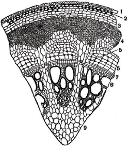
Fig. 65. Cross-section of a one-year-old stem branch of Dutchman's pipe:
1 - epidermis, 2 - collenchyma, 3 - primary cortex parenchyma, 4 - sclerenchyma,
5 - phloem, 6 - xylem, 7 - fascicular cambium, 8 - interfascicular cambium, 9 - pith
Under low magnification, the sieve tubes of the phloem are barely visible, whereas the large pitted Vessels of the xylem stand out prominently.
These thin sections can also be examined under high magnification. The multi-layered cuticle is clearly visible on the epidermis. Chloroplasts, typical of parenchymal tissue cells, are prominent in the cortex. Closer to the epidermis lies the collenchyma, featuring angular thickenings. The mechanical ring, individual fibers, pits, and areas of intercellular substance between the fibers are clearly discernible. Within the vascular bundles, the walls of spiral and pitted vessels, as well as tracheids and wood fibers (libriform), are easily distinguished. Sieve tubes are less conspicuous and can be identified by their companion cells. Both fascicular and interfascicular cambia are clearly visible.
The anatomical structure of a multi-year-old stem of Dutchman's pipe is more complex. This becomes apparent when comparing anatomical sections of one-year-old and older stems. Microscopic examination of perennial stem slides clearly reveals the changes occurring in the epidermis. The cells underlying the epidermis increase in volume, causing the growing Tissues to first stretch the epidermis and eventually rupture it in certain areas. Beneath the remaining epidermal patches, a new protective tissue—periderm (cork)—develops. Its cells are square-shaped with unevenly thickened walls, resulting in folds and wrinkles that are clearly visible on the stem surface. Cork cells are dead, and their walls are impregnated with suberin. The formation of cork is crucial because it is impermeable to both water and gases.
With the formation of cork, The connection between the epidermal cells and other living tissues is severed, causing the epidermis to die. On its inner side, the cork borders a strip of living cells containing granular cytoplasm and nuclei. This is the phellogen, or cork cambium, a secondary meristematic tissue. The phellogen produces cork cells outward. Beneath the phellogen lie collenchyma cells containing cytoplasm, followed by cortical parenchyma cells containing calcium oxalate crystals in the form of druses. Among the parenchymal cells, many store significant amounts of starch, the presence of which is easily detected using iodine tests. Mechanical fibers are well represented; their continuous ring is broken into separate segments interspersed with ground parenchyma cells. Such ruptures occur As a result of the vigorous radial expansion of the stem's central cylinder. Further inward lie parenchymal tissue cells, some containing calcium oxalate crystals, though the majority accumulate starch.
Finally, the phloem is encountered, in which rows of sieve tubes are clearly discernible; they appear light-colored and are separated by strips of parenchymal cells.
These cells function in the Transport of Assimilates and yield a typical iodine reaction, indicating the presence of starch within them. The elements of primary stem structure—epidermis, collenchyma, cortical parenchyma, and the mechanical ring—originate from the primary meristem. In contrast, the phloem elements arise from the secondary meristem, the cambium.
The central cylinder of the stem begins with the cambium. Inward from it lie the wood (xylem), pith, and medullary rays. Wood consists of wood parenchyma, vessels, and libriform (wood fibers). As perennial plants grow and develop, the ratio between the cortex and the central cylinder changes: the total mass of the cortex decreases, whereas the mass of the wood increases significantly.
Longitudinal sections from fresh material of Dutchman's pipe are difficult to obtain. To examine and study the anatomical structure in longitudinal sections, a small piece of a perennial branch is split in half with a sharp knife, crude sections are razor-cut and transferred to test tubes containing chromic acid or a 10% potassium hydroxide solution. The contents are boiled over an alcohol burner for 5–8 minutes, after which the liquid is poured onto a glass slide. The sections are retrieved, arranged, straightened with dissecting needles, and examined under low Microscope magnification.
Under the microscope, parenchymatous cork cells will be clearly visible. Due to the boiling process, the substances impregnating the walls of the cork cells dissolve, causing these walls to thin. The preparations clearly display collenchyma cells and fibrous sclerenchyma cells, the latter having heavily thickened walls with pits on their lateral surfaces. Among the vessels, large pitted vessels with visible pits, spiral vessels, tracheids, and parenchymal tissue cells stand out. Bordered pits can also be observed in such preparations.
Slide. Stem structure of dog rose (Rosa sp.)
Perennial plants (shrubs and trees) are characterized by the early developmental stage at which the xylem forms a continuous ring. Consequently, the young, growing twigs of various trees and shrubs exhibit a stem structure that differs very little from that of their older, multi-year-old regions. Independent vascular bundles here appear only as leaf traces. This structure is also characteristic of later stages of growth and development in herbaceous plants. For example, the stem Anatomy of the sunflower illustrates how a continuous xylem ring is formed, whereas in young, growing stem regions, the xylem initially consists of separate, disjointed bundles. These changes arise from various factors, namely: the expansion of primary bundles, the formation of new elements resulting from The activity of the interfascicular cambium, and the growth of the xylem ring formed during the Cytology/cytology/16.html">Early stages of Plant Growth and Development.
The stem anatomy of shrubs is identical to that of trees. The primary difference is that shrubs have a less developed cortex, whereas trees have a less developed ground parenchymal tissue. A convenient subject for studying shrub stem anatomy is the dog rose, which is common in our forests and brushwood. To study the stem anatomy of the dog rose, cross and longitudinal sections are prepared. These thin sections are placed in a drop of water and examined under low and high microscope magnification.
The Characteristic Features of dog rose stem anatomy are as follows. Its stem features a highly developed pith surrounded externally by wood and cortex. Three distinct groups of cells can be identified within the dog rose pith. The first group comprises thin-walled parenchymal cells. Initially, these cells are rounded in shape, but later, as the plant grows and external pressure increases, they assume the shape of spherical triangles. Pits are present in the walls of these cells.
The COMPOSITION OF THE Cell contents in the first group of pith cells changes over time. In early spring, they are almost empty, but at the beginning of the growing season, driven by enhanced photosynthetic activity, starch accumulates within the cells, reaching a maximum volume in autumn. Throughout the year, this stored starch undergoes complex transformations, converting into sugars and Other Compounds. These conversions are closely linked to metabolic pathways. Pronounced transformations occur in early spring, prior to leaf formation. During this period, starch is converted into sugars and transported to the growing PARTS OF THE plant. Thus, in the stem of the dog rose, the storage function typically performed by wood parenchyma is instead carried out by the pith cells.
The second group of cells in the rosehip pith consists of small cells with thickened walls. These cells perform a mechanical function and are arranged in groups among the larger pith cells.
A third group of small cells with dense yellowish contents is also distinguished in the rosehip pith. These are secretory cells that accumulate waste products that do not directly participate in metabolic processes.
Characteristic structural features are also inherent in the rosehip bark. Here, the epidermis (Fig. 66) has layers of cuticle. The epidermal cells contain cytoplasm, cell sap, and have a bright pink color, which is especially typical of young shoots. This coloration is caused by the presence of anthocyanins in The Cell sap.
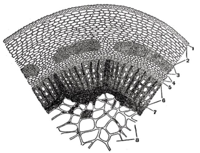
Fig. 66. Cross-section of a rosehip stem:
1 - epidermis, 2 - cortical parenchyma, 3 - bundles of bast fibers, 4 - phloem (sieve tubes), 5 - cambium, 6 - secondary xylem, 7 - primary xylem, 8 - pith
Beneath the epidermis lies the cortical parenchyma, which consists of living collenchyma cells whose walls are thickened not at the corners, but uniformly around the entire perimeter, and parenchyma underlying the collenchyma. The parenchyma cells contain chloroplasts and inclusions. Groups of bast fibers with heavily thickened walls lie behind the parenchyma. They lack cell contents and are dead cells whose cavities appear black under a microscope. Parenchyma cells run between the specified groups of fibers, separating them such that each group of bast fibers corresponds to a single section of sieve tubes, which are also separated by parenchyma cells. The layers of parenchyma cells resemble triangles with acute angles lying between the sieve tubes near the cambium, and a broad base situated between the bast fibers closer to the periphery.
In older branches, In addition to the aforementioned elements, periderm (cork) develops in the bark. It arises from the cork cambium (phellogen), which originates from the parenchyma cells located between the collenchyma and the epidermis. The cork cambium cells divide by partitions parallel to the surface. The resulting cells grow to normal size, their walls become impregnated with suberin and turn brown, forming a tissue of rectangular cells that become suberized and die off.
A characteristic feature of the xylem structure is the highly developed libriform (wood fibers), which forms layers between the vessels and consists of fibrous cells. The walls of the libriform cells are thickened to such an extent that only small lumens remain within the cell cavities.
The libriform forms a dense ring within the xylem, which is intersected by pith rays. Primary pith rays consist of large cells; they extend into the bark as well, dividing the sieve tubes into separate sections. Secondary pith rays consist of smaller cells and reach only the cambial layer (Fig. 66).
Slide. Cross-section of an elderberry stem (Sambucus nigra L.)
Cross-sections of an elderberry stem provide a clear illustration of the secondary protective tissue—the periderm (cork). Examining elderberry branches of various ages, even without a microscope, reveals that cork begins to form on annual branches. This phenomenon is easily observed because the green color of the branches changes to gray, and subsequently they turn brown. These changes occur as a result of
the epidermis dying off, sloughing away, and being replaced by newly formed cork cells. Cross-sections of a young elderberry branch, even at low magnification, allow the details of the stem structure to be studied and establish that its exterior is covered by an epidermis consisting of slightly elongated cells. The upper cell walls are thickened and covered with a cuticle. Some epidermal cells are ruptured and die off, while others remain living, possessing cytoplasmic contents and chloroplasts.
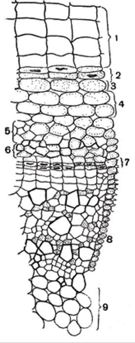
Рис. 67. Поперечний зріз стебла бузини:
1 - корок, 2 - фелоген, 3 - фелодерма, 4 - пластинчаста коленхіма, 5 - паренхіма кори, 6 - луб, 7 - камбіальні клітини, 8 - ксилема, 9 - серцевина
Beneath the epidermis lies the periderm (cork) (Fig. 67), which consists of thin-walled small cells arranged in regular radial rows. Cork cells stain red with Sudan III, similarly to the cuticle. This is due to the fact that the walls of the cork cells are impregnated with fat-like substances. Cork cells originate from the cork cambium, or phellogen, which is clearly visible in cross-sections; they are living, thin-walled, and rich in cell contents. The cork cambium produces not only cork cells, which are located outward toward the periphery and quickly die off, but also living tissue cells located inward—the phelloderm. The combination of cork cambium, cork, and phelloderm cells is called the periderm.
In the elderberry stem cross-section, lamellar collenchyma lies beneath the periderm. In the Initial Stages of stem development, collenchyma lies directly beneath the epidermis, but after the Formation of the periderm, it shifts to deeper layers of the bark. In the cells of the lamellar collenchyma, Cell wall thickening is observed not at the corners, but solely on the tangential sides, i.e., parallel to the surface. The radial walls remain unthickened. A layer of cortical parenchyma lies beneath the collenchyma, its cells containing cytoplasmic contents and chloroplasts. Mechanical tissue in the bark is poorly developed. It consists of small patches of bast fibers. Located behind the cortical parenchyma is the phloem — sieve tubes and companion cells — followed by a layer of cambial cells.
The central part of the stem consists of the xylem and the pith. The xylem contains various vessels and libriform fibers. The pith occupies a large volume and consists of thin-walled parenchymatous cells. Pith cells are empty and die off early. Lenticels are present On the surface of the elderberry stem.
Slide. Cross-section of a pine stem (Pinus silvestris L.)
In their anatomical structure, conifers differ from deciduous plants. The main difference is that the xylem of conifers lacks vessels. The sole elements conducting water and mineral nutrients are tracheids, which feature typical bordered pits. All conifers contain resin ducts in both the xylem and the bark.
The best subject for studying the stem structure of conifers is the pine. To study its structure, branches are harvested in advance
at 4–5 years of age, cut into small pieces, and placed in a mixture of alcohol and glycerin to soften the wood. Even without a microscope, cross-sections of a five-year-old pine branch reveal that its exterior is covered with brown cork. A brown pith is situated at the center of the stem. The xylem, colored white, lies between the pith and the cork. In pine, the xylem and phloem do not form separate vascular-fibrous bundles; instead, continuous rings of xylem and phloem are clearly visible, penetrated by primary pith rays. Annual rings forming concentric circles can be discerned even without a microscope.
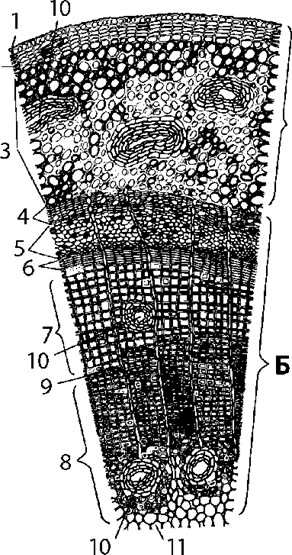
Рис. 68. Поперечний зріз стебла сосни:
A – cortex; B – central cylinder:
1 – epidermis, 2 – periderm, 3 – cortical parenchyma, 4 – primary phloem, 5 – secondary phloem, 6 – cambium, 7 – second-year xylem ring, 8 – first-year xylem ring, 9 – medullary ray, 10 – resin canal, 11 – pith
Make thin transverse sections and place them on a microscope slide in a drop of chlorozinc iodine. Before preparing the sections, ensure that the trimmed surface of the end face, prepared for cutting with a razor blade, is perpendicular to the longitudinal axis of the branch, as only then can a proper transverse section be obtained. Several sections should be made. Some of them may pass through the wood and pith, while others pass through the bark. To prevent the sections from tearing, The surface of the branch should be moistened with water to keep it damp. Examining such sections under low and high microscope magnification (Fig. 68), one can first establish that the peripheral part of the stem is brown, while the central part is white.
In the peripheral Regions of the bark, the bark (rhytidome) is visible, appearing as scales, with individual parts peeling off and falling away. Beneath the bark lies a relatively thick layer of primary cortex, consisting of parenchymatous cells. Individual resin canals are located in this part. Next comes the secondary cortex—the bast (phloem), where thin medullary rays are noticeable. They resemble wavy, curved, dark-colored thin lines. The section shows that the medullary rays originate from the xylem. They play an important physiological role in the plant's life by ensuring the horizontal transport of nutrients. The bast is divided into layers by concentric rows of cells containing starch grains and calcium oxalate crystals. In the outer layers of the phloem, specific groups of stone cells with heavily thickened walls can be observed. The contents of these cells are brown. Beyond the phloem lies a narrow strip of elongated cambial cells, followed by the xylem, in which annual rings are noticeable. The boundary between the xylem and cambium is especially distinct in autumn when cambial activity ceases. In spring, this distinction is not apparent, as thin-walled wood is formed with the onset of cambial activity. The xylem is penetrated by medullary rays consisting of cell strips stretching from the pith to the periphery. Such is the general anatomical STRUCTURE OF THE pine stem. To study the anatomical structure of the pine stem in greater depth, transverse sections are prepared separately through the bark and the wood. Thin sections are placed on a microscope slide in a drop of chlorozinc iodine and examined under low and high microscope magnification.
tubes, and beneath them the periderm.
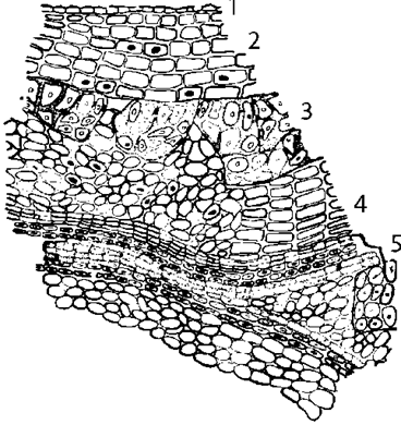
Fig. 69. Transverse section of a pine stem (bark):
1 – epidermis, 2 – parenchymatous cells of the primary cortex, 3 – stone cells, 4 – sieve tubes, 5 – periderm
First, we examine the transverse section through the bark (Fig. 69). The surface of the stem is covered with an epidermis, beneath which lie several layers of parenchymatous cells containing resin and stone cells, which occur in large groups. Within this parenchyma, there are resin canals lined on the inside with a layer of living cells that secrete resin. The resin canals form a cohesive interconnected system throughout the primary cortex. Beyond the areas of stone cells lie the sieve
Sections can also be treated with phloroglucinol and hydrochloric acid, an iodine-potassium iodide solution, or an aqueous solution of aniline blue. Different tissues will stain under The Influence of these Reagents on various preparations. For instance, Treatment with phloroglucinol and hydrochloric acid stains lignified cells cherry-red. Under the microscope, it can be established that the periderm (cork) in pine is heterogeneous, consisting of thin-walled and thick-walled cells. The thin-walled cork cells remain almost colorless, whereas the thick-walled ones are stained cherry-red. The thick-walled cells have lignified walls and lie in almost regular layers. This arrangement facilitates the rapid tearing of the outer bark. Several periderms are visible in the pine bark on a transverse section; on its outer surface, remnants of the browned, already dead primary cortex can be observed.
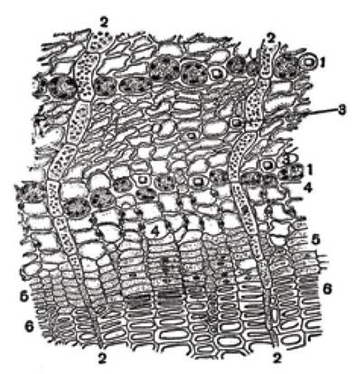
Fig. 70. Transverse section of a pine stem (through the bast):
1 – rows of dark brown cells, 2 – medullary rays, 3 – parenchymatous cells with crystals, 4 – sieve tubes, 5 – cambium, 6 – wood
The phloem in pine (Fig. 70) is characterized by the absence of sclerenchyma, i.e., bast fibers. It consists of sieve tubes and parenchymatous cells. Some of these cells contain cytoplasmic contents and starch, while others contain calcium oxalate crystals. Companion cells are absent. The phloem layers are radially intersected by wavy medullary rays, and transversely by rows of dark brown cells. The sieve tubes are four-sided in shape and arranged in rows. Their walls are slightly thickened and yellowish, and the lumina of the tubes contain fine-grained protein matter concentrated near the walls. Isolated parenchymatous cells containing crystals occur among the sieve tubes. Where the phloem borders the cambium, sieve plates with noticeable thickenings can be seen on the radial walls; these stain bluish-purple with aniline blue. This substance is callose. The cambium consists of several layers of cells that are very narrow, flattened, slightly elongated, with a granular cytoplasmic and nuclear content, and delicate, very thin walls.
The medullary rays passing through the phloem consist of radially elongated cells filled with cytoplasm, containing a Nucleus and starch grains.
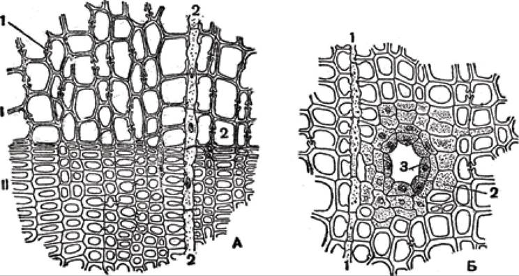
Fig. 71. Transverse section of a pine stem (through the wood):
A: I – spring wood; II – autumn wood: 1 – bordered pits, 2 – medullary ray; B: 1 – medullary ray, 2 – epithelium, 3 – resin duct
As already mentioned, the wood of pine is constructed solely of tracheids, as conifers lack vessels. Tracheids simultaneously perform two Functions: conducting water and dissolved substances, and providing mechanical strength to the stem. The presence of tracheids can be verified on transverse sections made exclusively through the stem wood when viewed in a drop of water under a microscope at high magnification. The tracheids composing the wood, or xylem, are structured differently depending on whether they originate in spring or autumn, which leads to the formation of so-called annual rings. Under the microscope, it is clearly noticeable that spring tracheids mostly consist of rectangular, wide, and thin-walled cells (Fig. 71, A). Autumn tracheids consist of narrow, thick-walled cells (Fig. 71, B). In each annual ring, it is difficult to sharply demarcate the spring xylem from the autumn xylem because the transition between them is gradual, but one annual ring is sharply demarcated from another. Bordered pits are located on the radial walls of the tracheids. In a transverse section, such pits resemble two bidentate forks with their Teeth turned toward each other.
The space between the teeth is intersected by a thin membrane with a lens-shaped thickening in the middle, called the torus. Many bordered pits can be seen in the spring wood. In the late xylem, the tracheids appear radially flattened, their lumen is poorly developed, the walls are thickened, and bordered pits are either absent or very rare.
It is believed that spring and autumn tracheids perform different functions: spring tracheids serve primarily for The transport of water and dissolved substances (physiological function), whereas the function of autumn tracheids is mechanical, similar to libriform fibers. Through maceration and The Use of appropriate reagents, it has been established that the walls of tracheids consist of primary, secondary, and tertiary layers.
The degree of cell wall lignification decreases from the primary layer toward the tertiary layer.
Resin canals can be observed in the xylem (Fig. 71, B), but they are smaller than those in the primary cortex. The resin canals are surrounded by an epithelium, or lining cells. A Second layer of living cells containing cytoplasmic contents and starch is located right here.
Slide. Longitudinal radial section of a pine stem (Pinus silvestris L.)
Take a small piece of a three- or four-year-old pine branch and split it in half with a sharp knife. Then smooth the cut surface and prepare a longitudinal radial section. To do this, place the split piece of the pine stem horizontally and make a transverse notch at some distance from its ends. Starting from one end of the notch to the other, prepare several radial sections calculated to pass perpendicularly through the annual rings. Transfer the prepared sections to a microscope slide in a drop of iodine-potassium iodide solution or aniline sulfate and examine them under a microscope. These reagents are used to confirm the lignification of the walls, which stain yellow (with aniline sulfate).
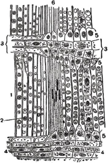
Fig. 72. Longitudinal (radial) section of a pine stem:
1 - tracheids, 2 - bordered pits in tracheids, 3 - medullary ray, 4 - central cells of the medullary ray, 5 - albuminous cells of the medullary ray, 6 - cambium
The preparations (Fig. 72) clearly demonstrate tracheids, which consist of elongated cells. Their structure within an annual ring is uneven: spring tracheids are wide, whereas autumn tracheids are narrow. In some areas of the sections, their tapered ends can be observed. Early wood tracheids feature numerous large bordered pits on their radial walls, resembling light concentric patches. By adjusting the micrometric screw of the microscope, one can observe that each pit typically contains not one, but two circles, increasing in size from the center toward the periphery. Late tracheids differ slightly in structure compared to early ones. They are narrower, have fewer bordered pits, and the pits themselves are smaller than those in spring tracheids. Due to their shape and thickened walls, tracheid cells resemble sclerenchymatous fibers, specifically libriform fibers, which they substitute here. Longitudinal radial sections pass through the medullary rays, which appear as broad strips intersecting the tracheids. Each medullary ray consists of several rows of cells containing albuminous and central cells. The albuminous cells are located along the edges of the medullary ray; their inner walls are uneven and irregularly dentate, featuring small bordered pits where they abut the tracheids. These cells are referred to as tracheidal cells. They conduct water and dissolved Mineral Substances horizontally from one layer to another. Albuminous cells contain cytoplasm and a nucleus.
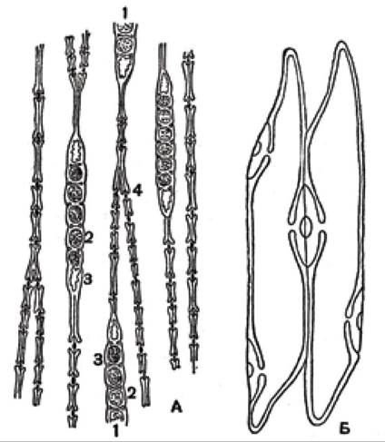
Fig. 73. Section of a pine stem:
A - longitudinal (tangential) section: 1 - medullary ray, 2 - central cells, 3 - albuminous cells of the medullary ray, 4 - end of the tracheid; B - bordered pits between two adjacent tracheids
The central cells contain cytoplasmic content, a nucleus, and starch grains. The pits in these cells are simple and appear as large light patches. Resin ducts may occasionally appear in the sections. The walls of the resin duct do not stain with aniline sulfate.
The tangential section of the pine stem is prepared so that it does not pass through the center and remains perpendicular to the medullary rays. The prepared sections are placed in a drop of phloroglucinol with hydrochloric acid, which stains lignified walls containing Lignin red. As seen in the section (Fig. 73), the medullary rays are cross-sectioned, appearing as short single- or multi-layered regions. Tracheids are visible at the ends of the ray, while living cells containing Starch and other inclusions are found in the middle. The starch-containing cells are loosely packed, with large intercellular spaces between them. Pits are visible in the living cells, appearing as thin membranes between the thickened areas of The cell wall. Tracheids with bordered pits are cross-sectioned along their lateral walls, which explains why they look similar to those in transverse sections. Cross-sectioned resin ducts are frequently encountered in tangential sections.
Preparation. Structure of the linden stem (Tilia cordata L.)
Sections are prepared from one-year-old and three- or four-year-old branches. To obtain them, knot-free branches with a well-developed cork layer are selected. The branches should be harvested in autumn, after growth has ceased and processes related to reserve nutrient accumulation have been completed. The harvested material is stored in a mixture of alcohol and glycerin.
Sections are cut through a portion of the stem from a branch that is first split in half; the sections are then placed on a microscope slide in a drop of water or glycerin and examined under low magnification. The sections (Fig. 74) show that the stem is externally covered by a periderm. The outer cells of the periderm are darker than the inner ones. Beneath the periderm lies the primary cortex, where parenchymal cells containing cytoplasm, starch, and chloroplasts are visible closer to the periphery. These are collenchyma cells; they have white, glossy, thickened walls.
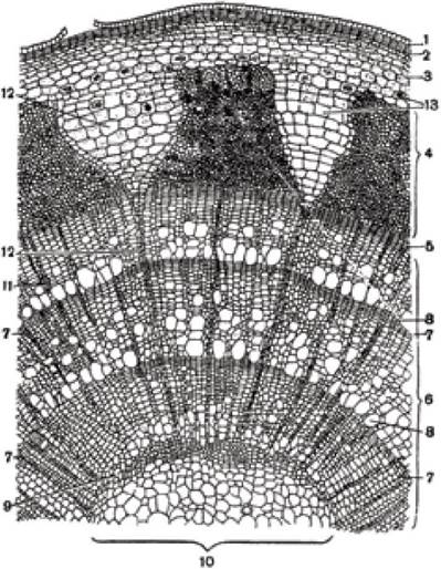
Fig. 74. Transverse section of a three-year-old linden branch:
1 - epidermis, 2 - periderm, 3 - primary cortex, 4 - bast (phloem), 5 - cambium, 6 - annual wood rings, 7 - annual ring boundary,
8 - large vessels, 9 - primary xylem, 10 - pith, 11 - secondary medullary rays, 12 - primary medullary rays, 13 - druses
Beneath the collenchyma lies the parenchyma of the primary cortex, the cells of which have thin walls. Some parenchymal cells are empty, while others contain cytoplasm, chloroplasts, and calcium oxalate crystals in the form of druses.
Adjacent to the cortical parenchyma is the phloem, divided into regions that expand from the periphery toward the center. This phloem arrangement resembles trapezoids in which bast fibers alternate with sieve tubes and phloem parenchyma cells. Medullary rays pass between the phloem regions, forming triangles with their bases at the periphery. The oldest parts of the phloem are located in the narrowest part of the phloem region, whereas the youngest ones adjoin the cambium. Each phloem region contains glossy yellow strips of cells. These are sclerenchyma cells known as bast fibers, or hard bast. They possess thickened walls, and the cell cavity is darkly stained. In addition to thick-walled elongated cells, thin-walled cells are noticeable in the phloem, representing the soft bast, which consists of sieve tubes and companion cells.
The phloem is separated from the xylem by a thin layer of cambium cells. Cambium cells have thin walls that rupture easily, which is why the entire outer part of the stem—including the periderm, primary cortex, and secondary phloem with medullary rays—can be easily peeled off. Collectively, these tissues are conventionally referred to as the bark.
The cambium appears as a strip of small cells flattened in the radial direction. All cambium cells contain cytoplasmic content and nuclei. They divide by tangential walls, thereby giving rise to secondary xylem and secondary phloem. Considerably more secondary xylem is always produced than phloem, which explains why the central part of the stem is larger in volume than the peripheral part.
In transverse section, the xylem, or wood, consists of regions separated from one another by primary medullary rays. The following components can be distinguished within the xylem: vessels, tracheids, libriform fibers, wood parenchyma, and medullary rays.
Medullary rays consist of living, polygonal, slightly elongated cells containing cytoplasmic content, nuclei, and starch.
Medullary rays are radially arranged and subdivided into Primary and secondary. Primary medullary rays extend from the pith to the cortex, having originated during the early stages of SHOOT development from the primary meristem. Secondary medullary rays arise later from the cambium and therefore do not reach the pith.
The entire xylem is divided into annual rings, in which elements are arranged in a specific pattern: thin-walled elements occupy the inner part of the annual ring, while thick-walled elements occupy the outer part. Each annual ring also exhibits a gradual transition from thin-walled to thick-walled elements. Large, thin-walled elements are formed in spring, whereas small, thick-walled ones are formed in autumn. This makes it possible to determine the age of a plant by counting the rings on transverse stem sections of perennial trees. The width of the annual rings varies depending on the conditions of the growing season.
Annual rings cannot be distinguished in the phloem; instead, only the hard bast strips can be counted. Their number is twice that of the wood rings, indicating that linden forms two strips of hard bast during a single growing season.
Xylem elements have a diverse structure. Vessels are made up of hollow, dead cells whose walls are pierced by pits. The cell walls of vessels feature uneven thickening, allowing them to be classified into spiral, annular, pitted, and other types. In addition to vessels and tracheids, the xylem includes wood parenchyma cells and libriform fibers. Wood parenchyma cells are living and filled with cellular contents, whereas libriform cells are dead.
The central part of the stem is occupied by the pith. It consists of large, polygonal cells that die off early and become filled with air. Interspersed among these large cells are smaller ones containing a brownish substance; the exact function of this group of cells remains unclear. The walls of the large cells feature pits, which are not discernible in the smaller ones. In the regions of the pith adjacent to the primary xylem, all cells are noticeably smaller.
The constituent elements of the xylem perform distinct functions. Vessels and tracheids conduct water and mineral nutrients, while also providing structural strength to the stem. Wood parenchyma cells store reserve nutrients such as sugars, fats, and other compounds. Medullary rays serve a dual function: they store reserve nutrients and facilitate the horizontal transport of plastic (assimilated) substances.
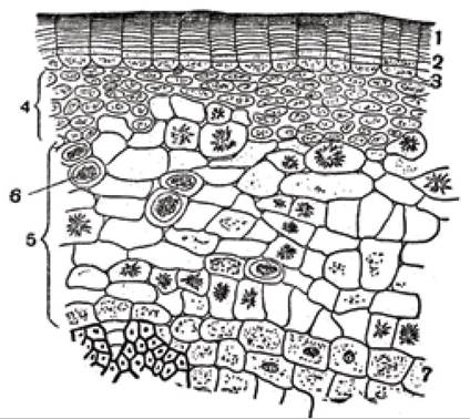
Fig. 75. Cross section of linden bark:
1 - cork, 2 - phellogen, 3 - phelloderm, 4 - collenchyma, 5 - thin-walled parenchyma, 6 - idioblasts, 7 - starch-storing cells
The bark of a linden stem has a complex structure that can be observed in cross sections (Fig. 75). The periphery is occupied by the cork, which consists of narrow cells: the outer cells are dark, while the inner ones are light. Cork cells contain a brownish substance. Beneath the cork lies the phellogen,
which consists of elongated cells containing fine-grained contents. Toward the center from the phellogen lie the phelloderm cells with thickened walls. Internally, these cells contain cytoplasm and chloroplasts. Next comes the collenchyma, which is composed of rounded cells with significantly thickened, non-lignified, living walls containing cytoplasm, chloroplasts, and starch grains. The collenchyma cells abut the thin-walled parenchyma, which contains scattered idioblasts. The parenchymal cells extend all the way to the bast (phloem).
Due to cambial activity, the phloem expands from the inside toward the periphery. The newly formed elements exert pressure on the surrounding parts of the bark, causing its cells to be compressed and deformed, especially those whose walls remain unthickened. The primary cortex is separated from the phloem by a layer of starch-storing cells. Hard and Soft bast differ in their structure. Hard bast consists of bast fibers whose walls are so heavily thickened and lignified that the cell lumen appears merely as a tiny dot (Fig. 76).
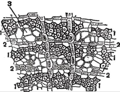
Fig. 76. Cross section of linden bast:
1 - hard bast, 2 - soft bast, 3 - sieve tubes
The fibers of the hard bast lie close together, forming strips that extend across the bundle between the medullary rays. The function of the hard bast is mechanical: it protects the soft bast, through whose cells plastic substances are transported. The soft bast consists of two elements: thin-walled parenchymal cells that are tangentially flattened and contain cytoplasmic contents (bast parenchyma), and polygonal, hollow cells with unthickened walls (sieve tubes). Also clearly visible are companion cells—small cells containing a granular substance and nuclei.
Slide. Longitudinal section of a linden stem (Tilia cordata L.)
To prepare tangential sections, a three- or four-year-old linden branch is taken, and thin longitudinal sections are sliced with a sharp razor to include the bark as well. The prepared sections are placed on a glass slide in a drop of water, chlor-zinc-iodine solution, aniline sulfate, or phloroglucinol with hydrochloric acid, and examined under low and high microscope magnification. The slides (Fig. 77) clearly show a periderm layer at the edges of the section containing lenticels. This is followed by the primary cortex, which consists of outer layers of collenchyma and deeper layers of parenchymal cells containing druses.
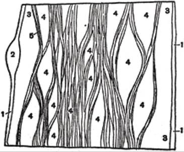
Fig. 77. Surface longitudinal tangential section of a linden stem (diagram):
1 - periderm, 2 - lenticel, 3 - primary cortex, 4 - medullary rays, 5 - thick-walled bast
Further inward lie bundles of thick-walled bast, the cells of which stain red with phloroglucinol and hydrochloric acid, and yellow with chlor-zinc-iodine or aniline sulfate. In tangential view, the medullary rays resemble loops. In very thin, deeper tangential sections (Fig. 78), one can observe uniseriate rays, whose cells are rich in starch, alongside multiseriate medullary rays. Bast parenchyma cells may be empty with pitted walls, contain crystals, or possess granular contents and nuclei.
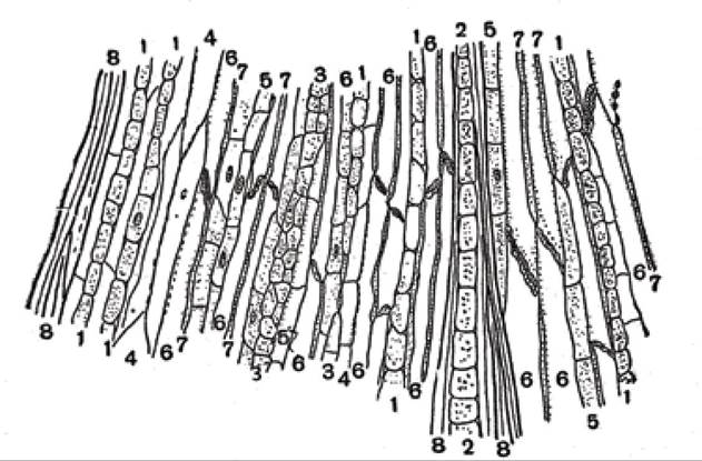
Fig. 78. Deeper tangential section of linden branch bast:
1 - uniseriate medullary ray, 2, 3 - multiseriate medullary rays, 4 - crystal-bearing parenchyma cells, 5 - living bast parenchyma cells, 6 - sieve tubes, 7 - companion cells, 8 - thick-walled bast
The slide reveals long, nearly empty cells with oblique sieve plates. Adjacent to them are narrow cells containing cytoplasm and nuclei—these are sieve tubes with companion cells. Highly prominent are the thick-walled cells with shiny walls, representing the thick-walled bast.
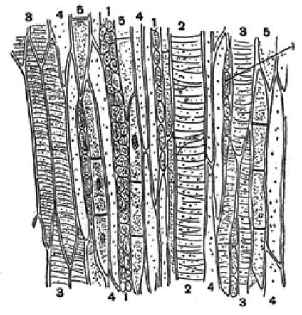
Fig. 79. Longitudinal tangential section of linden wood:
1 - pith rays, 2 - porous vessels, 3 - tracheid, 4 - libriform fibers, 5 - wood parenchyma
By moving the slide, one can examine individual xylem elements in a tangential section (Fig. 79), specifically spiral and pitted vessels. Tracheids appear as tapered fibers with pointed ends, featuring spiral thickenings and bordered pits in their walls. The pointed ends of the tracheids interlock with one another. Libriform fibers appear as long, narrow fibers with hollow interiors and slit-like pits. Wood parenchyma consists of cells with thick transverse partitions containing living contents and starch. Pith rays are composed of parenchymal cells containing cytoplasmic contents and starch. Among them, there are uniseriate and multiseriate, short and long rays.
Slide. Radial section of a linden stem (Tilia cordata L.)
All structural elements can be observed in radial sections (Fig. 80). Cork cells are visible on the periderm surface, with a shape very similar to that seen in cross-sections. These are followed by the phellogen and phelloderm. Behind them lie collenchyma cells, which appear elongated in radial sections. Crystal-bearing cells occur within the thin-walled parenchyma. Next, the starch sheath cells are discernible, followed by the hard bast (phloem fibers). The cells of the hard bast appear as fibers with very thick walls and almost completely lack cell contents. Sieve tubes and companion cells, phloem parenchyma cells, and crystal-bearing cells are located between the strands of hard bast.
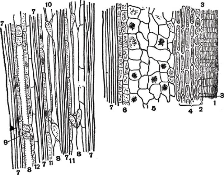
Fig. 80. Longitudinal radial section of a linden stem (through the phloem):
1 - cork, 2 - phellogen, 3 - phelloderm, 4 - collenchyma, 5 - thin-walled parenchyma, 6 - starch sheath, 7 - hard bast, 8 - sieve tubes, 9 - sieve plates, 10 - companion cells, 11 - phloem parenchyma, 12 - crystal-bearing cells
By moving the slide, one can examine the cambium and the xylem region of the stem. The cambium consists of narrow, elongated, thin-walled cells. It borders the wood, the elements of which vary depending on the thickness of the section and the specific area under the microscope's field of view. The xylem contains the following elements: pith rays, wood parenchyma, libriform fibers with thickened walls, tracheids with pointed ends, and vessels with bordered pits and spiral thickenings.
Large and small parenchymal cells can be observed in the pith. The large parenchymal cells are empty with thin walls. Arranged in longitudinal rows between the large cells are short cells with thick cellulosic walls containing a yellowish-brown substance. Similar cells are adjacent to the starch parenchyma, which consists of small cells with cytoplasmic contents lying just outside the pith. These are followed by one or two rows of elongated cells that also contain granular contents, after which the primary xylem begins.
Slide. Cross-section of an oak stem (Quercus robur L.)
To study the anatomical structure of the oak stem, one-year-old as well as two- or three-year-old branches are used. The anatomical structure is first examined in young, one-year-old branches, and subsequently in older ones. Sections are cut from green twigs so as to capture the entire surface of the end face, i.e., in cross-section. The prepared sections are placed on a glass slide in a drop of water and examined first under low and then under high magnification. Under the microscope, the outer tissue—the epidermis, collenchyma, and beneath it, the cortical parenchyma—is clearly visible. The cells of the cortical parenchyma and collenchyma are living, containing cytoplasmic contents and chloroplasts. The bast fibers in oak consist of elongated cells with thickened walls. The bast fibers are divided into areas by bands of phloem parenchyma. Sieve tubes and companion cells are clearly noticeable in the phloem. Located beyond the phloem is the cambium, which consists of a band
of elongated, thin-walled cells containing living contents and a nucleus. Situated beyond the cambium is the xylem, which forms the bulk of the central cylinder.
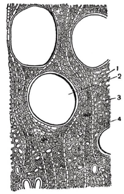
Fig. 81. Cross-section of an oak branch (through the wood):
1 - tracheids, 2 - porous vessels, 3 - pith ray, 4 - wood fibers (libriform)
In the wood, or xylem, porous vessels (Fig. 81), tracheids, and fibrous libriform cells, which provide mechanical strength to the stem, are visible. The wood is intersected by pith rays, which ensure the radial transport of nutrients across the stem. When preparing sections from young oak branches, the razor blade (iron) reacts with the Tannins present in the cell sap, resulting in the formation of ink. Tannins impregnate the cell walls of the collenchyma, parenchyma, cambium, and pith rays.
In preparations made from two- or three-year-old branches, the epidermis gradually disappears and is replaced by the periderm. Here, one can observe collenchyma, parenchyma, and regular layers of cork. On older branches, the peeling of cork is observed, which is eventually replaced by rhytidome (bark). Its formation is driven by the activity of the cork cambium, which successively dies off in the outer layers of the cortex and forms deeper within, thereby generating new areas of cork cells.
Instead, even deeper layers of cork cambium emerge, functioning in the same manner. As a result, the bark becomes permeated with layers of cork tissue, between which Tissues of the primary and secondary cortex are located. Oak bark is characterized by exceptional durability because its cells become lignified. In addition, it contains numerous stone cells (sclereids), whose walls are heavily thickened and possess almost no lumen. The walls of the stone cells are pierced by numerous branched pit canals.
Oak bark is distinguished by the abundant development of mechanical tissues within it. These include thick-walled bast fibers with lustrous walls and thin-walled bast fibers.
Wide and narrow pith rays, consisting predominantly of cells with granular contents, are noticeable in the xylem and phloem. The main xylem elements observable under the microscope are vessels, tracheids, wood parenchyma, and libriform fibers. As the plant grows, the volume of the pith does not increase compared to one-year-old branches, but its cell walls become lignified, and by the second and third years, they die off. As oak vessels age, they become occluded by ballast substances and tyloses, while the function of transporting water and plastic metabolites is carried out by young vessels located close to the cambium in the peripheral layers of the xylem—the sapwood. Consequently, the stem exhibits a central heartwood, the cells of which gradually die off, giving it a dark coloration.
Slide. Cross-section of a birch stem (Betula sp.)
A 5-6-year-old birch branch is taken, and cross-sections are made. The prepared sections are placed on a microscope slide and examined under high and low magnification. The following parts can be distinguished: the cortex, the central cylinder, and the pith, which consists of rounded cells with thickened and lignified walls. The pith has an angular shape and projects with its sharp points into the primary wood.
At the periphery of older birch twigs, the epidermis is replaced by a whitish tissue known as birch bark (outer bark/bark paper). It may also have a pinkish hue. This outer bark is formed by the phellogen and is laid down in regular annual layers. Its stratification is explained by the differential activity of the phellogen in spring and autumn. In spring, the phellogen produces regular rows of thin-walled, wide cells containing fine-grained contents. In autumn, by contrast, narrow cells are formed that are flattened in the radial direction toward the stem. The granular Contents of the wide spring cells are insoluble in water but soluble in alcohol. These contents consist of granules representing an organic substance—betulin—which imparts the white color to the birch bark. The layered formation of the outer bark causes it to exfoliate every two years. In the process, the outer thin-walled cells rupture, releasing a white powdery betulin.
In older parts of the stem, the outer birch bark is replaced by rhytidome (bark). Even to the naked eye, one can detect strips of periderm featuring light (spring) and dark (autumn) layers within it. Wide areas of the rhytidome consist of cortical parenchyma cells whose walls have become suberized and lignified. Dense structures composed of stone cells are noticeable among the parenchymal cells.
In the cross-section of young branches prior to the formation of the outer birch bark, the epidermis is noticeable, which is quickly replaced by the periderm, including cork (Fig. 82). Beneath the periderm lies parenchymal tissue containing groups of stone cells and bast fibers.
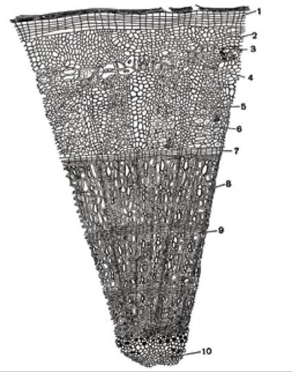
Fig. 82. Cross-section of a birch stem:
1 - periderm, 2 - parenchyma tissue,
3 - stone cells, 4 - bast fibers, 5 - phloem, 6 - medullary ray, 7 - cambium, 8 - spring wood, 9 - autumn wood, 10 - pith
Sieve tubes and companion cells (phloem) are typical, though they do not form distinct areas, and bast fibers are not visible here. In the birch bark, groups of stone cells perform the function of mechanical tissue.
In the xylem, which consists of spring and autumn wood, vessels with lignified pitted walls are clearly visible even in cross-section, especially where the vessels are in contact with one another. The vessels are evenly distributed within the libriform tissue, which occupies a significant portion of the xylem. Medullary rays consist of elongated cells and extend from the pith to the bark. The cambium divides the stem into a central cylinder and a peripheral phloem region.
Slide. Cross-section of the rhizome of bracken fern (Pteridium aquilinum L.)
Thin cross-sections are prepared from a fresh or fixed bracken fern rhizome, placed in a drop of water, and examined under a microscope at low magnification. The rhizome is externally covered by the epidermis. When treated with phloroglucinol, its cells stain a rust color. The outer walls of the epidermis are covered with a cuticle. Beneath the epidermis lie cork cells whose walls are impregnated with suberin. These are followed by yellowish-brown parenchymal cells that occupy a large volume. Noticeable intercellular spaces shaped like triangles are visible in the fundamental parenchymal tissue. The cells of the ground tissue are rounded and thin-walled. Their cytoplasmic contents and starch grains are clearly visible.
Amidst the ground tissue lie darkly stained areas of fibrous cells and vascular bundles of various shapes.
In the central part of the vascular bundles, clusters of large irregular rings corresponding to water-conducting elements can be observed. Slit-like pits are visible in places on the walls of these elements. At the points of contact between the conducting elements, middle lamellae are visible, or they are separated by small parenchymal cells containing dense cytoplasmic contents and starch grains.
Sieve tubes are located around the xylem elements. They form one or two rows and have large lumens. Phloem parenchyma cells adjoin the sieve tubes; they contain proteinaceous matter and stain yellow with iodine. These tissues are surrounded by a single layer of starch-storing cells, followed by the endodermis, which consists of small cells with thickened walls and performs a mechanical function.
The mechanical tissue has a characteristic structure. Its regions are separated from the vascular bundles. The mechanical tissue forms flat arches with their concave sides facing each other, or they fuse into a horseshoe-shaped area. The conducting elements are located inside the horseshoe, as well as partially outside it along the sides.
Comparing ferns with other plants, one can conclude that each bundle corresponds to the entire ring of vascular bundles characteristic of dicotyledonous plants. As noted earlier, each fern bundle is surrounded by a single-layered ring of starch-storing parenchyma and a ring of mechanical elements—the endodermis.
Slide. Longitudinal section of the rhizome of bracken fern (Pteridium aquilinum L.)
Thin longitudinal sections are prepared from a fresh or fixed bracken fern rhizome, placed in a drop of water, and examined under a microscope.
In the longitudinal section of the bracken fern rhizome, The structure of individual elements can be observed: the epidermis and parenchymal cells, which are longitudinally elongated and have slanting ends. This cell arrangement has a mechanical significance, presumably replacing collenchyma, which is absent here. The ground tissue consists of rounded cells containing cytoplasm and starch.
The mechanical tissue resembles bast fibers in shape, but its cells are shorter, have pointed ends, and their walls are perforated with pits. Tracheids of considerable size with slanting and tapered ends are clearly visible within the vascular bundles. Their lateral walls exhibit scalariform thickening. Upon treatment with phloroglucinol and hydrochloric acid, the tracheid walls stain red, indicating that they have lignified. The pits here are bordered.
Sieve tubes stain purple with chlor-zinc-iodine. They feature numerous oval or elliptical sieve plates. Cells of the conducting parenchyma are clustered near the tracheids and sieve tubes, containing cytoplasmic contents and starch. Each bundle is surrounded by a starch-storing and mechanical ring.
Last update: 07/08/2026
Editorial and Educational Adaptation: This material has been compiled based on the primary/original source text. The project team performed an editorial review, corrected technical inaccuracies, structured sections, and adapted the content for an educational format.
What was processed:
- elimination of formatting defects (OCR errors, structural breaks, corrupted characters);
- editorial organization of content;
- standardization of terminology in accordance with academic sources;
- verification of factual statements against the original source text.
All mentions of the author, publication year, and origin of the primary text have been preserved in accordance with the source.