Plant Anatomy: Workshop - Paniuta O.O. 2019
Topic 3. Structure of Vegetative Organs
Laboratory Work No. 12. Structure of Dicotyledonous Plant Stems
Objective: to study the Structural Features of the stem in dicotyledonous plants.
Materials and equipment: light microscopes, Glass slides and cover slips, dissecting needles, forceps, glass rods, filter paper, distilled Water, chlor-zinc-iodine, aniline sulfate, phloroglucinol with Hydrochloric acid, iodine in potassium iodide, glycerin, plant material.
Slide. Cross-section of a pumpkin stem (Cucurbita pepo L.)
Thin sections are cut from a fresh or preserved pumpkin stem and treated with chlor-zinc-iodine for a more detailed study. This Treatment is necessary to better distinguish between different Tissues. One or two drops of chlor-zinc-iodine are placed on a glass slide, the sections are immersed in it for two minutes, and then covered with a cover slip. Such preparations can be examined even without a Microscope if the glass slide is placed on white paper. The sections reveal a central cavity that forms large hollows in the stem between the vascular bundles. Most of the section will be stained reddish-purple, against the Background of which certain areas appear significantly lighter. These areas constitute the conducting tissue, or vascular-fibrous bundles. The section clearly shows openings—vessels through which water and mineral nutrients move from the ROOT to the leaves. They form part of the xylem. Sieve tubes, through which organic nutrients move from the leaves to the root, have a matte appearance. These are elements of the phloem. Even without a microscope, the arrangement of the vascular bundles in two circles is visible. The part of the stem located outside the vascular bundles is called the primary cortex, and the part toward the center from the bundles is the pith. Pith rays lie between the vascular bundles.
Class="center">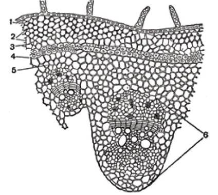
Fig. 60. Cross-section of a pumpkin stem:
1 - epidermis, 2 - collenchyma, 3 - primary cortex parenchyma, 4 - sclerenchyma,
5 - fundamental parenchyma, 6 - vascular-fibrous bundle
Afterward, even thinner sections must be prepared for examination under high magnification of a microscope. Such an examination reveals (Fig. 60) that the outer surface of the stem is covered by an epidermis bearing linear multicellular hairs and papillae. The hairs terminate in small conical Cells. If the preparations are made from a young stem, the Hair cells are living and contain Cytoplasm, nuclei, and METABOLISM/14.html">Chloroplasts. The epidermis stains yellow with chlor-zinc-iodine. The outer walls of the epidermis are impregnated with cutin and wax, which prevents water permeability. The film formed On the surface of the epidermis is called the cuticle. Beneath the epidermis lies the mechanical tissue, collenchyma, which consists of several layers of cells arranged in discrete patches. The collenchyma cells are filled with living contents. They border the living parenchymal tissue, behind which lies a strip of sclerenchyma—a mechanical tissue whose cells have lignified walls and stain yellow with iodine. Sclerenchyma cells are dead; they lack cytoplasmic contents, have uniformly thickened walls, and fit tightly against one another.
Beyond the sclerenchyma are the Cells of the fundamental parenchyma with thin walls, cytoplasmic contents, and abundant starch. Pores are visible in The Cell walls under a microscope. Vascular-fibrous bundles are embedded within the fundamental parenchyma.
Slide. Longitudinal radial section of a pumpkin stem (Cucurbita pepo L.)
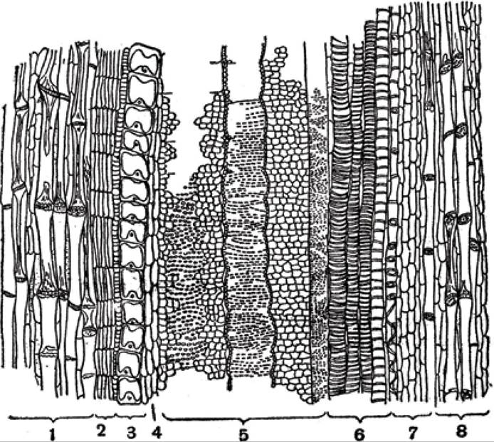
Fig. 61. Cytology/practical/54.html">Longitudinal section of a pumpkin stem:
1 - outer phloem, 2 - cambium,
3 - developing vessel, 4 - wood parenchyma, 5 - secondary xylem, 6 - primary xylem, 7 - parenchymal cells, 8 - inner phloem
A piece of the pumpkin stem is split to expose the inner parts, and the cut surface is trimmed smooth with a razor blade. Preparations are made so as to capture the main Tissues of the stem. The prepared sections are placed in a drop of water, a solution of chlor-zinc-iodine, or aniline sulfate, and examined under low and high magnification. Different tissues stain differently with these Reagents. Under low magnification, the microscope reveals the epidermis with its cuticle and the collenchyma cells, which have a regular cylindrical shape with horizontal cross-walls. In longitudinal section, the fundamental parenchyma differs little in cell shape from what is observed in cross-sections (Fig. 61).
Of particular interest here is the sclerenchyma, which consists of long fibers with thickened cell walls. The longitudinal walls feature Pores in the form of tiny dots.
The wood contains various types of vessels that are easily distinguished by treating the sections with appropriate reagents, such as aniline sulfate, which turns them yellow. Here, annular, spiral, and pitted vessels can be observed. The walls of larger vessels exhibit a complex network of thickenings and pits, each surrounded by a ring of less-thickened wall material. Treatment of the preparations with phloroglucinol and hydrochloric acid shows that the bulk of the vessel walls are either unlignified or only slightly lignified. This is why a bright magenta coloration appears only where the walls are significantly thickened. The cambium can be observed in longitudinal section. Its cells are elongated longitudinally, with rounded corners. A Nucleus is clearly visible in each cell.
To get a better look at the sieve tubes, a new preparation is made and treated with chlor-zinc-iodine, which stains the sieve tube walls purple and their internal contents yellow.
It can be seen that individual sieve tube members are cylindrical and slightly widened at the level of the transverse partitions. The partitions are arranged horizontally or obliquely relative to their long axis. In the absence of callose, the partitions are thin, and pores are evenly distributed across their entire surface. When callose is present, uniform thickenings appear on both sides of the primary partition, making it look like a series of closely packed cylinders with rounded ends. The Structure of the lateral walls is also interesting. Their longitudinal sections occasionally appear beaded due to pores piercing the walls. Preparations may reveal pores in the walls that feature partition-like plates, each pierced by tiny openings resembling a miniature sieve.
Through these lateral sieves, organic nutrients contained in the sieve tubes are transported to parenchymal cells, cambial cells, and companion cells. This plays a crucial role in metabolism and ensures the supply of organic nutrients to the living cells of the stem.
Companion cells are located adjacent to the sieve tubes and have tapered ends. Nuclei and cytoplasmic contents are clearly visible in these cells.
Thus, the phloem and xylem in the pumpkin stem consist of various elements. The xylem includes vessels, tracheids, wood or mechanical fibers located between the vessels, and parenchymal cells.
The phloem comprises sieve tubes, companion cells, bast fibers, and parenchymal cells located between the sieve tubes. These elements are situated outward from the cambium.
Slide. Cross-section of a sunflower stem (Helianthus annuus L.)
The preparations are made from the stems of young plants, which are pre-fixed. Transverse sections are cut from the stem and placed in a drop of water or glycerin. These sections can be examined using a hand lens. Even under low magnification, darkish spots are visible on the sections—these are vascular-fiber bundles arranged in a circle near the stem surface. Such sections provide a schematic Overview of the stem's structure.
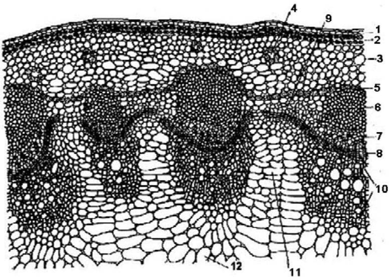
Fig. 62. Transverse section of a sunflower stem:
1 - epidermis, 2 - collenchyma, 3 - cortical parenchyma, 4 - resin duct,
5 - endodermis (starch sheath), 6 - sclerenchyma, 7 - phloem, 8 - fascicular cambium, 9 - interfascicular cambium, 10 - xylem, 11 - pith ray parenchyma, 12 - pith parenchyma
To study the Anatomical Structure in greater detail, a piece of the stem is split in half with a knife, after which several thin sections are prepared. For better observation, the sections are treated with various reagents to examine specific tissues in each. These reagents include: an iodine solution in potassium iodide, phloroglucinol with hydrochloric acid, aniline sulfate, etc. Under a microscope, even at low magnification, it is noticeable that the outer surface of the stem is covered by the epidermis (Fig. 62), bearing hairs of various sizes. Beneath the epidermis lies the collenchyma, which consists of cells with thickened walls. Collenchyma cells are living and contain cytoplasm, a nucleus, chloroplasts, and inclusions. Next is the parenchymatous tissue, composed of small and large thin-walled cells. The starch sheath borders the central cylinder; it consists of cells filled with living contents and starch. The central cylinder is made up of vascular bundles and the pith. Each bundle consists of phloem, located toward the periphery, and xylem, lying toward the center. Between them is the fascicular cambium, and interfascicular cambium is also visible. The bundles vary in size, with larger ones alternating with smaller ones. Sclerenchyma develops externally in front of each large bundle. The center of the stem is occupied by the pith, which consists of large, polygonal, thin-walled cells. In the cortex and the central cylinder, distinct dark-colored areas can be observed—these are resin canals.
Under high magnification, thin sections treated with appropriate reagents (Fig. 63) allow for a more detailed examination of the preparations, as well as sketching and labeling individual tissues. Such magnification reveals that the epidermis consists of small, slightly elongated cells. The cuticle on the outer walls of the epidermis is poorly developed. Multicellular, rough, and bristly hairs are clearly visible, situated on small epidermal outgrowths. The collenchyma consists of several layers of cells with white, shiny walls. These cells contain cytoplasm, a nucleus, chloroplasts, and starch grains. Toward the center of the stem, the collenchyma gradually transitions into the thin-walled parenchyma of the primary cortex.
Chloroplasts are visible within it.
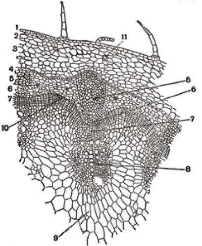
Fig. 63. Part of the transverse section of a sunflower stem:
1 - epidermis, 2 - lamellar collenchyma, 3 - thin-walled parenchyma of the primary cortex, 4 - starch sheath, 5 - sclerenchyma, 6 - phloem, 7 - cambium, 8 - xylem, 9 - pith, 10 - interfascicular cambium, 11 - resin ducts
The starch sheath is formed by a single layer of thin-walled cells rich in starch grains. It lies directly against the strands of sclerenchyma, being separated from them only in places by thin-walled cells.
Sclerenchyma cells have highly thickened walls that react positively with phloroglucinol and hydrochloric acid, as well as with aniline sulfate. In the first case, they stain red, and In the second, yellow. These reactions indicate that the cell walls of the sclerenchyma are lignified.
Vascular bundles are located beyond the sclerenchyma. Positioned externally and directly adjacent to the sclerenchyma is the phloem, in which sieve tubes and companion cells with granular contents are clearly visible. The cambial strip between the phloem and xylem within the vascular bundles is not sharply demarcated. The cambium consists of narrow, small, thin-walled cells filled with dense cytoplasm. Division of cambial cells produces elements of secondary xylem and phloem. When the preparation is treated with phloroglucinol and hydrochloric acid or aniline sulfate, the phloem and cambium show no reaction, meaning their coloration does not change. This indicates that their cell walls are not lignified.
Located behind the cambium is the secondary wood, or xylem, which consists of various elements, predominantly large scalariform or pitted vessels.
Secondary xylem is adjacent to primary xylem, which originates from the procambium. Primary xylem comprises tracheids, parenchyma, and annular and spiral vessels. They are arranged in a pattern resembling outstretched fingers of a hand.
The STRUCTURE OF THE interfascicular cambium deserves attention. It is easily observed by slightly moving the preparation under the microscope so that the center of the field of view lies between two vascular bundles. Under the microscope, it is visible that the interfascicular cambium consists of several layers of slightly flattened, thin-walled cells with cavities filled with granular cytoplasm. The interfascicular cambium contains cells that do not yet differ from others, as they have not yet transformed into phloem or xylem elements. One can also observe The formation of new vascular bundles from the interfascicular cambium. This phenomenon is especially noticeable in cells lying toward the periphery, where sieve tubes and companion cells can be discerned.
The central part of the stem is occupied by the pith, which consists of thin-walled parenchymatous cells. In the sunflower stem, the size of the pith cells increases toward the center, while those adjacent to the xylem are smaller. In a young stem, pith cells contain cytoplasmic contents, which disappear as the stem grows and develops. The walls of the pith cells are white and appear glittering under the microscope. They form a so-called loose parenchyma composed of cells with large intercellular spaces.
Resin canals are present in the sunflower stem. They are located in the primary cortex and the central cylinder and possess a typical structure: a cavity surrounded by small epithelial cells. If the sections are prepared from alcohol-preserved material, the resinous substances filling the cavity of the canals dissolve in the alcohol.
Preparation: Longitudinal section of a sunflower stem (Helianthus annus L.)
Longitudinal sections are also prepared from a thin young stem. A small piece is cut off and split in half with a knife. The cut surface is then smoothed with a sharp razor to ensure that microscopic sectioning captures all elements of the various tissues. Radial sections are prepared from the split half. It is also necessary to obtain sections that pass through the primary xylem. Because these sections are wide and do not fit entirely within the microscope's field of view, the preparation must be moved from the periphery to the center (or vice versa) to study the structure on a longitudinal section. This makes it possible to thoroughly examine the entire complex set of Structural elements of the sunflower stem.
Moving the preparation from the pith to the periphery (Fig. 64) reveals that the central part of the stem consists of pith formed by polygonal parenchymatous cells with very thin walls. The pith is directly adjacent to the primary xylem, where primary wood parenchyma and various vessels—specifically annular, narrow spiral, wide spiral, and pitted vessels—can be observed. Wood parenchyma cells are scattered among the vessels; they are wide-walled and contain finely granular contents.
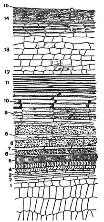
Fig. 64. Longitudinal section of a sunflower stem:
1 - pith, 2 - primary wood parenchyma, 3 - annular vessel, 4 - narrow spiral vessels, 5 - wide spiral vessels, 6 - pitted vessels, 7 - wood parenchyma, 8 - libriform, 9 - cambium, 10 - phloem, 11 - sclerenchyma, 12 - endodermis, 13 - thin-walled parenchyma, 14 - collenchyma, 15 - epidermis
Beyond the porous vessels lies libriform tissue, which consists of elongated, fiber-like cells. These cells possess heavily thickened walls and lack cellular contents; narrow slit-like pits are visible on their walls, and the fiber ends are pointed. Libriform tissue is of great importance, serving a mechanical function by imparting strength to the stem.
Internal to the libriform tissue lies the cambium, composed of relatively short cells with thin, delicate walls. The cells contain granular cytoplasmic contents and elongated nuclei. Located inward from the cambium is the phloem, featuring distinct sieve tubes with sieve plates on their transverse walls. A brownish callose substance is also observable here. The sieve tubes are built of short segments whose length decreases from the periphery toward the cambium, indicating that primary phloem elements are smaller than secondary ones.
Positioned more peripherally relative to the sieve tubes are the bast (phloem) fibers. These cells have thickened walls and tapered ends; lacking any internal contents, they are dead cells. Much like the libriform tissue, bast fibers perform a mechanical function.
Beyond the sclerenchyma lies the endodermis, if starch grains are absent, or alternatively the starch sheath, whose cells are packed with starch. These are followed by cells of the thin-walled parenchyma and collenchyma. In the collenchyma cells, the longitudinal walls are thickened and appear glossy under a microscope, whereas the transverse walls are mostly thin. The cells of both the thin-walled parenchyma and collenchyma contain cytoplasm, a nucleus, chloroplasts, and starch grains. Externally, the stem is covered by the epidermis, which consists of a single layer of cells with thin walls. The cells contain protoplasts, and hairs (trichomes) are visible on the epidermal surface.
Last update: 07/08/2026
Editorial and Educational Adaptation: This material has been compiled based on the primary/original source text. The project team performed an editorial review, corrected technical inaccuracies, structured sections, and adapted the content for an educational format.
What was processed:
- elimination of formatting defects (OCR errors, structural breaks, corrupted characters);
- editorial organization of content;
- standardization of terminology in accordance with academic sources;
- verification of factual statements against the original source text.
All mentions of the author, publication year, and origin of the primary text have been preserved in accordance with the source.