MODERN BOTANY — P. RAVEN — 1990
CHAPTER IV. DIVERSITY
CHAPTER 14. UNICELLULAR PROTISTS: WATER MOLDS, SLIME MOLDS, CHYTRIDIOMYCETES, AND UNICELLULAR ALGAE
Characteristics of Phyla
This chapter examines four phyla of heterotrophs and three phyla of autotrophs. The heterotrophs are extremely diverse. Among them, chytridiomycetes and oomycetes possess coenocytic (multinucleate, but not partitioned by cross-walls) hyphae, which in some oomycetes can be long and extensively branched. Oomycetes produce motile asexual spores (zoospores) with two flagella—one smooth and one tinsel-like (see Fig. 2-26), whereas the zoospores of chytridiomycetes have a single posterior smooth flagellum. Myxomycetes spend most of their life cycle, when not reproducing, as a flowing mass of Cytoplasm. Acrasiomycetes are amoeboid organisms that do not form flagellated Cells at any stage of their life cycle. The Characteristic Features of these heterotrophic phyla and autotrophic protists are summarized in Table 14-1.
The latter also vary considerably in flagellar Structure (when present) and biochemical traits, particularly in pigment composition, storage products, and Cell wall components. These features (including those for organisms discussed in Ch. 15) are presented in Table 14-1. The names of some algal phyla derive from the coloration of their predominant accessory pigments, which mask the green color of chlorophylls. During Photosynthesis, accessory pigments channel the energy of the light they absorb primarily into Photosystem II. A wide variety of storage products is found among these organisms; in most cases, these are various CARBOHYDRATES, frequently accompanied by Lipids. The heterotrophic origin of autotrophic protists is indicated by the fact that certain genera of chrysophytes, dinoflagellates, and euglenoids regularly engulf solid food particles, just like representatives of many phyla that did not acquire symbiotic METABOLISM/14.html">Chloroplasts in the course of evolution.
Class="center">Table 14-1. Comparative characteristics of 10 protist phyla
Phylum |
Number of species |
Storage carbohydrates |
Flagella |
Cell wall components |
Habitat |
|
475 |
None |
2; smooth and tinsel; Cellulose only in reproductive cells |
Aquatic or moisture-dependent |
|||
Chytridiomycota (chytridiomycetes) |
750 |
None |
Glycogen |
1 posterior smooth; reproductive cells only |
Chitin, other polymers |
Aquatic |
Acrasiomycota (cellular slime molds) |
70 |
None |
Glycogen |
None (amoeboid movement) |
None |
Terrestrial |
Myxomycota (plasmodial slime molds) |
450 |
None |
Glycogen |
2 smooth; reproductive cells only |
None |
Terrestrial |
Chrysophyta (chrysophytes) |
6650 |
Chlorophylls a and c, carotenoids, including fucoxanthin |
Chrysolaminarin |
0, 1, or 2; apical, smooth or tinsel, equal or unequal |
None or cellulose, some with silica scales; silica in diatoms |
Marine and freshwater |
Pyrrhophyta (dinoflagellates) |
1100 |
Chlorophylls a and c, carotenoids, including peridinin |
Starch |
0 or 2 lateral tinsel |
Cellulose, other substances |
Marine and freshwater |
Phaeophyta (brown Algae)1 |
1500 |
Chlorophylls a and c, carotenoids, including fucoxanthin |
Laminarin, mannitol |
2 lateral, tinsel directed forward, smooth backward; reproductive cells only |
Cellulosic matrix with alginic acids (Polysaccharides) |
Almost all marine; very abundant in cold oceans |
Rhodophyta (red algae)1 |
4000 |
Chlorophyll a, carotenoids, phycobilins |
Starch |
None |
Cellulose, pectic substances, calcium carbonate in many |
Marine, some freshwater, many tropical species |
Chlorophyta (green algae)1 |
7000 |
Chlorophylls a and b, carotenoids |
Starch |
0, 2, or more; apical or lateral, equal, smooth |
Polysaccharides, sometimes cellulose |
Mostly freshwater, some marine |
Euglenophyta (euglenoids) |
800 |
Chlorophylls a and b, carotenoids |
Paramylon |
1 to 3, apical, tinsel (with a single row of lateral hairs) |
No cell wall; possess a protein pellicle |
Mostly freshwater |
1 Phyla consisting mainly or entirely of Multicellular Organisms; described in Ch. 15.
Various protist groups accumulate very distinct Introduction/36.html">Carbohydrates and lipids As a result of photosynthesis. In green algae, starch accumulates within the chloroplasts (as in their plant descendants); in all other groups, reserve substances are deposited in the cytoplasm. In red algae and dinoflagellates, starch is stored in this manner, whereas in brown algae, the reserve is the polyglucoside laminarin, which features different bonds between glucose residues. In other phyla containing chlorophyll c, the primary storage product is chrysolaminarin, a more polymerized form of laminarin. Many brown algae also accumulate mannitol, a sugar alcohol derivative of mannose. Euglenoids synthesize paramylon (a polysaccharide with a spiral configuration), which sharply distinguishes them from other phyla.
Phylum Oomycota
The phylum Oomycota, comprising approximately 475 species, is a very distinct group of protists. The Cell walls of these organisms are composed primarily of cellulose or cellulose-like polymers, which sharply differentiates them from Fungi. The Chromosomes of oomycetes resemble those of most eukaryotes and bear no resemblance to the highly condensed chromosomes of fungi. Meiosis and mitosis are analogous to these processes in other eukaryotes; centrioles are present. This phylum encompasses forms ranging from unicellular to extensively branched coenocytic filamentous types.
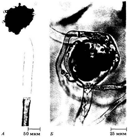
Many species of oomycetes can reproduce both sexually and asexually. Sexual reproduction, oogamy, involves The formation of an oogonium containing numerous egg cells, and an antheridium containing multiple male nuclei (Fig. 14-4). Syngamy leads to the formation of a thick-walled zygote (oospore), which serves as a resting spore—a structure that gave the phylum its name. As already mentioned, asexual reproduction in oomycetes occurs via motile zoospores bearing two flagella: one smooth and one tinsel-like.
Fig. 14-4. Achlya ambisexualis, a water mold with sexual and asexual reproduction. A. Empty sporangium with zoospores forming cysts at its opening (a distinctive feature of Achlya). B. Sex Organs. Fertilization tubes of the antheridium are shown penetrating through the oogonium wall toward the egg cells.
A major group of oomycetes consists of aquatic organisms. Their representatives, known as water molds, abound in fresh water and can be easily isolated from it. Many of them are saprotrophs, but parasites are also known, including those causing diseases in fish and their eggs.
In some water molds (e.g., Saprolegnia, Fig. 14-5), the sexual process occurs through the formation of male and female sex organs on the same individual, i.e., these organisms are homothallic. Other species (e.g., in the genus Achlya, Fig. 14-4) are heterothallic, meaning that male and female sex organs develop on different specimens, or if on the same one, genetic self-fertilization is impossible. Both Saprolegnia and Achlya can reproduce both asexually and sexually.
Fig. 14-5. Life Cycle of the oomycete Saprolegnia. The mycelium of this water mold is diploid. Reproduction is predominantly asexual. Biflagellate zoospores emerge from the zoosporangium, swim for some time, and then encyst. Each ultimately gives rise to a secondary zoospore, which also encysts and then germinates, giving rise to a new mycelium. During sexual reproduction, oogonia and antheridia form on the same hypha. Meiosis takes place inside them. Oogonia are enlarged cells in which several spherical egg cells are formed. Antheridia develop from the tips of other filaments of the same individual and contain numerous male nuclei. Upon maturation, the antheridia grow toward the oogonia and form penetrating fertilization tubes. Male nuclei migrate through these tubes toward the female nuclei and fuse with them, resulting in the formation of a thick-walled zygote—the oospore. Upon germination, it develops into a hypha, which then forms a zoosporangium, and the cycle begins anew.

Another group of oomycetes is predominantly terrestrial, although these organisms also form motile zoospores in the presence of free liquid water. This includes the order Peronosporales, several representatives of which are of major economic importance. As C. J. Alexopoulos noted: "At least two of them have had a hand—or, more precisely, hyphae—in shaping the economic history of a substantial part of humanity."
The species Plasmopara viticola causes a disease known as downy mildew of grapevine. This disease was accidentally introduced into France in the late 1870s on American grapevines imported for their resistance to other diseases, and it soon threatened the entire winemaking industry of the country. Control of downy mildew was ultimately achieved through a combination of serendipity and careful observation. Farmers around Médoc customarily sprayed their roadside vineyards with an unpalatable mixture of copper sulfate and lime to deter passersby from picking the clusters. A professor from the University of Bordeaux, studying the mildew problem, noticed that such plants remained healthy. After consulting with vineyard owners, he prepared a special chemical mixture termed Bordeaux mixture, which has been widely used since 1882. This was the first chemical agent employed to combat plant diseases.
Another economically significant representative of oomycetes is the genus Phytophthora (meaning "plant destroyer" in Greek), which includes 35 species and is a particularly virulent plant pathogen because it severely affects many agricultural crops on a large scale, including cacao, pineapples, tomatoes, rubber trees, papaya, onions, strawberries, apples, soybeans, tobacco, and citrus fruits. A well-known representative of this genus is Phytophthora cinnamomi, a soil-dwelling Organism that destroyed or ruined millions of avocado trees in southern California and elsewhere several years ago. It has also wiped out tens of thousands of hectares of valuable eucalyptus plantations in Australia. Zoospores of P. cinnamomi are attracted to susceptible plants by chemical secretions from their roots. This oomycete forms resistant spores that can persist for up to six years in damp soil. Intensive breeding programs are currently underway to develop varieties of avocados and other crops resistant to it.
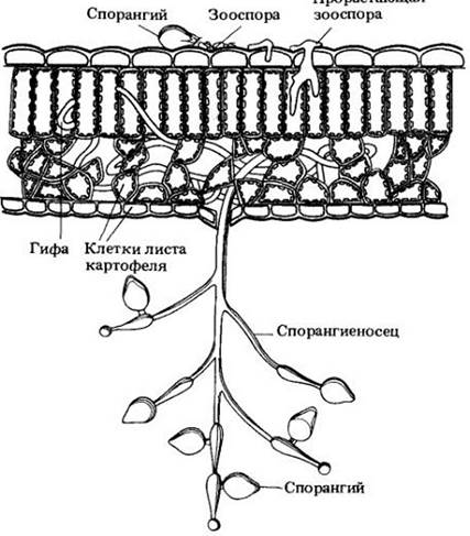
However, the most "notorious" species is Phytophthora infestans (Fig. 14-6), the causal agent of late blight of potato, which triggered the severe famine in Ireland in 1846–1847. The country's population, which had grown from 4.5 to 8.5 million between 1800 and 1845, dropped to 6.5 million by 1851 as a result of this disaster. About 800,000 residents died of starvation, while the rest emigrated, mainly to the United States. Virtually all potato crops in Ireland perished within a single week in the summer of 1846, spelling catastrophe for local peasants who subsisted almost entirely on potatoes at a rate of 4 to 6 kg per day per adult (a ration necessary to obtain sufficient protein for normal physiological function).
Fig. 14-6. Phytophthora infestans, the causal agent of late blight of potato. Leaf cells are shown in gray. In the Presence of water at low temperatures, zoospores emerge from sporangia and swim to the site of germination (as shown here), or the sporangia themselves germinate by producing a germ tube.
Another member of this division—blue mold (Peronospora hyoscyami)—caused approximately a quarter of a billion dollars in damage to tobacco crops in the United States and Canada in 1979. This species spreads via multinucleate spores that are easily carried by the air. An increase in the frequency of such epidemics can be expected in the future as the genetic diversity of many crops continues to narrow. This issue will be discussed in Chapter 30.
Phylum Chytridiomycota
Chytrids, comprising about 750 species, live primarily in water. These organisms are extraordinarily diverse in form, The Nature of their sexual processes, and their life cycles. Their cell walls are composed mainly of chitin, although other polymers are also present. Mitosis and Meiosis, as far as is known, are analogous to those occurring in oomycetes. All chytrids are coenocytic, though several septa appear upon maturation.
The primary characteristic feature of chytrids is the single, smooth, posterior flagellum of their motile cells (zoospores and Gametes). This feature distinguishes them from other protists and indicates an evolutionary relationship among the various forms grouped within this phylum.
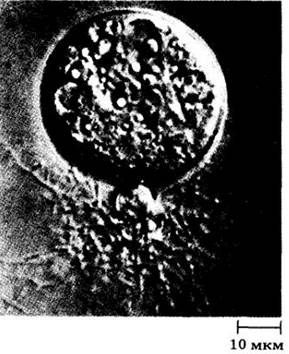
Some chytrids are simple unicellular organisms that do not form a mycelium. In these species, the entire body becomes a reproductive structure at the appropriate time. Others possess slender rhizoids for attachment to the substrate (Fig. 14-7). Various species of chytrids parasitize algae, aquatic oomycetes, spores, pollen grains, and other parts of vascular plants; others are saprobic, living, for example, on dead insects.
Fig. 14-7. Chytridium confervae, a widespread chytrid (imaged using Nomarski differential Interference contrast Microscopy). The fine, downward-extending rhizoids are clearly visible
The genus Coelomomyces comprises obligate parasites of mosquito larvae and other dipterans. Its life cycle resembles that of rust fungi because it involves alternating hosts—small aquatic copepod crustaceans and insect larvae. Coelomomyces is currently being studied as a potential biological control agent for mosquitoes.
The structure and reproduction of other chytrids are much more complex. Consider, for example, The life cycle of the genus Allomyces. Some of its species exhibit an alternation of isomorphic generations (see Fig. 14-8), whereas others show heteromorphic generations, where Haploid and Diploid individuals differ in appearance. Morphology/12.html">ALTERNATION OF GENERATIONS is characteristic of plants and many algae, but among other living creatures, it is found only in the genus Allomyces, in another closely related chytrid genus (see Fig. 10-10), and in a very large number of heterotrophic protists not covered in this book. In terms of morphology, physiology, developmental cycle, and sexual Hormones, Allomyces is one of the most thoroughly studied protists.
Fig. 14-8. The chytrid Allomyces arbusculus, whose life cycle is shown in the diagram, exhibits an alternation of isomorphic generations. Haploid and diploid individuals are indistinguishable until the formation of reproductive organs begins. On the haploid individuals, roughly equal numbers of colorless female and orange male gametangia are produced. Male gametes are attracted by the hormone sirenin, which is secreted by the female gametes that are twice as large. The zygote loses its flagella and germinates, giving rise to the diploid organism. This sporophyte produces Two Types of sporangia: (1) asexual—colorless, thin-walled structures that release diploid zoospores, which in turn germinate back into the diploid generation; and (2) sexual—thick-walled, reddish-brown structures capable of withstanding harsh environmental conditions. After a dormant period, meiosis occurs within them, producing haploid zoospores. These zoospores develop into haploid individuals, which, upon maturation, form gametangia

Phylum Acrasiomycota
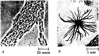
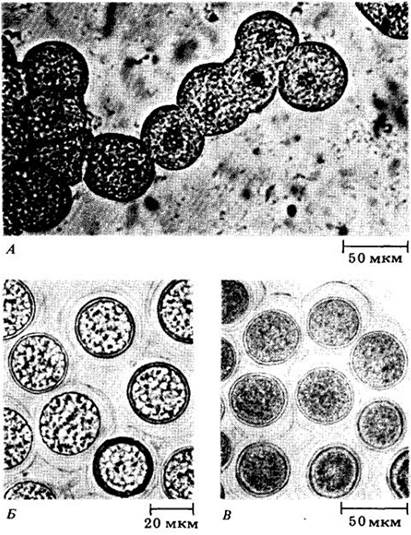
Cellular slime molds—a group of 65 species classified into several genera—may be more closely related to ROOT-like amoebae (phylum Rhizopoda) than to other organisms. Unlike fungi, their cell wall is rich in cellulose, and mitosis is accompanied by The breakdown of the nuclear envelope; centrioles are present. One representative of this division, Dictyostelium discoideum, was discussed in Chapter 8 as a model for cellular differentiation. In this and other species of acrasids, amoebae (myxamoebae) retain their individuality even when aggregating at a certain stage of the life cycle to form a "slimy" pseudoplasmodium (Fig. 14-9). Myxamoebae can also encyst individually (forming microcysts) and thus survive brief periods of drought or starvation in this form.
Fig. 14-9. Dictyostelium discoideum. A. Aggregation of myxamoebae. Note that each amoeba maintains its individuality. The arrow indicates the direction of the myxamoebal stream. B. General view of A large number of aggregated amoebae under low magnification
Asexual reproduction via spores is common among cellular slime molds. Sexual reproduction involving macrocysts is also frequently observed. In Dictyostelium discoideum, for example, macrocysts are somewhat flattened, irregularly rounded to elliptical, multicellular structures 25–50 µm in diameter (Fig. 14-10). During their formation, pairs of haploid amoebae first fuse to produce zygotes, which then attract and engulf neighboring amoebae. The resulting aggregates are smaller than those formed during pseudoplasmodium ("slug") formation and tend to be rounded rather than elongated in shape. Each macrocyst is surrounded by a thin membrane secreted by the amoebae. Later, the entire group of cells within the membrane becomes encased in a thick, cellulose-rich cell wall. Within the macrocyst, the zygote (the only diploid cell in the life cycle) undergoes meiosis followed by several mitotic divisions before germination, resulting in the formation and release of new haploid amoebae.
Fig. 14-10. The process of macrocyst formation in Dictyostelium mucoroides. A. Each zygote, or giant cell, begins to engulf surrounding amoebae. B. The giant cells have engulfed all the amoebae and become enclosed in a cellulose wall. C. Mature macrocysts; at this stage, their contents appear homogeneous
Cellular slime molds were once considered rare. In 1933, K. Raper discovered Dictyostelium discoideum; his subsequent research on this organism drew widespread attention to the group. It is now known to include common inhabitants of most soils, particularly forest litter, where they feed on Bacteria. Like many groups of organisms, acrasids are more richly represented in the tropics than in the temperate zone. Cellular slime molds are easily isolated and cultured, making them excellent experimental models for studying cellular differentiation and molecular biology (see pp. 124–125).
In 1982, D. Waddell of Princeton University described an unusual property of a predatory acrasid cultured in the laboratory, Dictyostelium caveatum, which he isolated from bat guano in Blanchard Springs Cave, Arkansas. The amoebae of this species formed joint aggregates with amoebae of all tested cellular slime mold species, suppressed their development, and consumed them, ultimately forming fruiting bodies consisting exclusively of cells of their own species.
Phylum Myxomycota
This phylum unites plasmodial slime molds, or myxomycetes—totaling about 450 species—which apparently have no direct phylogenetic ties to cellular slime molds or other protist groups. Under favorable conditions, myxomycetes exist as a thin, flowing mass of protoplasm that moves like an amoeba. Lacking a cell wall, this "naked" protoplasm is called a plasmodium. As they move, plasmodia engulf and digest bacteria, Yeast cells, fungal spores, and small fragments of decaying PLANT AND ANIMAL matter. The successful cultivation of plasmodia in media lacking particulate food particles suggests that they are also capable of absorptive Nutrition.
A plasmodium can reach a mass of 20–30 g, but because it spreads in an extremely thin layer, this quantity can cover a surface area of several square meters (see Fig. 10-5A). It contains numerous nuclei that are not separated by cell walls. As the plasmodium grows, these nuclei undergo repeated synchronous divisions. Centrioles are present, and mitosis is typical, though the chromosomes are very small.
As a rule, a moving plasmodium is fan-shaped, featuring flowing protoplasmic strands that are thicker at its base and branch and taper toward the advancing margin. These strands consist of slightly gelated protoplasm through which the more fluid fraction flows rapidly. The leading edge of the plasmodium consists of an ultra-thin film of gel, separated from the substrate only by The Plasma Membrane and a mucous layer of unknown chemical composition.
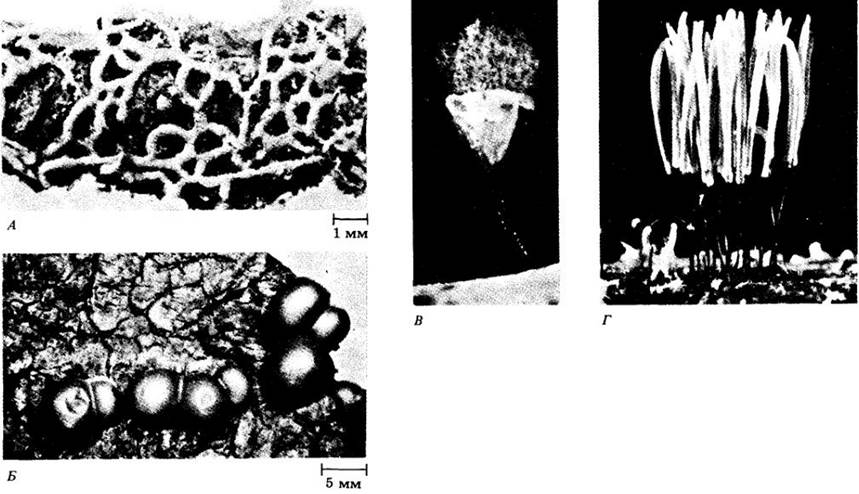
Plasmodial growth continues as long as moisture and food are abundant. Typically, if either of these factors becomes scarce, the plasmodium migrates away from the "feeding site." It may then be observed crossing roads, lawns, climbing trees, and appearing in other unexpected places. In many species, a stationary plasmodium divides into numerous small mounds of uniform volume and size, suggesting that their formation is chemically controlled by the plasmodium itself. Each mound develops into a mature sporangium, usually atop a stalk; this sporangium often has a complex shape (Fig. 14-11C, D). Meiosis occurs within the young diploid spores after they separate from one another and form a wall. This results in four nuclei, three of which degenerate, leaving a spore with a single haploid nucleus. In some myxomycetes, individual sporangia are not formed; instead, the plasmodium may develop either into a plasmodiocarp (Fig. 14-11A), which retains the branching shape of the plasmodium, or into an aethalium (Fig. 14-11B), which is a large cushion-like mass representing essentially a single giant sporangium.
Myxomycete spores are resistant to extreme environmental conditions, and some of them are capable of germinating after being stored in a laboratory for 60 years. Thus, spore formation here ensures not only genetic recombination, but also survival through unfavorable periods.
Fig. 14-11. Spore-producing structures of myxomycetes. A. Plasmodiocarp in Hemitrichia serpula. B. Aethalia of Lycogala on tree bark. C. Sporangium of Arcyria cineria. D. Sporangia of Stemonitis splendens
Under suitable conditions, the spores open, releasing a protoplast that remains amoeboid or develops one or two smooth flagella. These two forms can easily transform into one another. Amoebae feed on bacteria and organic matter, reproducing mitotically. If food sources are depleted or conditions are unfavorable, they can stop moving and round up, secreting a thin wall to form a microcyst. These microcysts remain viable for a year or more and resume activity when conditions improve.
Following a growth period, the amoebae develop into a plasmodium. This process is regulated by a variety of factors, including cell age, environmental conditions, amoeba population density, and chemical stimuli whose role is analogous to that described for the cellular slime mold Dictyostelium discoideum (see p. 124 — 0125). One way a plasmodium forms is through the fusion of gametes, which are typically genetically distinct and originate from different haploid spores. Essentially, these are simply amoebae or flagellated cells taking on a new role. In some species and strains, a plasmodium may also arise from a single amoeba; in this case, it is typically haploid, just like the ancestral amoeba.
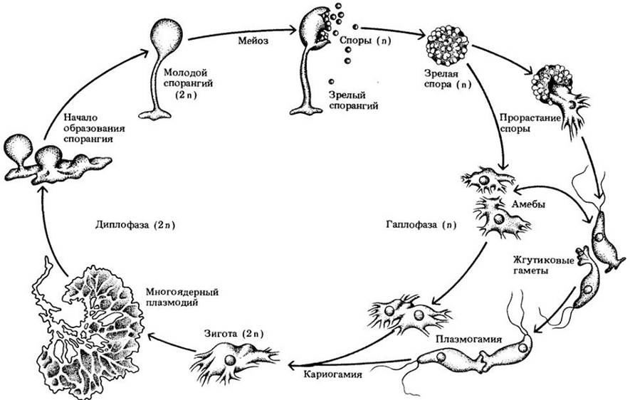
The life cycle of a typical plasmodial slime mold is shown in Fig. 14-12.
Fig. 14-12. Life cycle of a typical myxomycete. Sexual reproduction in plasmodial slime molds involves three distinct stages: plasmogamy, karyogamy, and meiosis. Plasmogamy consists of the union of two protoplasts, placing two haploid nuclei within a single cell. Karyogamy is the fusion of these two nuclei, leading to the formation of a diploid zygote and the initiation of the so-called diplophase of the life cycle. The plasmodium is a multinucleate, freely flowing mass of protoplasm capable of passing through silk fabric or filter paper with virtually no alteration. Meiosis restores the haploid state, and the haplophase of the cycle begins
Phylum Chrysophyta
Chrysophytes are autotrophic, unicellular organisms abundantly distributed in fresh and marine waters worldwide. They contain chlorophylls a and c, whose color is often masked (in golden algae and diatoms) by the accessory carotenoid pigment fucoxanthin. The reserve carbohydrate of chrysophytes is called chrysolaminarin; it frequently accumulates as a large grain near the posterior end of the cell. Chrysophyte cells may lack a cell wall or possess one; in the latter case, it is composed primarily of cellulose and covered with protruding scales or, in many Representatives of the phylum, a silica frustule. Members of one of the three chrysophyte classes, Xanthophyceae, contain the pigment vaucheriaxanthin, which is related to fucoxanthin. Organisms of this class are typically a bright yellowish-green.
Chrysophyte chloroplasts are biochemically and structurally analogous to those of brown algae (see Ch. 15) and dinoflagellates. It can be hypothesized that these Organelles in all three groups originated from the same bacterial form. To date, bacteria with precisely identical characteristics have not been discovered, but Heliobacterium chlorum, a species described in 1983 at Indiana University, may be close to the precursors of chloroplasts. This is an obligate anaerobic, brownish-colored bacterium capable of Nitrogen Fixation. Another biochemical similarity between chrysophytes and brown algae is the storage of the polysaccharide laminarin (or its more polymerized form, chrysolaminarin) outside the chloroplasts; in addition, brown algae accumulate mannitol.
The unequal flagella of chrysophytes and brown algae are similar (see Fig. 2-26). Opinions have been expressed regarding the possibility of combining these organisms into a single phylum.
The chloroplasts of dinoflagellates (phylum Pyrrhophyta) are biochemically and structurally analogous to those found in chrysophytes and brown algae. However, dinoflagellates differ from these groups in many other respects, making a common ancestor seem highly unlikely. The similarity among the three phyla is likely explained by the chloroplasts of both evolutionary lineages having originated from the same group of bacteria. No other direct interrelationships exist between them.
Class Chrysophyceae
This largest class of chrysophytes—the golden algae—includes about 500 species (Fig. 14-13). Until recently, it was considered a predominantly freshwater group, but it has turned out that golden algae make a significant contribution to the productivity of marine plankton, especially nanoplankton.
Fig. 14-13. Scanning Cytology/cytology/93.html">ELECTRON MICROGRAPHS OF two genera of marine golden algae. A. Distephanus speculum, a cold-water species with a siliceous Skeleton inside the amoeboid protoplast containing numerous small chloroplasts. B. A species of the genus Gephyrocapsa, which includes extremely small members of the group known as coccolithophores. Although these organisms are quite abundant in the nanoplankton, they are so small that they cannot be caught with a standard plankton net. They also dissolve in acidic fixatives, making them difficult to detect and study
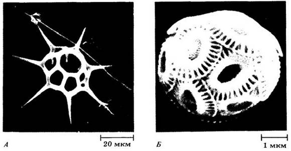
Many golden algae lack a distinct cell wall, but possess siliceous scales or skeletal elements that can be external or internal and are often intricately structured. Most members of this class are unicellular forms with flagella, although some lack flagella and sometimes resemble amoebae. These amoeboid cells differ from rhizopods only by the presence of chloroplasts, and the two groups may be closely related. Indeed, some golden algae capture bacteria and other organic particles. Species of this group typically have one or two large chloroplasts per cell; a voluminous chrysolaminarin granule frequently accumulates near the posterior end of the cell. Reproduction in most golden algae is asexual via zoospores. Some species form colonies.
Class Xanthophyceae
Organisms of this class (yellow-green algae), which includes 550 species, exhibit the characteristic pigmentation that gave the entire group its name. They are similar to golden Algae in the presence of chlorophyll c in many species, but differ in the absence of fucoxanthin. Yellow-green algae are predominantly non-motile entities, although some possess an amoeboid form or flagella, as do their gametes.
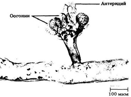
A widely known genus in this class is Vaucheria ("water felt")—a coenocytic, sparsely branched, filamentous alga. It reproduces both asexually by forming large, complex, multiflagellated zoospores, and sexually via oogamy (Fig. 14-14). Vaucheria is distributed in fresh, brackish, and marine waters. It can frequently be found on periodically flooded coastal mud.
Fig. 14-14. Vaucheria ("water felt"), a coenocytic filamentous representative of the chrysophytes. Vaucheria is an oogamous organism that forms oogonia and antheridia. The antheridium shown in the figure is empty
Class Bacillariophyceae
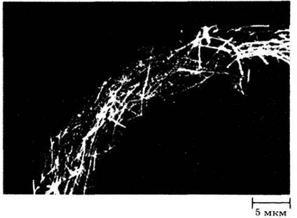
These organisms, known as diatoms, are mostly unicellular. They are an exceptionally vital component of the phytoplankton and a primary food source for freshwater and marine animals (Fig. 14-15). This class is estimated to comprise about 5,600 living species. If extinct forms are also taken into account, diatoms number at least 40,000 species, with their species diversity occasionally being immense within a very small area. For example, 369 species were identified in two small mud samples from the ocean near Beaufort, North Carolina. Most diatoms are part of the plankton, but some inhabit the bottom, or live on other algae and plants in both fresh and marine waters.
Fig. 14-15. Part of a fluffy aggregate formed by intertwined chains of two diatom species of the genus Rhizosolenia. Aggregates of this kind are often abundant in nutrient-poor oceanic waters; the algae shown in the photograph were collected from the central North Pacific. Symbiotic nitrogen-fixing bacteria inhabit the vacuoles of diatoms in large numbers. Such coexistence makes a very important contribution to the productivity of nutrient-poor waters
Diatoms differ from other chrysophytes in the absence of flagella (with the exception of male gametes in certain species) and in their unique frustules, or cell walls. These delicate bivalve frustules are composed of polymerized opaline silica (SiO2 x nH2O), with the Valves fitting one inside the other. The fine ornamentation of the frustules, used to identify diatom species, has traditionally been employed by microscopists to check the quality of optical instruments. Electron microscopy has shown that the fine striations on these frustules actually consist of numerous tiny, intricately shaped depressions, pores, or tubules that connect the living protoplasm inside the shells with the external environment (Fig. 14-16). The most characteristic feature of the diatom protoplast is its brownish Plastids containing chlorophylls $a$ and $c$, as well as fucoxanthin. Reproduction in diatoms is primarily asexual, occurring via Cell Division (Fig. 14-17).
Fig. 14-16. A. Some marine diatoms under the Light Microscope. Scanning electron micrographs of an Entogonia frustule (B) and the centric diatom Thalassiosira nordenskioeldii (C). D. The pennate diatom Pinnularia under the light microscope
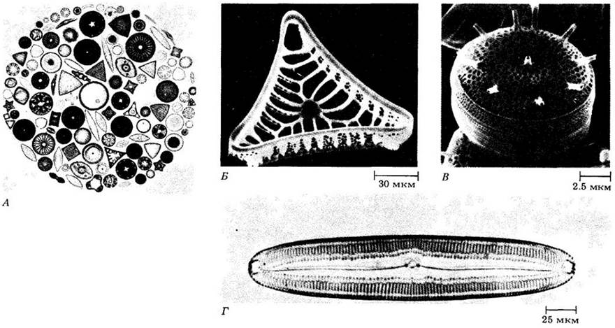
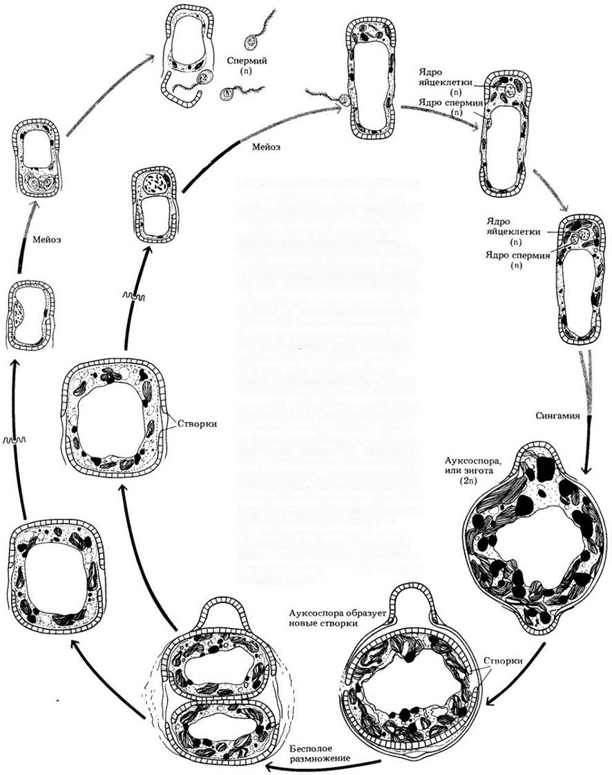
Fig. 14-17. Reproduction in diatoms is mainly asexual, through cell division. Each daughter cell (bottom left) receives one of the parental frustules (bottom right) and synthesizes the second one. The parental valve always forms the larger half (the "lid") of the silica "box", overlapping the sides of the new one. Thus, one cell from each daughter pair is typically smaller than the parent. In some species, the frustules enlarge through the expansion of the internal protoplasm, whereas in others they are more rigid. When individuals In the second case decrease in size to 30% of their maximum diameter, sexual reproduction may occur. Some cells function as male gametangia, producing four sperms via meiosis. Others act as female gametangia; three of the meiotic products degenerate, leaving only a single egg cell. Following syngamy (fertilization), an auxospore or zygote is formed, which grows to the maximum size characteristic of the species. The walls of auxospores often differ markedly from those of asexually reproducing individuals of the same species. Once mature, the auxospore divides, producing new frustules identical in every detail of their ornamentation to the original ones. The figure illustrates the Reproduction of a centric diatom
Based on Symmetry, two types of diatoms are distinguished: pennate (bilaterally symmetrical, Fig. 14-16, D) and centric (radially symmetrical, Fig. 14-16, C) forms. Centric diatoms are most abundant in marine waters. Their sexual process (when present) is oogamous. Male gametes may possess a single tinsel flagellum (the only flagellated cells in the diatom life cycle). In pennate diatoms, sexual reproduction is isogamous, with both gametes lacking flagella.
Although most diatom species are autotrophic, some can become heterotrophic and survive by absorbing organic carbon. These are predominantly pennate diatoms inhabiting relatively shallow areas of the sea floor. A few species are obligate heterotrophs, lacking chlorophyll and incapable of photosynthesis. Conversely, certain diatoms, lacking the characteristic frustules of the class, live symbiotically within large marine Protozoans (foraminiferans), supplying their host with organic carbon.
Despite the lack of flagella and other locomotor organelles, many species of pennate diatoms are motile. This movement is associated with finely regulated secretion of substances in response to a wide variety of Physical and Chemical stimuli. Apparently, a so-called raphe runs along the middle axis of the valves in all motile diatoms, which essentially consists of two pores connected to a thin, intricately structured slit in the frustule. Many non-motile diatoms attach to one another by their frustules, forming long chains (Fig. 14-15). The raphe as a locomotor adaptation likely evolved through the modification of apical pores that secreted substances to bind non-motile diatoms into such chains.
A diatom moves in response to external mechanical stimuli, light, heat, and toxins with the aid of contractile bundles adjacent to the raphe. Their contraction causes dehydrated crystalline bodies to move toward the reservoirs near the raphe pores. From there, these crystalline bodies are expelled into the pores, where they absorb water and swell, transforming into twisted fibrils. The fibrils move along the raphe until they contact a surface, instantly adhering to any substrate and subsequently contracting. If the object to which they have attached is sufficiently large, the diatom is pulled toward it, leaving a trail of secreted substance much like a snail leaving a slime trail. If the object is small, it is displaced along the raphe while the diatom remains stationary. However, motile diatoms are typically at rest. They can travel only a limited distance because the supply of crystalline bodies required for locomotion is limited at any given time.
The siliceous frustules of diatoms have accumulated over millions of years, forming a fine-grained, loose powder known as diatomaceous earth, which is used as an abrasive for fine polishing, as well as for filtration and extraction. It is estimated that 1 cm3 of diatomaceous earth contains about 4.6 million such frustules. In the Santa Maria oil basin (California), subterranean deposits of diatomaceous earth reach a thickness of 900 m, and near Lompoc (California), 270,000 tons of it are mined annually for industrial purposes.
Diatoms become abundant in deposits dating back about 100 million years, i.e., in the Cretaceous period. Many fossil species are identical to modern ones, indicating an extraordinary stability of representatives of this class throughout geological history.
Division Pyrrhophyta
Most of these organisms, referred to as dinoflagellates, are unicellular biflagellated forms (see Table 14-1). They comprise over 1,000 species, many of which are abundant and highly productive components of marine plankton, alongside freshwater representatives. In dinoflagellates, the flagella run within grooves, with one encircling the body like a girdle and the other positioned perpendicularly to the first. The beating of the flagella within the grooves causes the individual to spin like a top during movement. The girdle flagellum is ribbon-shaped. Numerous non-motile forms also occur, sometimes lacking flagella altogether. Some dinoflagellate genera ingest solid food particles, thereby obtaining their required nutrition partially or entirely.
Many dinoflagellates possess a bizarre shape with rigid cellulose plates forming The cell wall (the theca), often resembling a strange helmet or a piece of ancient armor (Figs. 14-18, 14-19). The plates of this wall are located in vacuoles beneath the plasma membrane rather than outside it, unlike the cell walls of most algae.
Fig. 14-18. The "armor" of some dinoflagellates consists of cellulose plates lying within vacuoles beneath the plasma membrane. In genera that appear "naked", these vacuoles may or may not contain cellulose plates
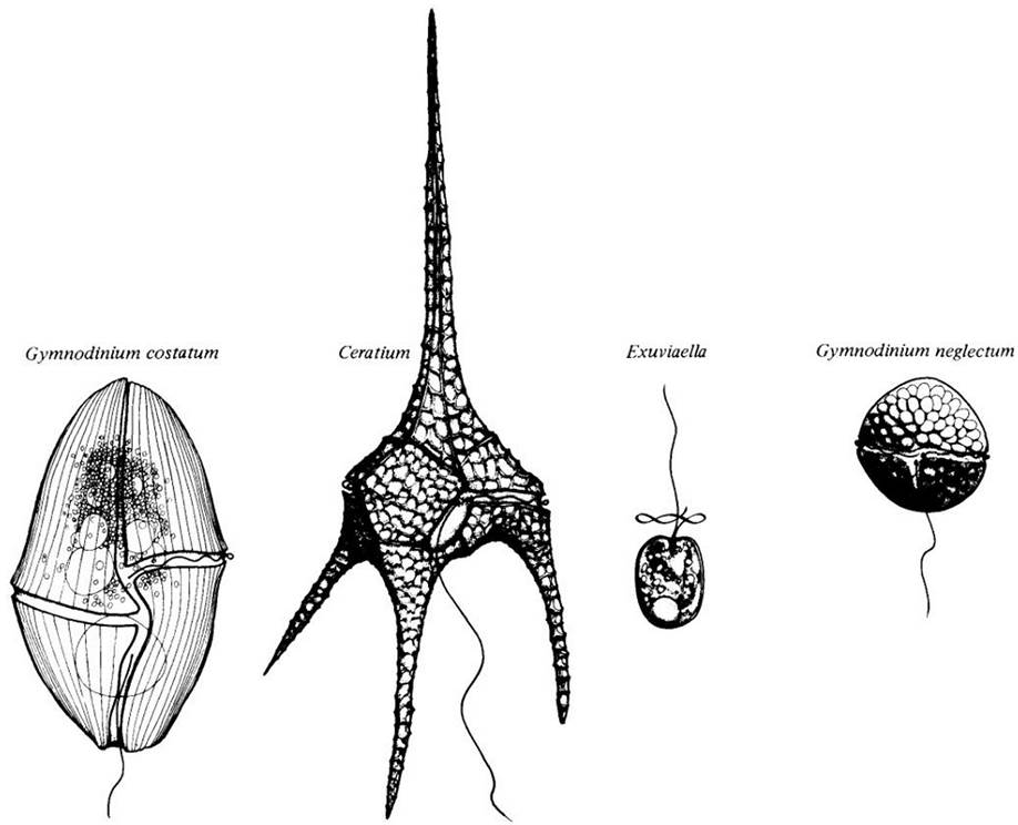
Fig. 14-19. A. Ceratium tripos, a dinoflagellate with a bizarre frustule. B. Noctiluca scintillans, a bioluminescent marine dinoflagellate. C. Gonyaulax polyedra, a dinoflagellate responsible for spectacular "red tides" off the coast of southern California
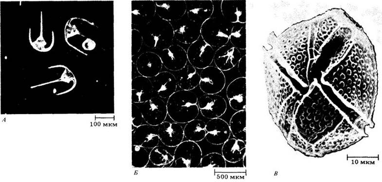
Dinoflagellates generally contain chlorophylls $a$ and $c$, which are usually masked by carotenoids, including perinin (peridinin), which is closely related to fucoxanthin. Their chloroplasts, as mentioned above, likely originated from the same bacterial group as those of chrysophytes and brown algae, but in all other respects they differ sharply from representatives of those divisions.
The reserve carbohydrate of dinoflagellates is starch. Some species lack chlorophyll and are heterotrophic, but their structure clearly indicates an affinity with other members of this division. Even autotrophs typically have a strong requirement for vitamin B12 (like many diatoms), meaning their autotrophic status comes with at least this caveat. Some dinoflagellates are capable of engulfing other cells. It is hypothesized that colorless forms obtain food in precisely this manner, as well as by absorbing small organic particles.
Cyanobacteria are common symbionts of dinoflagellates, which in turn establish symbiotic relationships with numerous other organisms, including Sponges, jellyfish, sea anemones, tunicates, corals, octopuses and squids, gastropods, turbellarians, and certain protists. In giant bivalve Mollusks of the family Tridacnidae, the dorsal part of the inner mantle surface may appear dark chocolate-brown due to the presence of symbiotic dinoflagellates. Functioning as symbionts, they lose their theca and appear as golden spherical cells called zooxanthellae (Fig. 14-20).
Fig. 14-20. Zooxanthellae, a symbiotic form of dinoflagellates shown here within a coral tentacle, make a significant contribution to coral reef productivity
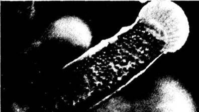
Zooxanthellae primarily provide photosynthetic productivity that enables coral reefs to thrive in nutrient-poor tropical waters. Coral Tissues can harbor up to 30,000 symbiotic dinoflagellates per 1 mm3, predominantly within the cells lining the polyp's digestive cavity. Because algae require light for photosynthesis, corals that host them typically grow in ocean depths no greater than 60 m. Many Structural Features of corals are related to the light-capturing properties of various geometric configurations. A similar phenomenon can be observed in tree canopies, where the branching pattern maximizes the exposure of leaves to sunlight.
Dinoflagellates also play a vital role in human life. In the winter and spring of 1974, the west coast of Florida was devastated by a massive "red tide," the 25th such event since 1844. Hundreds of thousands of dead fish littered the beaches, and millions of dollars in tourism revenue were lost. Such phenomena are triggered by unusual population blooms of dinoflagellates that turn the seawater red or brown, known as a water "bloom" (Fig. 14-21). These dinoflagellates are consumed not only by fish—which may suffer direct poisoning—but also by bivalves, particularly mussels, which generally remain unaffected. Bivalves accumulate and concentrate the toxins synthesized by the algae and, depending on the Nature of the poison, pose a hazard to humans who consume them. In the fall of 1972, a red tide was observed for the first time along the New England coast, stretching from Maine to Cape Cod. Consequently, 26 people were poisoned by shellfish that had fed on Gonyaulux excavata. The public uproar was so severe that shellfish harvesting in Massachusetts plummeted by a third over the next four years. Red tides occur periodically. During the largest outbreak following 1972, commercial shellfish harvesting along the entire coast of Maine was officially banned from mid-August to mid-October 1980, costing the seafood industry $7 million in lost revenue from oysters, mussels, and other bivalves.
Fig. 14-21. Prychodiscus brevis, an unarmored dinoflagellate responsible for red tides off the coast of Florida. The curved transverse flagellum lies within a groove encircling the organism. The longitudinal flagellum (only partly visible) extends from the cell center downward to the left. The apical depression is a distinguishing feature of the genus Ptychodiscus.
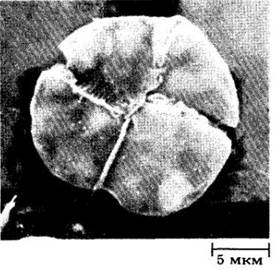
The toxins produced by certain dinoflagellates, such as Gonyaulax catenella, are exceptionally potent neurotoxins. The chemical nature and biological activity of most of these compounds are relatively well understood. Conversely, the triggers for red tides remain poorly understood. Nutrient and trace metal concentrations, sewage runoff, water salinity and Temperature, winds, light, and numerous other factors likely contribute to these events. The Cyclical Nature of these blooms is underscored by the observation that if conditions deteriorate during a bloom, dinoflagellates can shed their flagella and form resting cysts that sink to the bottom, remaining dormant until conditions improve once again.
The primary mode of reproduction in dinoflagellates is longitudinal fission, in which each daughter cell receives one of the flagella and a portion of the theca, and subsequently synthesizes the missing components through a highly complex sequence (see "Mitosis in Dinoflagellates," p. 231). Some non-motile forms produce zoospores. Occasionally, only the zoospores exhibit a typical dinoflagellate structure, whereas mature individuals lack flagella and may unite into filaments. A sexual process has also been described for A number of dinoflagellates, predominantly isogamy, and occasionally anisogamy.
Phylum Euglenophyta
More than 800 species of euglenoids are known, living primarily in freshwater, especially in organic-rich habitats (Table 14-1). Their length ranges from 10 to 500 µm, and their shapes vary widely. All are unicellular, with the exception of the colonial genus Colacium.
As previously discussed, the similarities between the chloroplasts of euglenoids and green algae suggest that these photosynthetic organelles evolved independently in both groups from a bacterial ancestor, possibly similar to Prochloron (see Figs. 11-2, 11-17). In other respects, the two phyla differ sharply. Among approximately 40 euglenoid genera, about one-third possess chloroplasts containing chlorophylls a and b along with several carotenoids.
Euglenoids store carbohydrates in the form of paramylon, a polysaccharide found in no other group of organisms. Like all autotrophs except green algae and plants, this reserve substance is formed outside the chloroplasts. Among chloroplast-lacking euglenoids, some absorb organic matter while others ingest it. As already mentioned, representatives of this phylum are essentially flagellated protozoans that acquired chloroplasts during the course of evolution; perhaps the two groups should be combined.
Euglenoids reproduce by fission, during which individual cells retain their motility. The nuclear envelope does not break down during mitosis—a feature shared with most green algae, dinoflagellates, many fungi, and certain Ciliates. Centrioles function as basal bodies and form the typical mitotic spindle within the nuclear envelope. Chromosomes remain condensed throughout interphase and mitosis, much like in dinoflagellates. Sexual reproduction is unknown.
The phylum is named after the widespread genus Euglena, many species of which have an elongated shape (Fig. 14-22). Their cells are complexly organized and contain numerous small chloroplasts. A prominent, long flagellum bearing very fine hairs emerges from the anterior end, alongside a short flagellum that does not extend beyond a specialized pocket (see below). The long flagellum typically extends ahead of the cell like a fishing line.
Fig. 14-22. A. Euglena, showing two paramylon storage bodies and the nature of the pellicle. B. Ultrastructure of Euglena as revealed by electron microscopy.
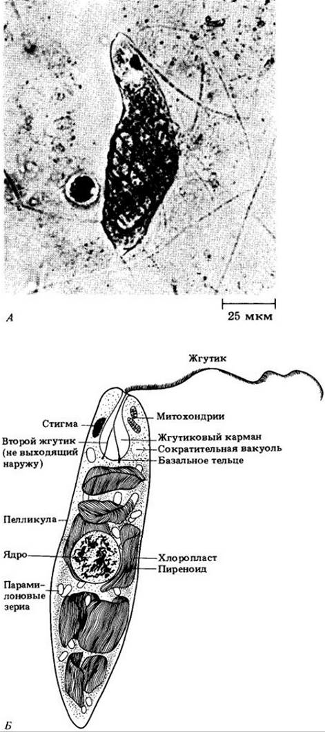
In Euglena, the flagella are anchored at the base of a flask-shaped flagellar pocket (or reservoir) at the anterior end of the cell. A contractile vacuole collects excess water from throughout the cell and discharges it into this same pocket. The cell is bounded by a plasma membrane, beneath which run a series of helical, interconnected protein strips. Together with the membrane, these form a structure known as the pellicle. Unlike the rigid cell walls of plants, the flexible pellicle allows Euglena to change shape, providing an alternative mode of locomotion for benthic, mud-dwelling forms.
When a culture of Euglena is placed near a window receiving sunlight, the cells form a distinct green aggregation in the water, migrating dynamically toward the brightest (yet not excessively bright) region as lighting conditions shift. If the light is too intense, euglenoids avoid it. Their ability to orient toward light is likely mediated by two specialized structures: the stigma (eyespot) and an associated photoreceptor represented by a Swelling at the Base of the flagellum. Depending on the organism's orientation relative to the light source, differential "shading" of the photoreceptor by the pigmented stigma presumably determines the direction of movement. Among colorless euglenoids, only a few possess a stigma.
Certain species of Euglena can survive in the dark without photosynthesis, provided they are supplied with an organic carbon source, essential Vitamins, and minerals. When certain strains are kept in the light under appropriate temperatures and in nutrient-rich media, the cells can divide faster than their chloroplasts replicate, yielding non-photosynthetic offspring that can nevertheless persist indefinitely in a suitable environment. In essence, euglenoids are protozoans with unstable chloroplasts.
Last update: 07/08/2026
Editorial and Educational Adaptation: This material has been compiled based on the primary/original source text. The project team performed an editorial review, corrected technical inaccuracies, structured sections, and adapted the content for an educational format.
What was processed:
- elimination of formatting defects (OCR errors, structural breaks, corrupted characters);
- editorial organization of content;
- standardization of terminology in accordance with academic sources;
- verification of factual statements against the original source text.
All mentions of the author, publication year, and origin of the primary text have been preserved in accordance with the source.