MODERN BOTANY - P. RAVEN - 1990
SECTION II. ENERGY AND LIVING CELLS
CHAPTER 7. PHOTOSYNTHESIS
Light Reactions
The Role of Pigments
The first step in converting light energy into chemical energy is the absorption of light. A pigment is a substance that absorbs visible light. Some pigments absorb light of all wavelengths and thus appear black; others absorb only certain wavelengths, while transmitting or reflecting the rest. Chlorophyll is the pigment responsible for the green color of leaves; it absorbs primarily violet, blue, and red light while reflecting green light (which is why it appears green).
The absorption spectrum of chlorophyll is shown in Fig. 7-5 (an absorption spectrum illustrates the fraction of light energy absorbed as a function of wavelength).
Evidence that chlorophyll is the primary pigment involved in Photosynthesis comes from the striking similarity between its absorption spectrum and the action spectrum of photosynthesis (Fig. 7-6). The action spectrum determines the relative effectiveness of different wavelengths of light in driving light-dependent processes such as photosynthesis, flowering, and phototropism (the bending of a plant toward light). The correspondence between a pigment's absorption spectrum and a process's action spectrum is generally taken as proof that the pigment drives that process (Fig. 7-7).
Class="center">Fig. 7-5. Absorption spectrum of chlorophyll a measured with a spectrophotometer. A beam of monochromatic light is directed at the sample, and the percentage of light absorbed by the test sample compared to a blank (control) is recorded. A special mirror reflects half of the light while transmitting the other half. A photoelectric Cell connected to an electronic device records the percentage of Light absorption across various wavelengths.

Fig. 7-6. The results of experiments conducted by T. W. Engelmann in 1882 made it possible to determine the action spectrum of photosynthesis in a filamentous alga. Much like modern researchers, Engelmann measured The rate of photosynthesis by monitoring O2 evolution. However, instruments sensitive enough to directly measure oxygen were not available at the time. Instead, he used oxygen-requiring Bacteria as indicators of oxygen concentration. He replaced the mirror and Diaphragm of a Light Microscope with a "microspectral apparatus" that produced fine beams of light of various wavelengths. Engelmann then placed this apparatus beneath the microscope over a Glass slide containing the specimen, aligning the filamentous alga parallel to the spectrum. The aerobic bacteria congregated primarily along the Regions of the algal filament illuminated by red and violet light. As can be seen, the action spectrum of photosynthesis in Engelmann's experiments matches the absorption spectrum of chlorophyll (see Fig. 7-5). He concluded that photosynthesis relies on light absorbed by chlorophyll. This is a classic example of what scientists call an "elegant" experiment—one that is both brilliantly and simply designed, providing a definitive answer to the research question.
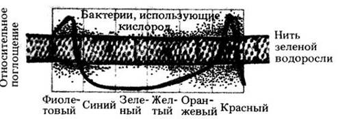
Fig. 7-7. The top curve represents the action spectrum of photosynthesis, while the lower curves show the absorption spectra of chlorophylls a and b, as well as carotenoids, within the chloroplast. The overlap of these spectra indicates that the light absorbed by chlorophylls a and b and carotenoids is utilized in photosynthesis.

When pigments absorb light and electrons are elevated to a higher energy level, several outcomes are possible: (1) the electron's energy is dissipated as heat; (2) it is immediately re-emitted as light of a longer wavelength (a phenomenon known as fluorescence, whereas delayed light emission is called phosphorescence); or (3) the energy is captured in the form of chemical bonds, which is what happens during photosynthesis.
When chlorophyll molecules are extracted and illuminated in solution, they fluoresce. In other words, the pigment molecules absorb light energy, causing electrons to jump to a higher energy level and then drop back down, releasing most of the absorbed energy as light. Light absorbed by isolated chlorophyll molecules cannot be converted into any other form of energy useful to living systems. Chlorophyll is capable of transforming light energy into chemical energy only when associated with specific Proteins located in the thylakoids.
The pigments involved in photosynthesis include chlorophylls, carotenoids, and phycobilins.
Several forms of chlorophyll have been discovered, differing in their molecular Structure. Chlorophyll a (Fig. 7-8) is found in all photosynthetic eukaryotes and cyanobacteria. It is believed to be the primary pigment driving photosynthesis in these organisms.
Vascular plants, mosses, green Algae, and euglenoids also contain chlorophyll b. This accessory pigment broadens THE SPECTRUM OF light absorbed during photosynthesis.
When a chlorophyll b molecule absorbs light, the excited electron transfers its energy to chlorophyll a, which subsequently converts it into chemical bond energy during photosynthesis. Because chlorophyll b absorbs wavelengths of light different from those absorbed by chlorophyll a, it broadens the overall range of wavelengths that can be used for photosynthesis (Fig. 7-7). Chlorophyll b generally accounts for about 1/4 of the total chlorophyll content in the leaves of green plants.
In certain groups of algae, notably brown and diatom algae, chlorophyll c Functions instead of chlorophyll b. Photosynthetic bacteria (other than cyanobacteria) cannot utilize Water as an electron source and therefore do not release oxygen. They contain either bacteriochlorophyll (purple bacteria) or chlorobium chlorophyll (green sulfur bacteria). Chlorophylls b and c, as well as bacterial photosynthetic pigments, represent chemical variations of the core structure shown in Fig. 7-8.
Fig. 7-8. Chlorophyll a is a large molecule featuring a magnesium atom at the center of a porphyrin ring. Attached to the ring is a long, insoluble hydrocarbon tail that acts as an anchor, securing the molecule within the inner membranes of the chloroplast. Chlorophyll b differs from chlorophyll a by having a -CHO group in place of the -СН3 group highlighted in the diagram. The alternation of single and double bonds (known as conjugation) is characteristic of the porphyrin ring not only in chlorophylls but also in all other pigments. Note the structural similarity between the chlorophyll molecule shown here and the cytochrome molecule (Fig. 6-9).

Other types of pigments—carotenoids and phycobilins—also participate in energy conversion. The energy absorbed by accessory pigments must be transferred to chlorophyll a; these pigments cannot replace chlorophyll a in the photosynthetic process itself.
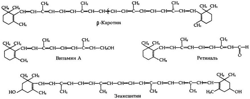
Carotenoids are red, orange, or yellow fat-soluble pigments found in METABOLISM/14.html">Chloroplasts and cyanobacteria. Like chlorophylls, chloroplast carotenoids are embedded in the thylakoid membranes. Chloroplasts typically contain two groups of carotenoids—carotenes and xanthophylls (the latter, unlike carotenes, contain oxygen). Beta-carotene, found in plants, serves as a primary source of vitamin A, which is essential for humans and animals (Fig. 7-9). In green leaves, carotenoids are masked by an Abundance of chlorophyll.
Fig. 7-9. A group of related carotenoids. Cleavage of the beta-carotene molecule at the indicated site yields two molecules of vitamin A. Oxidation of vitamin A yields retinal, a pigment involved in Vision. In carotenoids, conjugated bonds are located within the carbon chains. Zeaxanthin is the pigment responsible for the yellow color of corn kernels.
Phycobilins are characteristic of cyanobacteria and the chloroplasts of red algae. Unlike carotenoids, phycobilins are water-soluble.
Within chloroplasts (Fig. 7-1), chlorophyll and other pigments embedded in the thylakoids are organized into functional units called photosystems (Fig. 7-10). Each photosystem contains approximately 250 to 400 pigment molecules.
Fig. 7-10. Inner surface of a thylakoid; freeze-fracture replica. The particles embedded in the membranes are believed to represent the structural units of photosystems involved in light reactions.

All pigments in a photosystem can absorb photons (packets of light energy, see Appendix 1 to this chapter), but only a single chlorophyll molecule in a given photosystem can utilize the absorbed energy in photochemical reactions. This chlorophyll molecule is called the reaction center of the photosystem, while the other pigment molecules are called antenna pigments because they act like antennas to harvest light.
Light energy absorbed by a pigment molecule is transferred to another molecule until it reaches the reaction center, where a special chlorophyll a molecule is located. When this molecule absorbs light energy, electrons are elevated to a higher energy level and transferred to an acceptor molecule, initiating a flow of electrons. As a result, the chlorophyll molecule is oxidized and becomes positively charged.
There is Evidence for the existence of Two Types of photosystems. In Photosystem I, the reaction center is formed by a specific chlorophyll a molecule and is designated as P700 (P stands for pigment; 700 is the absorption optimum in nm). The reaction center of Photosystem II is also formed by a specific chlorophyll a molecule and is designated as P680 (absorption optimum at 680 nm).
Overall, both photosystems work synchronously and continuously. However, as will be shown below, photosystem I can function independently.
Model of Light Reactions
Fig. 7-11 illustrates the cooperative functioning of the Two Photosystems. According to this model, light energy enters photosystem II, where it is utilized by the P680 reaction center either directly or via pigment molecules. When P680 is excited, its excited energized electrons (in pairs) are transferred to an acceptor molecule (possibly a quinone), designated as Q due to its ability to quench the energy loss from fluorescence of excited P680. This is followed by a reaction that is not yet fully understood. The P680 molecule, having lost its electrons, can replace them with electrons from a water molecule. As water-derived electrons move to P680, the water molecule dissociates into protons and oxygen. This light-dependent oxidative cleavage of water molecules is termed photolysis. The Enzymes responsible for water photolysis are localized on the inner side of the thylakoid membrane. Thus, water photolysis contributes to the establishment of a proton gradient across the membrane. Manganese is an essential cofactor for the enzymes that mediate oxygen evolution.
Fig. 7-11. Non-cyclic electron flow and Photophosphorylation. This zigzag diagram (Z-scheme) shows The pathway of electron flow from water (bottom left) to NADP (top right) and the energetic relationships. To raise the energy of electrons from water to the level required to reduce NADP to NADPH2, electrons must be boosted twice (brown lines) by the action of light photons absorbed by photosystems I and II. Subsequently, the high-energy electron streams cascade down the pathways indicated by the black arrows. The process of photophosphorylation of ADP to form ATP is coupled with electron flow along the Electron Transport Chain linking photosystem II to photosystem I (Fig. 7-12). Ferredoxin serves as the electron donor for the reduction of NADP to NADPH2. In addition, ferredoxin performs other functions within the chloroplast: it donates electrons to enzymes involved in the Biosynthesis OF AMINO Acids and Fatty acids.
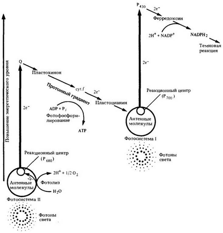
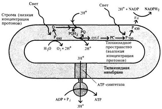
Electrons "cascade down" an electron transport chain to photosystem I. The components of this light-reaction electron transport chain resemble those of the Respiratory Electron Transport chain; the complex includes Cytochromes, iron-sulfur proteins, Quinones, as well as chlorophyll and the copper-containing protein plastocyanin. The electron transport chain between the photosystems is organized such that ATP can be generated from ADP and Pi, a process analogous to Oxidative Phosphorylation in Mitochondria. In chloroplasts, this process is called photophosphorylation (Fig. 7-12).
Fig. 7-12. Illustration of the Chemiosmotic Coupling hypothesis as The Mechanism of photophosphorylation. According to this hypothesis, protons are "pumped" across the thylakoid membrane from the stroma (external medium) into the interior (thylakoid lumen) by electron carriers positioned in a specific orientation within the membrane. The proton concentration in the thylakoid lumen increases partly due to water splitting and partly as a result of plastoquinone (PQ) oxidation on the inner surface of the membrane. As protons flow back down their gradient from the thylakoid lumen into the stroma, ADP is phosphorylated to ATP via ATP synthase. For every three protons passing through ATP synthase, one ATP molecule is synthesized. PC — plastocyanin; Fd — ferredoxin.
In photosystem I, light energy transfers electrons from P700 to the electron acceptor P430, which is likely an iron-sulfur protein. The next electron carrier is ferredoxin ("fer" denoting iron and "redoxin" emphasizing participation in redox reactions), which is also an iron-sulfur protein, though distinct from P430. Ferredoxin then transfers its electrons to the coenzyme NADP, which is reduced to NADPH2, while the P700 molecule is oxidized. The electrons lost by the P700 molecule are replaced by electrons from photosystem II.
Thus, in the light, electrons move from water to photosystems II and I and to NADP. This unidirectional flow of electrons from water to NADP is called non-cyclic electron flow; the accompanying ATP formation is termed non-cyclic photophosphorylation.
The change in Free energy (∆G) for the reaction
Н2О + NADP —> NADPH2 + 1/2О2
is 51 kcal/mol. The energy equivalent to 1 mole of light photons with a wavelength of 700 nm (the equivalent of 1 mole of photons is called an Einstein, E) is 40 kcal/E. Four photons are required to transfer two electrons to the NADPH2 level, i.e., 160 kcal. Approximately one-third of this Energy is stored in the form of NADPH2. The total energy yield of non-cyclic electron flow (involving 12 pairs of electrons passing from Н2О to NADP) amounts to 12 ATP and 12 NADPH2.
Cyclic Photophosphorylation
As already mentioned, photosystem I can operate independently of photosystem II (Fig. 7-13). In this process, known as cyclic electron flow, electrons are transferred from P700 to P430 upon illumination of photosystem I. Instead of proceeding to NADP, the electrons take an "alternative route" associated with both photosystems I and II, and then return to the reaction center of photosystem I. This generates ATP; because ATP synthesis is coupled with cyclic electron flow, it is referred to as cyclic photophosphorylation. This is believed to be the most primitive mechanism and evidently occurs in certain photosynthetic bacteria. Eukaryotic Cells are also capable of synthesizing ATP via Cyclic electron transport. However, this process does not involve the splitting of H2O, the evolution of O2, or The formation of NADPH2.
Fig. 7-13. Cyclic photophosphorylation occurs exclusively in photosystem I; ATP is generated from ADP in the same manner as shown in Fig. 7-12, but without oxygen evolution or NADP reduction.

It is believed that cyclic Electron Transport and photophosphorylation take place when The Cell has an abundant supply of the reducing agent in the form of NADPH2, yet requires additional ATP for other metabolic reactions.
Last update: 07/08/2026
Editorial and Educational Adaptation: This material has been compiled based on the primary/original source text. The project team performed an editorial review, corrected technical inaccuracies, structured sections, and adapted the content for an educational format.
What was processed:
- elimination of formatting defects (OCR errors, structural breaks, corrupted characters);
- editorial organization of content;
- standardization of terminology in accordance with academic sources;
- verification of factual statements against the original source text.
All mentions of the author, publication year, and origin of the primary text have been preserved in accordance with the source.