Human Biochemistry, Volume 2 - Murray R. 1993
Biochemistry of Intra- and Intercellular Communication
Characteristics of the Endocrine System
Hormone Receptors
General characteristics of Receptors
One of the most challenging problems encountered by researchers studying hormone-based communication systems is illustrated in Fig. 43.2. In the extracellular fluid, Hormones are present at very low concentrations—typically in the range of 10-15 to 10-19 mol/L. This is far below the levels of other structurally similar compounds (sterols, Amino Acids, Peptides, Proteins) and various other substances found in Blood at concentrations of 10-5 to 10-3 mol/L. Consequently, target Cells must distinguish a given hormone not only from Other Hormones present in trace amounts, but also from a vast array of Other Compounds present in 106- to 109-fold excess. This exceptionally high degree of selectivity is provided by specialized recognition molecules belonging to The Cell, known as receptors. The biological effect of hormones begins with their binding to specific receptors and typically ends with the dissociation of the hormone-receptor complex (in accordance with the principle that a reliable control system must possess a mechanism to terminate the action of an agent).
Class="center">
Fig. 43.2. Specificity and selectivity of Hormone Receptors. The extracellular fluid contains a multitude of diverse compounds, yet receptors recognize only a very few of them. Furthermore, receptors must select specific molecules from a Background of numerous other compounds present at much higher concentrations. The diagram shows that each cell may bear either a single type of receptor or multiple types.
A target cell is defined by its ability to selectively bind a given hormone using such a receptor, and radioactive ligands mimicking hormone binding are used to quantify this interaction. The study is conducted According to the following rules: 1) the Introduction of a radioactive label must not alter the biological activity of the Ligand; 2) ligand binding must be specific, i.e., The addition of an unlabeled agonist or antagonist should displace the label; 3) binding must be saturable; 4) binding must occur within the same concentration range as the expected biological response.
Recognition and Coupling Domains of the Receptor
All receptors, whether steroid or polypeptide, possess at least two functionally distinct domains (regions): the first domain (the recognition domain) binds the hormone, while the second generates a signal that couples hormone recognition to a specific intracellular process. Hormone binding by the receptor is based on the complementarity between the conformation of a region of the hormone molecule and a region of the receptor molecule. The degree of similarity, or fit, determines binding affinity, which is quantified by the affinity constant (K). If the relative K of a natural hormone is 1, that of other naturally occurring compounds ranges from 0 to 1. The absolute values of affinity, however, can differ by more than a trillion-fold. For certain receptors, ligands with a relative K > 1 have been synthesized and are utilized in studies of receptor biology.
Coupling (signal Transduction) is mediated by two primary mechanisms. Polypeptide and protein hormones, as well as catecholamines, bind to receptors located in The Plasma Membrane, thereby generating a signal that regulates various cellular Functions—typically by altering enzyme activity. Steroid and THYROID HORMONES interact with intracellular receptors, and the resulting complex generates the appropriate signal (see below).
The Amino acid sequences of these two domains have been identified for many polypeptide hormone receptors. By utilizing hormone analogs bearing substitutions of specific amino acids, one can alter both binding and biological activity. Steroid hormone receptors also possess at least two functional domains: one binds the hormone, while the other binds to a specific region of DNA. Recombinant DNA Methods are currently employed to study these receptors; as structural analysis demonstrates, the DNA-binding domains are highly homologous. Ultimately, The Essence of a receptor is defined by this dual function of binding and coupling, with the coupling between hormone binding and signal transmission (transduction)—the so-called receptor-effector coupling—serving as the initial step in the Amplification of the hormonal response. This dual function of the target cell receptor constitutes its fundamental difference from plasma carrier proteins, which bind the hormone but do not generate a signal.
Comparison of Receptor and Transport Proteins
There is a fundamental difference between the binding of hormones to receptors and their association with various transport proteins (carriers). This comparison is summarized in Table 43.1. The number of receptor molecules involved in ligand binding is on the order of several thousand per cell, and the binding itself is characterized by high affinity and specificity. Receptors are capable of recognizing and selecting specific compounds under concentration gradients of 106 to 107; at physiological hormone concentrations, this receptor binding is saturable. Hormone-receptor interactions depend on Temperature, pH, and salt concentrations in a manner characteristic of each hormone. Binding is driven by hydrophobic and electrostatic mechanisms and is therefore readily reversible, except in certain special cases.
In the Circulation, steroid and thyroid hormones exist as complexes with specific transport proteins. These proteins significantly outnumber intracellular receptor proteins, yet exhibit lower affinity and lower specificity for hormone binding. Transport proteins create a hormone reservoir in the blood, since in the bound state hormones are protected from METABOLISM and excretion. Biological activity is exclusively inherent to the unbound (free) hormone. Peptide and Protein hormones lack dedicated transport proteins in Blood Plasma, and consequently their half-Life in the bloodstream is much shorter (seconds or minutes) than that of Steroid Hormones (hours).
Table 43.1. Comparison of hormonal receptors and plasma hormone-transporting proteins
|
Property |
Receptors |
Plasma transport proteins |
|
Concentration |
Very low (thousands of molecules/cell) |
Very high (billions of molecules/μL) |
|
Binding affinity |
Very high (1011–109 mol/L) |
Low (10-7–10-5 mol/L) |
|
Binding specificity |
High |
Low |
|
Saturability at physiological hormone concentrations |
Yes |
No |
|
Binding reversibility |
Yes |
Yes |
|
Signal transduction |
Yes |
No |
Relationship Between Receptor Occupancy and Biological Effect
In many cases, the hormone concentration that results in receptor occupancy (occupation) virtually coincides with the concentration that elicits a biological response (Fig. 43.3, A). This holds true for all steroid hormones and A number of Peptide Hormones. This fact in itself is remarkable, especially considering the numerous steps separating The process of hormone binding and the complex response to it, such as enzyme induction, cell lysis, or Amino Acid Transport. However, in certain instances, a pronounced dissociation between these two processes occurs; the maximal biological response is achieved when only a few percent of the total receptor population are occupied (Fig. 43.3, B, effect 2). Receptors that do not participate in inducing the biological response are referred to as spare receptors.
Spare receptors have been identified through studies of the response to certain polypeptide hormones; it is believed that they serve both as a means of increasing target cell sensitivity to low hormone concentrations and as a receptor reservoir. THE CONCEPT OF spare receptors belongs to the category of working hypotheses and may be revised depending on which aspect of hormone action and in which tissue is being investigated. For example, granulosa cells exhibit excellent correlation between hormone binding and cAMP synthesis (when hormones activate adenylate cyclase, spare receptors are typically undetectable); at the same time, steroidogenesis in these cells (a cAMP-dependent process) occurs when fewer than 1% of the receptors are occupied (see effects 1 and 2 in Fig. 43.3, B). For the derepression of phosphoenolpyruvate carboxykinase Gene Transcription to occur in Liver cells, occupancy of substantially less than 1% of Insulin receptors is sufficient; on the other hand, thymocytes display a high degree of correlation between insulin binding and amino acid transport. Examples of dissociation between the level of receptor occupancy and the magnitude of the biological effect include The Influence of catecholamines on Muscle contraction, lipolysis, and ion transport. It is hypothesized that these ultimate biological responses result from a cascade amplification of hormone action. It has been found that the same cell exhibits different sensitivities to a hormone depending on which specific hormonal effect is being considered. Thus, in adipocytes, as Insulin Receptor occupancy progressively increases, there is a sequential activation of lipolysis, glucose oxidation, amino acid transport, and Protein Synthesis.
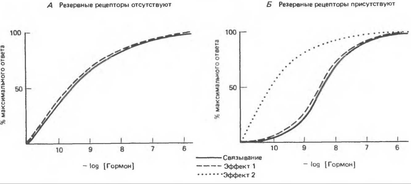
Fig. 43.3. Dependence of biological effect on hormone binding in the absence (A) or presence (B, effect 2) of spare receptors. In some cases, a biological effect may be tightly coupled to hormone binding by the tissue, whereas for another effect, the spare receptor phenomenon is manifested (cf. effects 1 and 2 in Fig. B).
Receptor Regulation
The number of receptors within a cell or on its surface is in a dynamic state: it is physiologically regulated and changes in disease states or under the influence of therapeutic agents. Receptors localized in the plasma membrane are the best understood in this regard. It has been demonstrated that their concentration and affinity for the hormone are regulated parameters. Changes in these parameters occur very rapidly and have a substantial impact on cellular sensitivity to the hormone. For instance, in cells exposed to β-adrenergic agonists, the activation of adenylate cyclase ceases and the biological response disappears within a certain timeframe (ranging from minutes to hours) upon subsequent re-addition of the agonist. Such desensitization is mediated by two mechanisms. The first involves the loss of receptors from the plasma membrane. This down-regulation is accomplished by receptor sequestration within the cell, i.e., their compartmentalization away from Other components of the cellular response system, specifically the regulatory and catalytic subunits of adenylate cyclase (see Ch. 44). Following removal of the agonist, receptors return to the cell surface, and hormone sensitivity is restored. The second mechanism of β-adrenergic system desensitization is Covalent Modification of receptors via phosphorylation. This is a cAMP-dependent process that is not associated with changes in receptor number or redistribution. As demonstrated by membrane reconstitution experiments (incorporating receptors into membranes previously devoid of them), phosphorylated receptors are incapable of activating cyclase, leading to the uncoupling of hormone binding from cell activation. Similar examples of physiological adaptation achieved through down-regulation of receptor number by a homologous hormone can be observed in the case of insulin, Glucagon, TRH, Growth Hormone, LH, FSH, and catecholamines. Certain hormones (angiotensin II and prolactin) up-regulate their own receptors. These changes in receptor number can occur very rapidly (over minutes or hours) and apparently serve as an important mechanism for regulating biological responses. The Effect of partial receptor loss on the biological response elicited by a given hormone concentration is determined by the presence or absence of spare receptors. Figure 43.4 illustrates how a 5-fold decrease in receptor number affects the concentration-response curve depending on this condition. In case A (spare receptors absent), the magnitude of the response reaches only 20% of the control; consequently, A change in Vmax occurs. In case B (spare receptors present), the maximal response is achieved, but at a much higher hormone concentration than in the control; this case is analogous to a change in Km.

Fig. 43.4. Effect of a 5-fold reduction in receptor number on the biological response in a system lacking (A) and containing (B) spare receptors.
Receptor Structure
The Acetylcholine Receptor is the best characterized, as it can be readily obtained in purified form due to its relatively abundant presence in the electric organ of the electric ray *Torpedo californica*. This receptor consists of four subunits: α2, β, γ, and δ. The two α-subunits bind acetylcholine. Site-Directed Mutagenesis has identified the Regions of the α-subunit involved in forming the transmembrane ion channel, which carries out The primary function of the acetylcholine receptor.
The Abundance of other receptors is extremely low, which has historically hindered their purification and analysis. Today, Genetic Engineering techniques make it possible to obtain sufficient amounts of material, leading to a surge in such research. It has been demonstrated that the insulin receptor is a heterotetramer (a2ß2) whose subunits are held together by multiple Disulfide Bonds. The extracellular a-subunit binds insulin, whereas the transmembrane ß-subunit is responsible for signal transduction, likely involving a Tyrosine kinase domain located in the cytoplasmic portion of the polypeptide. Receptors for insulin-like growth factor I (IGF-I), epidermal growth factor (EGF), and low-density Lipoproteins (LDLs) share a general structural similarity with the insulin receptor (see Fig. 51.16). Receptors for other polypeptide hormones are less well characterized; however, based on their sensitivity to various peptidases and Proteolytic Enzymes, they are believed to share a common protein component. In many cases, intact disulfide bonds, Phospholipids, and carbohydrate moieties appear to be essential for hormone binding.
Steroid hormone receptors are also proteins. While their functions have been extensively studied over recent years, their structural details are only now coming to light. Let us consider the glucocorticoid receptor as an example (Fig. 43.2). It comprises three functionally distinct domains: 1) a hormone-binding domain located in the C-terminal region of the polypeptide chain; 2) an adjacent DNA-binding domain; and 3) a specific region within the N-terminal half of the protein molecule, which is required for high-affinity binding to the target DNA sequence (and which contains the majority of the molecule's antigenic sites). The existence of these three functional domains has been confirmed through the analysis of receptors synthesized using recombinant DNA techniques. This modular architecture appears to be a general feature of various steroid hormone receptors, accompanied by a high degree of Amino Acid Sequence Homology in the corresponding regions. Another fascinating finding is the homology observed between this class of receptors and the v-erbA oncogene.
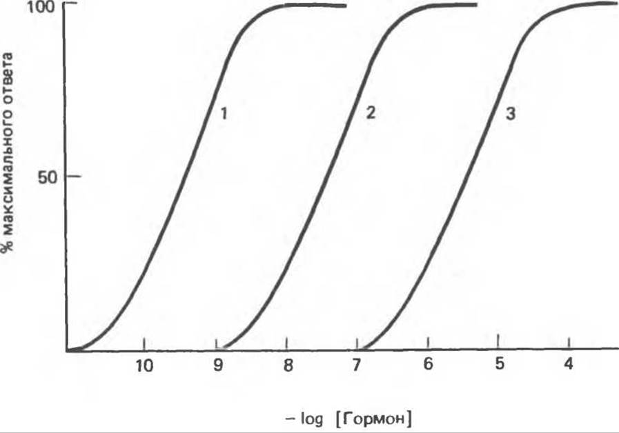
Fig. 43.5. Different hormones within the same class may vary in their potency. An identical biological response is achieved at different hormone concentrations.
The Agonist-Antagonist Concept
Hormonal chemical compounds can be divided into four groups based on their ability to elicit a biological response mediated by a specific hormone receptor: agonists, partial agonists, antagonists, and inactive compounds. This Classification has been thoroughly developed with respect to glucocorticoids (see Chapter 48).
Agonists are compounds capable of eliciting a maximal response, although the required concentrations may vary (Fig. 43.5 and Example A in Fig. 43.6). In Fig. 43.5, numbers 1, 2, and 3 can represent pork insulin, pork proinsulin, and guinea pig insulin, respectively. Across all tested systems, these insulin preparations elicited responses of approximately equal magnitude, but each at its own characteristic concentration. Similarly, numbers 1, 2, and 3 in this figure may designate dexamethasone, cortisol, and corticosterone (see Table 48.4).
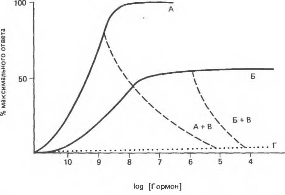
Fig. 43.6. Hormones can be categorized into the following groups: agonists (A), partial agonists (B), antagonists (A + B and B + V), and inactive agents (G).
Partial agonists elicit a diminished response even when administered at very high concentrations (see Fig. 43.6, B). Antagonists typically produce no intrinsic effect of their own, but completely inhibit the action of agonists and partial agonists (see examples A + B and B + V in Fig. 43.6). A large group of compounds structurally related to hormones exhibits neither intrinsic biological activity nor any effect on the action of Agonists and Antagonists. These are classified as inactive substances (Fig. 43.6, G).
Partial agonists frequently compete with agonists for receptor binding and activation, in which case they function as partial antagonists. The degree of inhibition of agonist activity exerted by partial or full antagonists depends on the concentration ratio of the respective Steroids. Typically, an antagonist causes inhibition at concentrations vastly exceeding those at which the agonist exerts its maximal effect. Such high concentrations are extremely rare in vitro, yet this phenomenon is widely exploited in The Study of glucocorticoid hormones under in vitro conditions.
Table 48.4 lists the steroids used in the studies that first suggested the dual function of the glucocorticoid receptor: namely, ligand binding and—via the resulting conformational change—DNA binding. This hypothesis implied that 1) agonists bind to the receptor, fully activate it, and elicit a maximal biological response; 2) partial agonists fully occupy the receptor but fail to fully activate it, thereby producing only a partial biological response; and 3) antagonists fully occupy the receptor, but the resulting complex is incapable of binding to DNA and thus does not directly trigger a biological response, although it successfully blocks the effect of agonists.
Once The Role of receptors in hormone action was elucidated, it became clear that receptor dysfunction could underlie a variety of pathological conditions. Table 43.2 outlines three main categories of such disorders. The first group encompasses pathologies caused by the generation of autoantibodies against specific hormone receptors. These Antibodies (of the IgG class) can block hormone binding (acanthosis nigricans with insulin resistance; asthma), mimic hormone binding (Graves' disease), or accelerate receptor turnover rates (myasthenia gravis).
The second group comprises diseases characterized by a failure to detect hormone binding to the receptor. Whether the receptors are genuinely absent in these cases or simply undetectable due to structural defects remains unknown, as standard receptor assays fundamentally rely on measuring hormone binding.
Table 43.2. Hormonal Receptors and Associated Diseases
|
Disease |
Receptor |
Nature of Defect |
|
Graves' disease (hyperthyroidism) |
TSH |
Antibodies stimulate the TSH receptor |
|
Insulin |
Antibodies block insulin binding to the receptor |
|
|
Acanthosis nigricans with insulin resistance |
||
|
Myasthenia gravis |
Acetylcholine |
Antibodies increase the turnover rate of the acetylcholine receptor |
|
Asthma |
ß-Adrenergic |
Antibodies block the binding of ß-adrenergic agents to the receptor |
|
Hereditary nephrogenic diabetes insipidus |
ADH |
Receptor deficiency |
|
Testicular feminization syndrome Pseudohypoparathyroidism |
Androgen PTH |
Receptor deficiency " " |
|
Vitamin D-resistant Rickets type II |
Calcitriol receptor |
" " |
|
Obesity |
Insulin |
Decreased hormone binding |
|
Type II Diabetes Mellitus [non-insulin-dependent diabetes mellitus (NIDDM)] |
" |
Same |
The third category consists of disorders caused by abnormal receptor regulation. Patients suffering from obesity or type II diabetes mellitus combined with obesity frequently exhibit glucose intolerance and insulin resistance despite elevated blood insulin levels. Such patients show a reduced number of insulin receptors (down-regulation) on target cells, such as adipocytes, hepatocytes, and myocytes. Weight loss in these patients leads to a gradual decline in blood insulin levels, an upregulation of receptor numbers, enhanced hormone sensitivity, and an amelioration of glucose intolerance. Recent studies on the molecular foundations of Cancer compellingly demonstrate that impaired coupling between growth factor receptors and effector mechanisms can drive the uncontrolled proliferation of malignant cells. These examples illustrate the wide spectrum of diseases stemming from pathologies of hormonal receptors.
Ginsberg В. Н. Synthesis and regulation of receptors for polypeptide hormones. Pages 59- 97. In: Biological Regulation and Development, Vol. 3B, Yamamoto K. (ed.). Plenum Press, 1985.
Granner D. K., Lee F. The multiple endocrine neoplasia syndromes, Chapter 76. In: Comprehensive Textbook of Oncology, Moosa A. R.. Robson M.C., SchimpffS. C. (ed.). Williams and Wilkins, 1984.
Mishina M. et al. Expression of functional acetylcholine receptor from cloned cDNAs. Nature, 1984, 307, 604.
Roth J.. Taylor S.I. Receptors for peptide hormones: Alternations in disease of humans, Annu. Rev. Physiol., 1982, 44, 639.
Roth J. et al. The evolutionary origins of hormones, Neurotransmitters. and other extracellular messengers. N. Engl. J. Med., 1982, 306, 523.
Sibley D. R.. Lefkowitz R. J. MOLECULAR MECHANISMS OF receptor desensitization using the ß-adrenergic receptor-coupled adenylate cyclase system as a model. Nature, 1985, 317, 124.
Last update: 06/08/2026
Editorial and Educational Adaptation: This material has been compiled based on the primary/original source text. The project team performed an editorial review, corrected technical inaccuracies, structured sections, and adapted the content for an educational format.
What was processed:
- elimination of formatting defects (OCR errors, structural breaks, corrupted characters);
- editorial organization of content;
- standardization of terminology in accordance with academic sources;
- verification of factual statements against the original source text.
All mentions of the author, publication year, and origin of the primary text have been preserved in accordance with the source.