Human Biochemistry Volume 1 - Murray R. 1993
Bioenergetics and Carbohydrate and Lipid Metabolism
Intermediary Metabolism
Localization of Metabolic Pathways
As shown in Fig. 2.3, Metabolic pathways can be studied at various Levels of Organization, which are conveniently divided into two main groups: 1) the Organ and tissue level—in this case, one can monitor the substrates entering a tissue and the metabolites leaving it, and describe the course of their transformations; 2) the subcellular level—each cellular organelle (e.g., mitochondrion) and each compartment (e.g., Cytosol) perform a specific biochemical role within the overall system of intracellular METABOLISM.
Intermediary Metabolism at the Tissue and organ Level
Amino Acids resulting from Protein Digestion and glucose produced by carbohydrate digestion enter the HEPATIC PORTAL VEIN upon absorption. Consequently, these metabolites, along with other Water-soluble digestion products, first reach the Liver (Fig. 16.5). The liver performs a crucial metabolic function: it regulates the concentrations of most Blood-borne metabolites, primarily glucose and amino acids. For example, excess glucose is either converted into Glycogen (Glycogenesis) or used for fat synthesis (Lipogenesis). Between meals, the liver maintains blood glucose levels within the physiological range through glycogen reserves (Glycogenolysis) or (along with the Kidneys) by converting non-carbohydrate metabolites such as lactate, glycerol, and amino acids into glucose (Gluconeogenesis). Maintaining an adequate blood glucose concentration is vital for A number of Tissues that rely exclusively on this monosaccharide as fuel (e.g., the Brain, erythrocytes). The liver is also the site of synthesis for major Plasma Proteins (such as albumin) and the deamination of excess amino acids; the resulting urea is transported via the bloodstream to the kidneys and excreted.
Skeletal Muscle uses glucose as fuel, converting it to lactate and CO2. Muscle glycogen is utilized as fuel During Muscle contraction. Muscle Proteins are synthesized from plasma amino acids within the Muscle tissue. Muscle accounts for about 50% of total body mass; thus, it represents a significant protein reservoir that can be mobilized to replenish plasma amino acids, particularly during periods of dietary deficiency.
Class="center">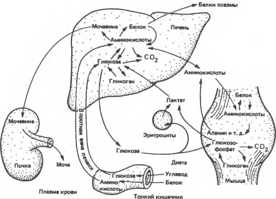
Fig. 16.5. Transport and metabolic fate of major CARBOHYDRATES, amino acids, and their metabolites. Note that the concentration of free glucose in muscle is low because, upon entering the muscle Cell, it is rapidly phosphorylated.
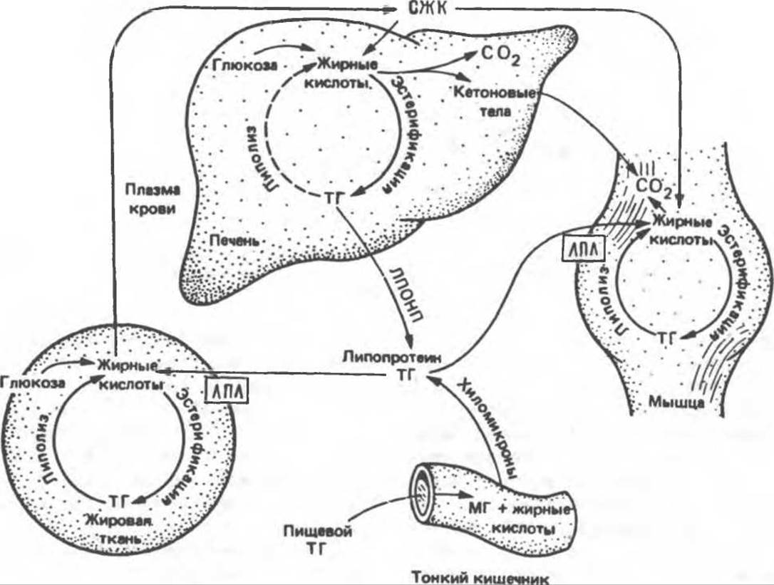
Fig. 16.6. Transport and metabolic fate of major lipid substrates and their metabolites. FFA — free fatty acid; LPL — lipoprotein lipase; MG — monoacylglycerol; TG — triacylglycerol; VLDL — very-low-density lipoprotein.

Fig. 16.7. Intracellular localization and integration of Major Metabolic Pathways in a hepatic parenchymal cell. AA → metabolism of one or more Essential Amino Acids; AA → metabolism of one or more non-essential amino acids.
Upon digestion, Lipids (Fig. 16.6) yield monoacylglycerols and Fatty acids. In intestinal Cells, they associate with proteins and are secreted first into The Lymphatic system and then into the Circulatory system, where they circulate as Lipoproteins known as chylomicrons. All hydrophobic, lipid-soluble digestion products (such as Cholesterol) are incorporated into lipoproteins, facilitating their transport between tissues in the aqueous environment of Blood Plasma. Unlike glucose and amino acids, chylomicron triacylglycerols are not taken up by the liver; instead, they are hydrolyzed in extrahepatic tissues by the enzyme lipoprotein lipase. The released Fatty acids are either incorporated into tissue lipids or oxidized and used as fuel. Another major source of long-chain fatty acids is their synthesis (lipogenesis) from carbohydrates, which occurs primarily in adipose tissue and the liver.
Adipose tissue triacylglycerol serves as the body's primary fuel reserve. Following its Hydrolysis (lipolysis), fatty acids are released and enter the bloodstream. Free fatty acids are subsequently taken up by most tissues (except the brain and erythrocytes), where they are either esterified to form acylglycerols or oxidized to CO2 to serve as fuel. The liver features two additional important metabolic pathways: 1. Excess triacylglycerols, formed either from fatty acids or via lipogenesis, are secreted into the bloodstream as very-low-density lipoproteins (VLDLs). These triacylglycerols then follow the same metabolic fate as chylomicrons. 2. Partial Oxidation of Fatty acids leads to the Formation of Ketone bodies (ketogenesis). Ketone Bodies are transported from the liver to extrahepatic tissues, where they act as another vital fuel resource.
Intermediary Metabolism at the Subcellular Level
The principal biochemical Functions of subcellular components and cellular Organelles are summarized in Table 2.4. However, because most cells perform specialized functions, certain metabolic pathways assume major importance while others remain practically unused. Figure 16.7 illustrates the major metabolic pathways, their interconnections, and their intracellular localization in hepatic parenchymal cells.
The Central Role of Mitochondria immediately becomes apparent, as carbohydrate, lipid, and amino acid metabolic pathways intersect there. Specifically, mitochondria house the Enzymes of The Citric Acid Cycle, the Respiratory Chain, ATP synthesis, fatty acid ß-Oxidation, and ketone body formation. They also serve as the collection point for keto acids (derived from AMINO ACID DEAMINATION), which are subsequently used to synthesize non-essential amino acids.
Glycolysis, the Pentose Phosphate Pathway, and fatty acid synthesis take place in the cytosol. Notably, during gluconeogenesis, even substances like lactate and Pyruvate, which are produced in the cytosol, must enter the mitochondria to be converted into oxaloacetate, from which glucose is subsequently synthesized.
Endoplasmic reticulum membranes contain the enzyme system for acylglycerol synthesis, while Ribosomes are responsible for Protein Synthesis.
It should be noted that The transport of metabolites—which vary in size, charge, and lipid solubility—across the membranes surrounding organelles involves highly complex mechanisms. Some of these were discussed in the context of the mitochondrial membrane (see Chapter 13); other mechanisms will be examined in subsequent chapters.
Last update: 06/08/2026
Editorial and Educational Adaptation: This material has been compiled based on the primary/original source text. The project team performed an editorial review, corrected technical inaccuracies, structured sections, and adapted the content for an educational format.
What was processed:
- elimination of formatting defects (OCR errors, structural breaks, corrupted characters);
- editorial organization of content;
- standardization of terminology in accordance with academic sources;
- verification of factual statements against the original source text.
All mentions of the author, publication year, and origin of the primary text have been preserved in accordance with the source.