Practical Protein Chemistry - A. Darbre 1989
Analytical Methods
Amino Acid Column Chromatography
Determination of Amino Acids
8.16.3.1. Post-Column derivatization. Prior to the 1970s, Amino Acids eluting from an ion-exchange column were detected via a reaction with ninhydrin, where mixing with the reagent took place in a post-column Reactor followed by passage of the solution through a heated coil. The intensity of the resulting color was measured first in one colorimeter at 570 nm and then in another, where the concentration of Proline and hydroxyproline was determined at 440 nm.
Class="center">
FIG. 8.4. Separation of amino acids by Ion-exchange Chromatography in a single-column system. Column (6×460 mm) packed with W-3 type resin to a height of 220 mm. Sodium citrate buffers: pH 3.25 (0.2 M Na+, with 1% propan-2-ol); pH 3.95 (0.4 M Na+); pH 6.4 (1.0 M Na+). Temperature 50–60 °C. Flow rate 44 mL/h (buffer), 22 mL/h (ninhydrin). Cuvette 12 mm. Full scale 2.0 A. Amino acid calibration mixture (50 nmol of each component in 100 µL). Reproduced with permission from Beckman.

FIG. 8.5. Single-column separation of amino acids (glycoprotein hydrolysate). Beckman Model 119Cl analyzer. Column (6×460 mm) packed with W-3H type resin to a height of 370 mm. Beckman sodium citrate buffers: pH 3.33 (0.20 M Na+); pH 4.25 (0.20 M Na+); pH 6.04 (1.0 M Na+). Temperature 30 and 60 °C. Flow rates 30 mL/h (buffer), 15 mL/h (ninhydrin). Cuvette 12 mm. Full scale 0.5 A. Sample volume 100 µL. Concentration (nmol) of each component is indicated in parentheses above the peak. Reproduced with permission from Beckman.
In 1972, fluorescamine was proposed as a selective fluorogenic reagent for primary amines [375], and somewhat later it began to be used for the modification of amino acids following their elution from an amino acid analyzer. The resulting detection sensitivity was 10 to 100 times higher than that of the ninhydrin method [21]. Measurements were performed using a fluorimeter with an excitation wavelength of 340 nm and emission at 450 nm. The widespread use of fluorescamine was hindered by its instability in aqueous media and high cost. These drawbacks are absent in o-phthalaldehyde (OPA), introduced into practice almost simultaneously with fluorescamine [320], which largely supplanted fluorescamine and has been used in amino acid analyzers by many researchers. The formation of a fluorescent derivative with OPA, having an excitation maximum at 360 nm and an emission maximum at 455 nm, occurs at pH 9–11. Lysine yields a weak fluorescence, which is enhanced by The addition of Brij reagent. Cysteine reacts only in the presence of iodoacetic acid [78], and cystine is best determined as cysteic acid following performic acid oxidation. Proline, hydroxyproline, and secondary amines react only after oxidation with chloramine-T, N-succinimide, or hypochlorite [33, 178, 321]. If the determination of proline and hydroxyproline is not required, it is undoubtedly advantageous to use OPA; the detection sensitivity for derivatives reaches 5 pmol, whereas for ninhydrin it is 100 pmol for both Primary and secondary amino groups. The mean relative standard deviation for amino acids is 6% (range 3–24%) [85].
More recent post-column Amino acid analysis Methods rely on fluorogens such as 7-chloro-4-nitrobenzo-2-oxa-1,3-diazole, which allows the detection of all amino acids, including proline and hydroxyproline, at the 1 pmol level [415], and 4-fluoro-7-nitrobenzo-2,1,3-oxadiazole [391], with a detection limit of 5 pmol for proline and 20 pmol for Other Amino Acids.
8.16.3.2. Pre-column modification. The application of post-column modification entails technical difficulties in mixing each separated amino acid in the eluate with the reagent solution under appropriate reaction conditions optimized for the process. Pre-column derivatization of amino acids is simpler to perform, and the resulting compounds can be separated by RP-HPLC; however, according to reported data, reproducibility is inferior to that achieved with the post-column method. The most thoroughly studied Amino Acid Derivatives—phenylthiohydantoins, the products of peptide degradation via the Edman method (Chapter 13)—have not been used directly for amino acid analysis, although several identification methods for them, including HPLC, have been detailed [186, 420]. Colored analogues of PTHs, namely dimethylaminoazobenzenethiohydantoins, are also separated within 30 min by HPLC [412] (see Chapter 14).
For a review on the pre-column modification of Amino Acids and Peptides followed by HPLC separation of their derivatives, see [360].
Dansyl Amino Acids. Thin-Layer Chromatography is widely used to identify dansyl amino acids in protein sequencing (Chapters 10 and 11). Recently, dansyl derivatives have been successfully separated on reverse-phase columns [15, 152, 167, 202, 330, 340, 363, 394, 403]. The detection sensitivity is 100 pmol for spectrophotometric determination, and a few femtomoles for fluorimetric [15, 202] or chemiluminescent determinations [166, 200].
Dansyl derivatives do not yield results comparable to those obtained with OPA in amino acid analysis. One of the drawbacks of Dansylation is that a single amino acid can produce multiple peaks, such as O-dansyltyrosine and O,N-didansyltyrosine alongside N-dansyltyrosine. An advantage, however, is The ability to identify proline and hydroxyproline simultaneously with other amino acids. It has been proposed [403] to use dansyl amino acid derivatives for peptide composition analysis with high sensitivity, as well as for the Quantitative determination of N- and C-terminal residues—specifically C-terminal amino acid amide residues cleaved enzymatically from amidated peptides at the 50 pmol level with UV detection [340]. D- and L-isomers of dansyl Amino acids have been separated by HPLC [209, 208].
o-Phthalaldehyde derivatives. o-Phthalaldehyde reacts with amino acids in the presence of ß-mercaptoethanol at 25 °C to form a substituted isoindole [341]. Compounds lacking an α-hydrogen atom yield a fluorescence signal amounting to <1% of the fluorescence of an equivalent amount of Glycine [83]; for cystine, this value is 3%, even when the reaction is carried out at 100 °C [84].
In the presence of mercaptoethanol, the reaction of OPA with primary amino groups goes to completion within 0.1–1.0 min, but the resulting derivatives of glycine, lysine, hydroxylysine, and phosphoserine have a half-life of 1–4 min [374]. The stability of OPA–2-mercaptoethanol amino acid derivatives has been investigated [80] and their HPLC separation accomplished within 22 min [79], demonstrating that product degradation can be minimized by rapid injection onto the column. The fluorescence intensity for lysine and hydroxylysine increases to 40–50% upon the addition of Brij-35 reagent to the reaction solution. Cystine can be quantified as cysteic acid and carboxymethylcysteine.
Secondary amino acids (proline and hydroxyproline) do not react directly with OPA, but upon oxidation with hypochlorite, they yield primary amino groups that can participate in the reaction. These Amino acids can also be determined after modification with 4-chloro-7-nitrobenzofuran (NBD) [376]. Excellent results have been achieved in the separation of OPA amino acid derivatives by HPLC with fluorimetric detection [79, 160, 161, 163, 187–189, 210, 219, 376].
The Use of electrochemical detectors opens up new possibilities not only for increasing the detection sensitivity, but also for enhancing the selectivity of detection for certain derivatives, such as basic amino acids [191].
The automation of fluorogenic compound assays places heightened demands on their stability, which OPA–mercaptoethanol–amino acid adducts fail to satisfy. OPA–ethanethiol derivatives exhibit a longer half-life. Although a 20–50% drop in fluorescence intensity over 6–8 h is observed for these compounds as well [342], their synthesis and subsequent separation have been successfully tested in an automated format for the analysis of plasma amino acids [111].
An automated routine method for analyzing OPA amino acid derivatives on an Altex Ultrasphere ODS column (250×4.6 mm, 5 µm particle size) with a Brownlee pre-column packed with C18 sorbent (5 µm) was described in 1984 [74]. This system allowed the analysis of over 2000 samples without changing the stationary phase.
OPA has been incorporated into automated amino acid sequencing programs on sequenators to block the α-amino groups of residual peptides when proline becomes the N-terminus in the sequence being determined [41].
Reagents for OPA modification.
OPA reagent [214]. Dissolve 62 g of boric acid and 25 g of KOH in 900 mL of Water. Adjust the pH to 10.4 with 45% KOH and dilute to 1 L. Filter through a 0.22 µm Millipore membrane and add 2 mL of mercaptoethanol, 6 mL of 30% (w/v) Brij-35, and 1.2 g of OPA dissolved in 15 mL of methanol. Store overnight in the dark under nitrogen at room temperature. The reagent is stable for 1 month under nitrogen at 4 °C. OPA has been used for the Determination of Amino acids at the 10 pmol level [214] and of phosphoamino acids (Section 8.11.2.3).
OPA–ethanethiol reagent [206]. Dissolve 100 mg of OPA in 5 mL of methanol. Add 50 µL of ethanethiol (ET) and 10 mL of 0.15 M sodium borate buffer (pH 10.5) containing 0.2% Brij-35 reagent. Purge the mixture with nitrogen and let stand in the dark for at least 16 h. To maintain stability, add 10 µL of ET to the reagent every 4 days (Section 8.12.2.3).
Pre-column OPA derivatization [187, 188].
Reagents. Dissolve 50 mg of o-phthalaldehyde in 1.25 mL of methanol and add 50 µL of 2-mercaptoethanol and 11.2 mL of 0.4 M borate buffer adjusted to pH 9.5 with 4 M NaOH. The reagent is stable for 1–2 weeks. Alternatively, Pierce reagent solution can be used; it contains OPA in borate buffer supplemented with Brij-35 reagent and 2-mercaptoethanol. The reagent is stable for 6 months at 4 °C.
Modification. Hydrolyze the protein in 6 M HCl, lyophilize, and dissolve the residue in water. If methanesulfonic acid was used for Hydrolysis, neutralize the hydrolyzate with 4 M NaOH. Dilute, if necessary, with 0.4 M borate buffer (pH 9.5) to a final concentration of each amino acid of <25 nmol/mL. Prior to hydrolysis, the protein may be oxidized or carboxymethylated.
Mix a 5–10 µL aliquot of the sample solution with 5 µL of the reagent solution. After one minute, add 20–100 µL of sodium acetate (pH 7.0) and immediately apply 20 µL to the column. 2-Aminoethanol serves as the internal standard.
Analysis of urine serum or CEREBROSPINAL FLUID. Mix 25 µL of the sample with 75 µL of acetonitrile, vortexing vigorously. Centrifuge at 1000 g for 2 min. Take a 10 µL aliquot for OPA derivatization.
HPLC. Columns packed with Ultrasphere ODS (45:240)×4.6 mm with 5 or 3 µm particle size and Microsorb C18 (100×4.6 mm) are used. A guard column (CO : PELL ODS sorbent, 40 µm) is required, particularly for C18 columns with 3 µm particles. Column length has virtually no effect on amino acid separation efficiency, so shorter columns (45–100 mm) are preferred to reduce analysis time. Achieving clear baseline separation of glycine and Threonine can sometimes be problematic. The reason is that columns even from the same manufacturer vary significantly. In some cases, glycine and threonine are completely resolved, whereas on other columns complete separation cannot be achieved even by varying the COMPOSITION OF THE elution system.
Measurements are performed using a fluorimeter equipped with a 9 µL flow Cell and filters for excitation at 305–395 nm and emission at 420–650 nm.
For gradient elution, two pre-degassed solutions are mixed:
Solution A. 0.1 M sodium acetate, pH 7.2 (for protein hydrolysates) or pH 6.4 (for physiological fluids).
Solution B. Absolute methanol.
A linear relationship between peak area and The amount of applied sample is maintained over the range from 500 fmol to several hundred pmol; at levels <5 pmol, significant deviations from linearity are observed for amino acids such as Serine, glycine, and Alanine due to Background impurity Interference. In such cases, background correction must be performed by subtracting blank run data. For Most amino acids, the sensitivity is 25–50 fmol, and the working range is 50 fmol–1 nmol. Fig. 8.6 shows the results of amino acid separation on a 3 µm Ultrasphere ODS column in 13.5 min. Elution profiles of vasopressin and Somatostatin hydrolysates under similar conditions are presented in Fig. 8.7.
Note. A mixture containing over 60 amino acids and amines was separated using pre-column derivatization with OPA. Recently, an analysis was performed in less than 7 min on a Perkin Elmer C18 column (4.6×32 mm), 3 µm particle size (Jones, personal communication). Cysteine was determined as its carboxymethyl derivative, and Methionine as methionine-S-oxide; proline and hydroxyproline were analyzed According to the method described in [376].

FIG. 8.6. Elution profile of an OPA-derivatized amino acid mixture. Each peak represents 40 pmol of substance. The HPLC system includes two pumps (Beckman, model 100A), a microprocessor gradient controller (Beckman, model 421), and a fluorimeter (Gilson, model 121) equipped with a 9 µL flow cell and filters with bandpass settings of 305–395 nm (excitation light) and 420–650 nm (emission). The emission filter is fitted with a 2 mm aperture Diaphragm. The fluorimeter scale range is 0.1 absorbance units (AUFS), with a 0.5 s time constant. Samples were injected onto the column using an injection valve (Beckman, model 210) equipped with a 20 µL sample loop. Chromatographic peaks were recorded and integrated using a C-RIA data system. An Ultrasphere ODS reverse-phase column (75×4.6 mm, 3 µm particles) was connected to a guard column (30×2.1 mm) packed with CO : PELL ODS sorbent (40 µm particles). The flow rate was 1.5 mL/min, and the initial column backpressure was 3100 psi. Kindly provided by Dr. W. N. Jones. The seventh peak from left to right is Asn.
Pre-column derivatization with 4-chloro-7-nitrobenzofurazan (NBD-Cl) [376] is used for modifying proline and hydroxyproline. Dilute an aliquot of the neutralized sample, if necessary, to a final proline concentration of ~20 nmol/mL. Mix equal volumes of 0.4 M borate buffer (pH 9.5) and an NBD-Cl solution in methanol (2 mg/mL). Heat at 60 °C for 5 min in a sealed vial. Stop the reaction by cooling to 0 °C. Apply an aliquot of the solution to the column.
HPLC. An Ultrasphere column (250×4.6 mm) with 5 µm particles is used.
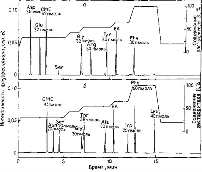
FIG. 8.7. Elution profiles of hydrolysates of two carboxymethylated Polypeptides. The amount of substance in each peak is expressed in picomoles. (a) Acid hydrolysate of vasopressin; the determined amino acid amounts are in good agreement with the known composition of the polypeptide: Asp, Glu, CMC2, Gly, Arg, Tyr, Phe; (b) enzymatic hydrolysate of somatostatin; expected composition of somatostatin based on the Amino Acid Sequence: CMC2, Asn, Ser, Gly, Thr2, Ala, Trp, Phe3, Lys2.
Solvent A. Tetrahydrofuran — 0.05 M sodium acetate (pH 6.6) (1:99).
Solvent B. Methanol.
Linear gradient from 30% to 45% B at a rate of 1.5% B per minute.
Flow rate 1.0 mL/min.
Detection at an excitation wavelength of 220 nm, emission up to 370 nm, and a sensitivity scale of 0.1 µA. NBD-proline and NBD-hydroxyproline adducts elute under these conditions in 6 min.
Phenylthiocarbamyl derivatives. The reaction of phenyl isothiocyanate (PITC) with amino acids has been studied in detail because it forms The basis of the Edman peptide degradation method, the End products of which are phenylthiohydantoins [104]. The first step of this reaction—the formation of phenylthiocarbamyl (PTC) derivatives—is used for the quantitative determination of amino acids in the so-called PICO-TAG™ analysis developed and introduced by Waters. PTC-Amino acids are stable, easily synthesized, and successfully identified after reverse-phase HPLC separation. Both primary and secondary amino groups of amino acids react, and all derivatives are separated simultaneously. Detection is performed using a UV detector at 254 nm, which is cheaper and simpler than using a fluorimeter for OPA derivatives. 35S-labeled PITC has also been used for the analysis [76, 151, 199].
Pre-column derivatization with phenyl isothiocyanate [151].
Reagents. PITC (in 1 mL ampoules sealed under vacuum), triethylamine (sequencing grade), amino acids (Pierce). Pyridine and triethylamine are distilled over ninhydrin and calcium hydride. HPLC-grade acetonitrile and methanol (Burdick & Jackson). Coupling buffer: acetonitrile — pyridine — triethylamine — water (10:5:2:3). Stable for one month at —5 °C. PTC-amino acids are stable when stored frozen at pH 6.8.
Derivatization. Evaporate the protein hydrolysate or amino acid solution to dryness in a small vial. Dissolve the residue in 100 µL of coupling buffer and evaporate to dryness using a rotary evaporator or a high-speed vacuum concentrator (Speed-Vac Concentrator). This pretreatment is necessary to remove traces of HCl and prevents the appearance of an extraneous peak on the chromatogram near Histidine.
Add 100 µL of coupling buffer and 5 µL of PITC. Let stand for 5 min at room temperature. Evaporate and dry under high vacuum (0.5–10-2 mm Hg). Re-dissolve the resulting PTC-amino acids in 250 µL of 0.05 M ammonium acetate buffer or a water — acetonitrile mixture (7:2). Inject 1–10 µL (100–1000 pmol of each amino acid) onto the reverse-phase column using an autosampler. An aliquot can be simultaneously analyzed on a standard amino acid analyzer to check the completeness of conversion into PTC derivatives.
HPLC. Columns (250×4.6 mm) packed with C3 and C18 stationary phases and 5 µm particles (Altex, Dupont, and IBM) are typically used.
Solvent systems:
A. 0.05 ammonium acetate (pH 6.8) acidified with phosphoric acid.
B. 0.1 M ammonium acetate (pH 6.8) in various Solvents:
1) acetonitrile — water (50:50);
2) methanol — water (80:20);
3) acetonitrile — methanol — water (44:10:46).
C. Acetonitrile — water (70:30).
Excellent separation of PTC-amino acids was achieved using a solvent gradient (see below) at a column temperature of 52 °C and a total analysis time of 30–40 min. Absorbance was measured in UV light at 254 nm:
|
Time, min |
Solvent |
content, % |
|
|
A |
B |
C |
|
|
0 |
100 |
0 |
0 |
|
15 |
85 |
15 |
0 |
|
30 |
50 |
50 |
0 |
|
30.1 |
0 |
0 |
100 |
|
40 |
100 |
0 |
0 |
The described Procedure is detailed in ref. [76]. Table 8.1 lists the molar extinction coefficients of PTC-amino acids (the latter are listed in order of their elution from the column).
The amino acid analysis procedure, termed the PICO∙TAG™ assay, involves treating several samples (up to 12 simultaneously) with an excess of PITC and removing volatile components under vacuum using a specially designed apparatus. A linear relationship between signal intensity and amount of substance is observed in the range of 5–1000 pmol. Process automation is possible. All Proteinogenic Amino Acids, including asparagine, glutamine, cysteic acid, carboxymethylcysteine, homoserine, methionine sulfoxide and methionine sulfone, hydroxyproline, and hydroxylysine, can be analyzed on a dedicated PICO-TAG™ column. The separation results for a standard amino acid mixture (Pierce Co.) at 250 pmol and 1 pmol (within 12 min) are shown in Figs. 8.8 and 8.9.
Table 8.1. Molar extinction coefficients of PTC-amino acids in solvent systems A, B (1), and C (see text) [151]
|
Amino acid |
Molar absorption at 254 nm (L/(mol·cm)) |
Amino acid |
Molar absorption at 254 nm (L/(mol·cm)) |
|
Ala |
15772 |
Arg |
13874 |
|
Glu |
14465 |
Pro |
15235 |
|
KM-Cys |
15500 |
Tyr |
16524 |
|
Hyp |
15420 |
Val |
16295 |
|
Ser |
14824 |
Met |
16716 |
|
Gly |
15326 |
Ile |
21375 |
|
His |
12645 |
Leu |
17069 |
|
Thr |
15152 |
Phe |
16293 |
|
Ala |
16316 |
Lys |
30224 |
Last update: 06/08/2026
Editorial and Educational Adaptation: This material has been compiled based on the primary/original source text. The project team performed an editorial review, corrected technical inaccuracies, structured sections, and adapted the content for an educational format.
What was processed:
- elimination of formatting defects (OCR errors, structural breaks, corrupted characters);
- editorial organization of content;
- standardization of terminology in accordance with academic sources;
- verification of factual statements against the original source text.
All mentions of the author, publication year, and origin of the primary text have been preserved in accordance with the source.