Biological Chemistry - Berezov T. T., Korovkin B. F. 1998
Metabolism of Simple Proteins
Intermediary Metabolism of Amino Acids in Tissues
Transamination of Amino Acids
Transamination refers to Reactions Involving the intermolecular transfer of an amino group (NH2—) from an amino acid to an a-keto acid without The intermediate formation of ammonia. Transamination reactions (formerly called "transamination proper") were first discovered in 1937 by Soviet scientists A.E. Braunshtein and M.G. Kritzman while studying the deamination of glutamic acid in Muscle tissue. It was observed that adding glutamic and pyruvic acids to a muscle homogenate yielded a-ketoglutaric acid and Alanine without any intermediate free ammonia; conversely, The addition of alanine and a-ketoglutaric acid led to The formation of pyruvic and glutamic acids, respectively.
Class="center">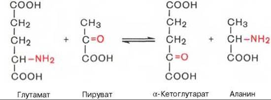
Transamination reactions are reversible and, as was later established, universal for All living organisms. These reactions proceed with the participation of specific Enzymes named aminotransferases (According to the modern Classification, aminotransferases or transaminases) by A.E. Braunshtein. Theoretically, transamination reactions can occur between any Amino Acid and keto acid; however, they proceed most intensively when one of the partners is a dicarboxylic amino or keto acid. The existence of Transamination reactions between monocarboxylic amino and keto acids has been proven in animal Tissues and microorganisms. The Donors of the NH2 group can also include not only a-, but also ß-, y-, and w-amino groups of various Amino Acids. Furthermore, A. Meister's laboratory demonstrated the transamination of glutamine and asparagine with keto acids in animal tissues.
Pyridoxal phosphate (a derivative of vitamin B6; see Chapter 5), which is a coenzyme of transaminases and reversibly converts into pyridoxamine phosphate during the reaction, plays an active role in The transfer of the amino group.
Mechanism of the transamination reaction. The general theory of the Mechanism of Enzymatic transamination was developed by Soviet scientists A.E. Braunshtein and M.M. Shemyakin. A similar mechanism was independently proposed by American biochemists E. Snell and D. Metzler. All transaminases (as well as amino acid Decarboxylases) contain the same coenzyme—pyridoxal phosphate. Transamination reactions share a common mechanism, with the Specificity of transaminases being ensured by their protein component. Transamination enzymes catalyze the transfer of the NH2 group not directly to the a-keto acid, but first to the coenzyme pyridoxal phosphate. The resulting intermediate compound (a Schiff base) undergoes intramolecular transformations (labilization of the a-hydrogen atom and redistribution of bond energy), leading to the release of the a-keto acid and pyridoxamine phosphate. In the second stage of the reaction, pyridoxamine phosphate reacts with any other a-keto acid, which through the same stages of intermediate compound formation (proceeding in the reverse direction) results in the synthesis of a new amino acid and the release of pyridoxal phosphate. Omitting the intermediate stages of Schiff base formation, both Stages of the transamination reaction can be represented by the general scheme:

A more detailed mechanism of transase action is shown in Fig. 12.3.
Since in all pyridoxal enzymes (including transaminases) the carbonyl group of the coenzyme (—CHO) is bound to the ε-amino group of a Lysine residue within the protein moiety, A.E. Braunshtein and E. Snell introduced the following addition to the classical mechanism of the transamination reaction. It was found that the interaction between the substrate, i.e., the L-amino acid (aspartate in the figure), and pyridoxal phosphate occurs not through Condensation with the release of a Water molecule, but via a substitution reaction in which the NH2 group of the substrate displaces the ε-NH2 group of lysine in the enzyme protein molecule, leading to the formation of a pyridoxal phosphate complex.
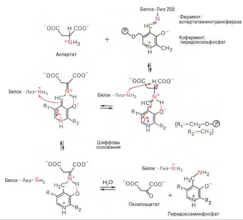
Fig. 12.3. MECHANISM OF ACTION of pyridoxal phosphate in aspartate aminotransferase.
The existence of this proposed transamination mechanism has been proven by various Methods, including spectral analysis techniques used to identify intermediate aldimine and ketimine derivatives of pyridoxal phosphate.
The Role of transaminases and transamination reactions in Amino acid METABOLISM.
The extremely widespread occurrence of transaminases in animal tissues, microorganisms, and plants, their high resistance to physical, chemical, and biological factors, absolute stereochemical specificity toward L-amino acids, and high catalytic activity in transamination processes have prompted detailed studies into the role of these enzymes in amino acid metabolism. It was noted earlier that at physiological pH values, The activity of L-Amino Acid Oxidase is sharply reduced. Taking this circumstance into account, along with the high reaction rate of transamination, A.E. Braunshtein hypothesized the existence of an indirect pathway for AMINO ACID DEAMINATION in animal tissues via transamination reactions, which he termed transdeamination. This hypothesis was also supported by data from H. Euler showing that among all Natural Amino Acids, only L-glutamic acid is deaminated at a high rate in animal tissues via a reaction catalyzed by highly active and specific Glutamate dehydrogenase.
According to this hypothesis, which has received experimental confirmation, all or nearly all natural amino acids (with the exception of Methionine) first react with a-ketoglutaric acid in a transamination reaction to form glutamic acid and the corresponding keto acid. The resulting glutamic acid then undergoes direct Oxidative Deamination catalyzed by glutamate dehydrogenase. The Mechanism of transdeamination can be schematically represented as follows:
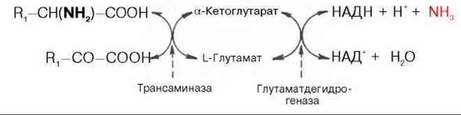
The overall reaction is as follows:
R1—CH(NH2)—COOH + НАД+ + H2O -> R—СО—СООН + НАДН2 + NH3.
Since both reactions (transamination and the deamination of glutamic acid) are reversible, conditions are created for the synthesis of essentially any amino acid, provided the Organism has the corresponding a-keto acids available. It is well known that animal and human organisms lack The ability to synthesize the carbon skeletons (a-keto acids) of so-called Essential Amino Acids; this ability is possessed only by plants and many microorganisms.

Fig. 12.4. Central role of L-amino acid transaminases and glutamate dehydrogenase in the Biosynthesis AND DEGRADATION of amino acids in animal tissues.
АМК - amino acids; а-КГ - a-ketoglutarate.
The mechanism by which natural Amino acids are synthesized in living organisms from a-keto acids and ammonia was termed transreamination by A.E. Braunshtein. Its essence lies in the reductive amination of a-ketoglutaric acid to form glutamic acid (catalyzed by NADP-dependent glutamate dehydrogenase operating in the synthetic direction), followed by the transamination of glutamate with any a-keto acid. This results in the Formation of the L-amino acid corresponding to the initial keto acid and the regeneration of a-ketoglutaric acid, which can then accept a new molecule of ammonia. The role of transamination reactions in both the deamination and BIOSYNTHESIS OF AMINO Acids can be represented by the following scheme:
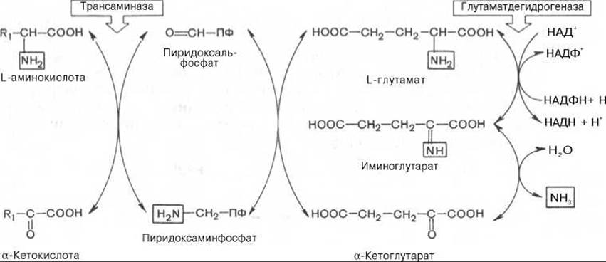
Thus, transaminases catalyze the glutamate dehydrogenase-mediated deamination of natural amino acids (black arrows) and The biosynthesis of amino acids (red arrows). A simplified representation of the role of these Key Enzymes of Nitrogen metabolism is shown in Fig. 12.4.
Evidence has been obtained for the existence in warm-blooded animals of yet another mechanism of indirect (mediated) deamination of L-amino acids, in which Glu, Asp, and AMP function as an NH2 group transfer system; the hydrolytic deamination of AMP leads to the formation of inosine monophosphate (IMP) and ammonia:

It is possible that in a similar system NAD participates as an intermediate NH2-group carrier instead of AMP.
Clinical significance of determining transaminase activity. The widespread distribution and high activity of transaminases in human Organs and tissues, combined with the relatively low activity levels of these enzymes in the Blood, have provided the rationale for measuring serum transaminase levels in various organic and functional disorders of different organs. For clinical purposes, the two most important transaminases are aspartate aminotransferase (AST) and alanine aminotransferase (ALT), which catalyze the following reversible reactions, respectively:

In the blood serum of healthy individuals, the activity of these transaminases is thousands of times lower than in parenchymal organs. Consequently, organic lesions in acute and chronic diseases accompanied by Cell destruction lead to the release of transaminases from the affected area into the bloodstream. For instance, as early as 3–5 hours after the onset of myocardial infarction, serum AST levels rise sharply (by a factor of 20–30). Peak activity for both blood transaminases occurs by the end of the first day, and within 2–3 days, under favorable clinical outcomes, serum transaminase levels return to normal. Conversely, in protracted cases or recurrent myocardial infarction, a new peak in enzyme activity is observed in the blood. This explains why the transaminase test is used clinically not only for Diagnosis, but also for prognosis and for monitoring Treatment efficacy*. In Liver cell damage, such as hepatitis, hypertransaminasemia is also observed (predominantly due to elevated ALT levels), but it tends to be more moderate and prolonged, with a slower rise in serum transaminase activity. In Various Forms of coronary insufficiency (angina pectoris, Heart defects, etc., excluding myocardial infarction), hypertransaminasemia is either absent or negligible. The determination of serum transase activity in heart diseases should be regarded as a differential diagnostic laboratory test. Furthermore, elevated serum transaminase levels have been noted in certain muscle diseases, particularly extensive trauma, limb gangrene, and progressive muscular dystrophy.
* Currently, for diagnostic purposes in internal medicine, reagent kits are widely used for the rapid (express) determination of transaminase activity in blood serum.
Transformations of a-keto acids. The a-keto acids formed during deamination and transdeamination undergo various transformations in animal tissues and can undergo re-transamination to yield the corresponding amino acid. This is the so-called synthetic pathway of transformation. Perfusion experiments using solutions of a-keto acids and ammonia through an isolated liver have demonstrated that the fluid flowing out of the liver indeed contains the L-amino acids corresponding to the initial keto acids. Furthermore,glycogenic, ketogenic, and oxidative pathways have been discovered, leading to the formation of glucose, Fatty acids, Ketone Bodies, and Components of the tricarboxylic acid (TCA) cycle, respectively. These processes can be represented in a generalized summary scheme:

The carbon skeletons of Amino acids can enter the TCA cycle via acetyl-CoA, Pyruvate, oxaloacetate, a-ketoglutarate, and succinyl-CoA. Five amino acids (Phe, Lys, Leu, Trp, Tyr) are considered "ketogenic" because they serve as precursors to ketone bodies, specifically acetoacetic acid, whereas most Other Amino Acids, designated as "glycogenic," act as a source of CARBOHYDRATES in the body, particularly glucose. Such de novo carbohydrate synthesis is enhanced in certain pathological conditions, such as Diabetes Mellitus, as well as during Hyperfunction of the adrenal cortex and the administration of glucocorticoids (see Chapter 8). However, the Classification of amino acids into "ketogenic" and "glycogenic" is somewhat arbitrary, since certain portions of the carbon atoms of Lys, Trp, Phe, and Tyr can also be incorporated into glucose precursors—for example, Phe and Tyr into fumarate. Leucine is the only truly "ketogenic" amino acid.
Last update: 06/08/2026
Editorial and Educational Adaptation: This material has been compiled based on the primary/original source text. The project team performed an editorial review, corrected technical inaccuracies, structured sections, and adapted the content for an educational format.
What was processed:
- elimination of formatting defects (OCR errors, structural breaks, corrupted characters);
- editorial organization of content;
- standardization of terminology in accordance with academic sources;
- verification of factual statements against the original source text.
All mentions of the author, publication year, and origin of the primary text have been preserved in accordance with the source.