Biological Chemistry - Berezov T. T., Korovkin B. F. 1998
Hormones
Prostaglandins
The term "Prostaglandins" was coined by U. von Euler, who first demonstrated that human semen and extracts from ram Seminal Vesicles contain substances that exhibit pronounced vasopressor activity and induce contraction of the uterine smooth Muscle. U. von Euler's hypothesis that these substances represent a specific secretion of the Prostate Gland (prostata) was not confirmed, as it is now established that they are present in all Organs and Tissues*. Nevertheless, this term has been retained in the literature (synonyms: prostatoglandins, prostaglandins).
* The only exception is the Thymus gland, upon which androgens exert a catabolic effect.
Over the past decade, prostaglandins and related biologically active compounds (Leukotrienes, prostacyclins, thromboxanes) have been the subject of intense research interest. This is because, In addition to their widespread distribution in tissues, they exert powerful pharmacological effects on numerous physiological Functions of the Organism, regulating renal hemodynamics, smooth muscle contractility, gastric secretory function, lipid and Water-Salt METABOLISM, and others. There is evidence that prostaglandins are probably not "true" Hormones, although some authors consider them "local hormones"; however, it has been shown that they modulate hormone action. The BIOLOGICAL EFFECTS OF prostaglandins appear to be mediated via Cyclic NUCLEOTIDES (see below).
Recently, The concepts of S. Bergström and co-workers have been confirmed, indicating that the precursors of all prostaglandins are polyunsaturated Fatty acids, particularly arachidonic acid (along with A number of its derivatives, dihomo-γ-linolenic and pentanoic acids, which in turn are formed in the body from linoleic and linolenic acids) (see Chapter 11). Following release from membrane phosphoglycerides (Phospholipids) under the action of specific phospholipases A (or C), arachidonic acid gives rise to prostaglandins and leukotrienes depending on the enzymatic pathway of conversion, According to the following scheme:
Class="center">
The first pathway was designated as the cyclooxygenase pathway of arachidonic acid metabolism, since the Initial Stages of prostaglandin synthesis are catalyzed by cyclooxygenase, more precisely prostaglandin-synthase (EC 1.14.99.1). Currently, data on The Biosynthesis of the main Prostanoids are well established (Fig. 8.3). The central chemical process of biosynthesis is the incorporation of molecular oxygen (two molecules) into The Structure of arachidonic acid, carried out by specific oxygenases that, in addition to oxidation, catalyze cyclization to form intermediate products—prostaglandin endoperoxides PG2[H2], designated as PGG2 and PGH2; under the action of prostaglandin isomerases, the latter are converted into primary prostaglandins. There are 2 classes of primary prostaglandins: ether-soluble prostaglandins PGE and phosphate buffer-soluble prostaglandins PGF. Each class is subdivided into subclasses: PGE1; PGE2; PGF1; PGF2, etc. Prostacyclins and thromboxanes are synthesized from these intermediates with the participation of Enzymes other than isomerases. The details of the prostanoid biosynthesis mechanism remain incompletely elucidated to date, as do the pathways of their oxidation to End products of Metabolism.
* Prostaglandins and the enzyme systems catalyzing their biosynthesis have not been detected only in human erythrocytes. It should be noted, however, that the highest concentrations of prostaglandins are found in organs and tissues belonging to the Reproductive System.

Fig. 8.3. Cyclooxygenase pathway of arachidonic acid metabolism.
R1 and R2 are side chains identical for all three prostaglandins. The symbol  denotes the blocking effect of the indicated substances.
Primary prostaglandins are synthesized in all Cells (except erythrocytes), act on the smooth Muscles of the digestive tract, reproductive and respiratory tissues, affect vascular tone, modulate The activity of Other Hormones, autonomously regulate nerve excitation, inflammatory processes (as mediators), and The rate of renal Blood flow; their biological action is mediated through The regulation of cAMP synthesis (see below).
Thromboxane A, particularly thromboxane A2 (TxA2), is synthesized predominantly in the Tissues of the Brain, Spleen, Lungs, Kidneys, as well as in platelets and inflammatory granuloma from PGH2 under the action of thromboxane synthase (see Fig. 8.3); the remaining thromboxanes are formed from TxA2. They induce platelet aggregation, thereby promoting thrombus formation, and furthermore exert the most potent vasoconstrictor action among all prostaglandins.
Prostacyclin (PGI2) is synthesized mainly in the vascular endothelium, Heart muscle, uterine tissue, and gastric mucosa. In contrast to thromboxane, it relaxes vascular smooth muscle fibers and induces platelet disaggregation, thereby promoting Fibrinolysis.
It is also important to highlight the specific Significance of the blood thromboxane-to-prostacyclin ratio, particularly TxA2/PGI2, for the physiological status of the organism. It has been found that patients predisposed to thrombosis exhibit a tendency toward a shift in the balance in favor of aggregation; conversely, patients suffering from uremia demonstrate platelet disaggregation. It has been suggested that the TxA2/PGI2 balance is crucial for the regulation of platelet function in vivo, cardiovascular Homeostasis, thrombotic disease, etc.
Fig. 8.3 also illustrates the pathways of prostanoid Catabolism. The initial stage of catabolism of "classical" prostaglandins is the stereospecific oxidation of the OH group at the 15th carbon atom, yielding the corresponding 15-keto derivative. The enzyme catalyzing this reaction, 15-hydroxyprostaglandin dehydrogenase, has been discovered in the Cytoplasm and requires NAD or NADP. Thromboxane is inactivated in vivo either via chemical Cleavage to thromboxane B2 or through oxidation by a dehydrogenase or reductase. Similarly, PGI2 (prostacyclin) rapidly disintegrates into 6-keto-PGF1α in vitro, whereas in vivo it is inactivated via oxidation by 15-hydroxyprostaglandin dehydrogenase to form 6,15-diketo-PGF1α.
The second pathway of arachidonic acid metabolism is the lipoxygenase pathway (Fig. 8.4), which differs in that it initiates the synthesis of yet another class of BIOLOGICALLY ACTIVE SUBSTANCES—leukotrienes. A characteristic structural feature of leukotrienes is that they lack a cyclic structure, although leukotrienes, like prostanoids, are composed of 20 carbon atoms. The structure of leukotrienes contains four double bonds; some of them form peptidolipid complexes with Glutathione or its constituent parts (leukotriene D can be further converted into leukotriene E by losing a Glycine residue). The main biological effects of leukotrienes are associated with inflammatory processes, allergic and immune reactions, anaphylaxis, and smooth muscle activity. Specifically, leukotrienes promote the contraction of airway and gastrointestinal smooth muscle, regulate vascular tone (exerting a vasoconstrictor effect), and stimulate coronary artery constriction. The Catabolic pathways of leukotrienes have not yet been fully established.
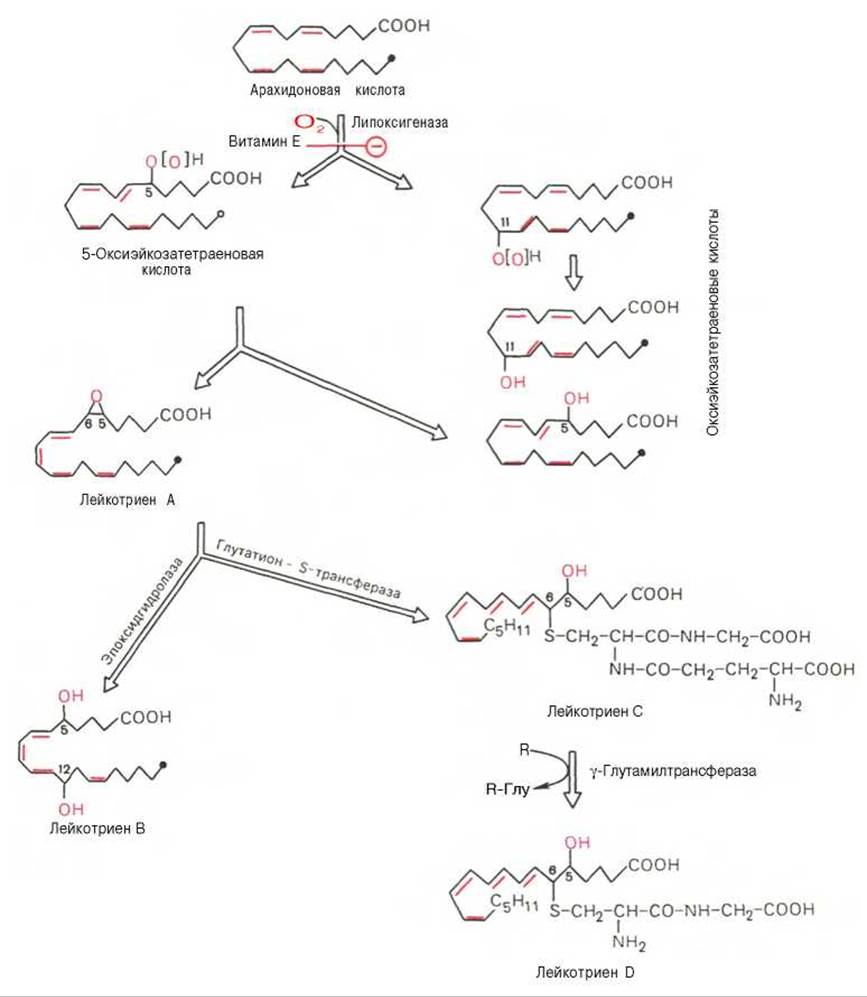
Fig. 8.4. Lipoxygenase pathway of arachidonic acid metabolism.
R is the glutamic acid residue acceptor. The symbol denotes the blocking effect of vitamin E.
Thus, owing to their widespread tissue distribution and high, versatile biological activity, prostaglandins (and prostanoids in general) and leukotrienes are finding increasingly broad application in medical practice as pharmaceutical agents. These circumstances stimulate further research both toward discovering novel prostanoids and toward the Chemical synthesis of their analogs with protected functional groups that are more stable upon administration into the organism.
Last update: 06/08/2026
Editorial and Educational Adaptation: This material has been compiled based on the primary/original source text. The project team performed an editorial review, corrected technical inaccuracies, structured sections, and adapted the content for an educational format.
What was processed:
- elimination of formatting defects (OCR errors, structural breaks, corrupted characters);
- editorial organization of content;
- standardization of terminology in accordance with academic sources;
- verification of factual statements against the original source text.
All mentions of the author, publication year, and origin of the primary text have been preserved in accordance with the source.