INVERTEBRATE ZOOLOGY IN THREE VOLUMES - BOOK 3 - G.I. Shcherbak - 1997
DEUTEROSTOMIA
PHYLUM ECHINODERMATA
Echinoderms are a highly distinctive animal group that appeared on Earth about 580 million years ago. Most of its species are now extinct (approximately 13,000 fossil species are known), while over 6,000 species exist today. All of them are exclusively marine benthic animals that are extremely sensitive to decreases in salinity. They inhabit the seas and oceans of all latitudes across the globe, ranging from the intertidal zone to the maximum depths of oceanic trenches.
Echinoderms are typically free-living animals with radial, mostly pentamerous Symmetry and elements of bilateral symmetry. Their bodies feature an oral side, which bears the Mouth opening, and an opposite aboral side. There is every reason to believe that radial symmetry is a secondary phenomenon, as evidenced by certain fossil forms, the bilateral symmetry of echinoderm larvae, and The Structure of several Organs in modern species.
The external body shape of echinoderms is quite unusual, taking the form of a flower, star, sphere, cucumber, and so on (Fig. 168). Sizes mostly range between 5 and 50 cm, although some smaller species exist, as well as those reaching several meters in length.
Class="center">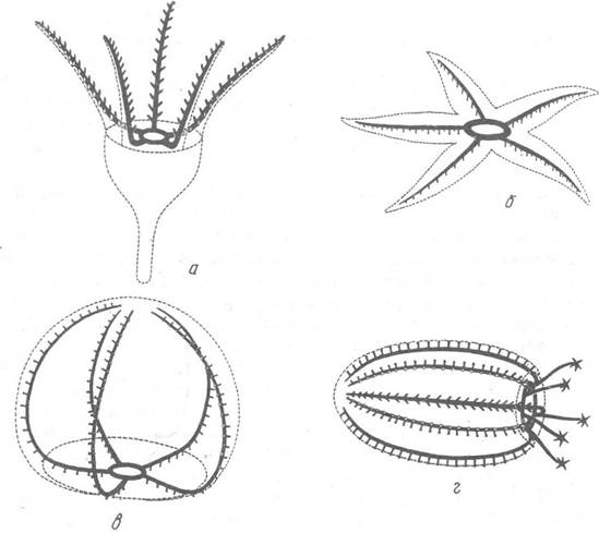
Fig. 168. Diagram of the body shape in echinoderms:
a — crinoids; b — sea stars; c — sea urchins; d — sea cucumbers
Representatives of the echinoderm classes differ significantly from one another; nevertheless, A number of features are common to all echinoderms and clearly set them apart from other animals.
Externally, echinoderms are covered by a single-layered ciliated epithelium (only sea cucumbers lack cilia), which contains numerous glandular Cells that secrete mucus, as well as sticky and poisonous secretions. Pigment cells are also present here, responsible for the diverse and striking coloration of many echinoderms. Beneath the epithelium lies a well-developed Connective Tissue layer (the cutis) of mesodermal origin, which is lined on the inside by a peritoneal epithelium delimiting the coelom. The internal Skeleton is formed within the plasma of the cells in the outer layer of the cutis as microscopic biycrystals consisting primarily of calcite with a minor organic component. These biycrystals grow, acquire a specific shape, and are extruded from the cells. Here they interconnect to form small plates of various shapes or fuse at their edges to create large spongy plates. Various outgrowths may form on their surface: spines (hence the name of the phylum), prickles, and specialized organs such as pedicellariae. Both simple and modified spines articulate with the skeleton via hemispherical socket joints, allowing for easy movement.
Musculature in most echinoderms is weakly developed; it is located primarily in the rays, where skeletal elements articulate with one another movably. Movable body outgrowths—such as tube feet, skeletal spines, and pedicellariae—are also equipped with Muscles. Only sea cucumbers possess a well-developed body wall musculature (cutaneomuscular sac). All echinoderm muscles are smooth.
Echinoderms are coelomate animals. Like other deuterostomes, their coelom develops as three pairs of coelomic pouches. In addition to the main body cavity, these pouches give rise to the canals of the ambulacral and perhaemal systems, the axial sinuses, the ampulla of the axial organ, and the genital strand.
The general body cavity achieves significant development in echinoderms, with two exceptions: in sea crinoids, its volume is greatly reduced due to the presence of numerous connective tissue strands, and in ophiuroids, due to The Development of specialized ectodermal bursal sacs that project into the body cavity. The coelom is lined with ciliated epithelium, which also covers The surface of all Internal Organs located within the coelom, and is filled with a transparent fluid whose composition is close to that of seawater, albeit with protein admixtures. This fluid also contains A large number of cellular elements (coelomocytes—14 types are distinguished) that participate in nutrient distribution, Respiration, excretion, and the defense of the Organism against Bacteria and parasites. Notably, it was experiments with sea star larvae that led I.I. Mechnikov to formulate The Theory of phagocytosis.
The body cavity also performs the mechanical function of providing structural support for the body walls, which lack a skeletal framework.
The most characteristic derivative of the coelom in phylum members is the ambulacral system. This is a system of thin-walled canals filled with fluid. Typically, the ambulacral system communicates with the external environment via a madreporite located on the body surface. Through numerous Pores in the madreporite, seawater passes via an ampulla into the stone canal (whose walls are impregnated with calcium carbonate), which connects to a ring canal surrounding the circumoral region of the digestive tract. Five radial canals extend blindly from the ring canal. Lateral branches of various structures extend in pairs from these radial canals to the muscular tube feet, or tentacles, which emerge on the exterior of the body. The walls of the canals consist of flagellated epithelium, connective tissue, a muscular layer, and an outer ciliated peritoneal epithelium. The details of the ambulacral system structure vary among representatives of different taxa.
The STRUCTURE OF THE Digestive System varies, and we will examine it in detail when studying each of the echinoderm classes.
Specialized excretory organs are absent. The organism is rid of Metabolic waste products by amoeboid cells present in the coelomic fluid, as well as in the circulatory and ambulacral systems. Cells laden with metabolic wastes are eliminated to the exterior through the body integuments or deposited in the connective tissue. The Chemical Nature of these products is not yet fully understood, but it is known that the excreta of echinoderms include Keratins and eratinin—compounds characteristic of vertebrate animals.
Characteristic of echinoderms is the perhaemal, or pseudohe haemal, system, which develops from isolated Regions of the general coelom; all its canals have walls lined with peritoneal epithelium. This system includes a circumoral ring canal lying between the ring ambulacral canal and the circumoral nerve ring, as well as five radial canals. It is closely associated with the Circulatory system and partially or entirely surrounds the latter. The perhaemal system contains the same fluid as the coelom, but with a slightly higher protein content. This system accompanies The Nervous system, underlying the nerve cords. It is believed to function in supplying them with nutrients and protecting them from deformation. Furthermore, the perhaemal system provides structural support for the circulatory system, which lacks walls of its own.
The CIRCULATORY SYSTEM OF echinoderms is characterized by a rather primitive Organization due to the absence of specialized musculature and Valves. There is no regular fluid Circulation within the circulatory system. The basis of the system consists of a circumoral lacunar ring, five radial lacunae, and the lacunae of the axial organ, intestine, and Gonads. The lacunae lie within the connective tissue and lack proper walls of their own. Only the dorsal and ventral intestinal Blood Vessels of sea cucumbers and sea urchins, which branch off from the circumoral ring, possess the character of true blood vessels. In structural detail, the circulatory system varies among representatives of different classes. The fluid of the circulatory system is compositionally similar to the coelomic and ambulacral fluids. The primary function of the circulatory system is The transport of nutrients, and only in sea cucumbers does it also perform a gas exchange function.
Specialized respiratory organs are weakly developed in echinoderms. These primarily include the respiratory trees (Water Lungs) of sea cucumbers, where intense gas exchange takes place, as well as the dermal branchiae (Skin gills) of sea urchins and sea stars. The thin-walled saclike bursae in ophiuroids mentioned above, the tube feet, and other thin-walled areas of the body surface also perform respiratory Functions. Individual cells possessing respiratory pigments, found within the coelomic and ambulacral fluids, participate in respiration.
The nervous system of echinoderms is quite peculiar: they lack a discrete central ganglion (Brain). It comprises three divisions or systems—the ectoneural, hyponeural, and apical (sometimes referred to as the endoneural or aboral system)—which are developed to varying degrees in representatives of different classes.
Each division consists of a nerve ring and radial nerve cords, the number of which corresponds to the number of radial ambulacral canals. In all echinoderms, the ectoneural nervous system is the best developed, positioned most superficially on the oral side of the body. In its simplest form, it appears as a diffuse subepidermal plexus with condensations of Nerve Cells and their processes forming ring and radial cords. Situated deeper within the body is the second, hyponeural system, which is also represented in nearly all echinoderms (except for sea urchins, in which it is greatly reduced or entirely absent).
On the aboral side lies the apical system, which achieves significant development in crinoids. In representatives of other echinoderm classes, it is more or less reduced or completely absent. The primary function of the ectoneural nervous system is sensory, while the other two are locomotory (regulating movement).
A distinctive feature of the Development of the echinoderm nervous system is The formation of nerve cells from ectodermal, mesodermal, and endodermal epithelium. The ectoneural nervous system is of ectodermal origin, the hyponeural and apical are mesodermal, and the nerve plexus present within the walls of the intestine is endodermal.
Echinoderms are highly sensitive to external stimuli, although they lack complex Sense Organs. Numerous sensory cells, or larger and smaller clusters of them located on the ambulacral feet, oral tentacles, skin, and other PARTS OF THE body, function as organs of Touch, smell, taste, and Vision.
Most echinoderms are dioecious animals. The gonads develop in close connection with the coelom. A genital sinus, in which a genital cord develops, extends from one of the coelomic rudiments. As it grows, the genital sinus forms the pouches of the gonads, while the genital cord forms the gonads themselves. These pouches fuse with the body wall, where genital pores are formed; these pores connect the gonads to the external environment and serve for the release of Gametes.
Echinoderms typically produce numerous small eggs with a low yolk content, which are released into the water where Fertilization takes place. Embryonic development mostly occurs in the water, though in some cases eggs are brooded in specialized brood chambers (in ophiuroids, holothurians, and sea urchins).
Cleavage in echinoderm eggs is total, more or less equal, and of the radial type. In rare cases, when the egg contains a large amount of yolk, cleavage approaches superficial cleavage (e.g., in the sea lily Isometra vivipara). In most echinoderms, the egg hatches into a free-swimming, ciliated blastula larva, marking the end of embryonic development. Gastrulation processes and the onset of Organogenesis take place during postembryonic development.
Even before the onset of gastrulation, mesenchyme cells migrate from the vegetal pole of the blastula into the blastocoel to later form the larval skeleton. The gastrula is formed through a combination of immigration and invagination; the gut rudiment (archenteron) appears as a narrow cylindrical invagination (Fig. 169, a, b). Following gastrulation, the blastopore shifts to the ventral side and transforms into the anus, while closer to the animal pole of the larva, an ectodermal invagination appears and connects with the anterior part of the entodermal gut rudiment, forming the mouth (secondary mouth, Fig. 169, c, d).

Fig. 169. Features of embryonic and postembryonic development in echinoderms:
a — immigration of mesenchyme cells into the blastocoel cavity; b — gastrula; c–d — Formation of the mouth and anus; e–g — successive stages of coelom development; 1 — larval mesenchyme cells; 2 — gut rudiment; 3 — blastopore; 4 — ectoderm; 5 — rudiment of the anterior coeloms; 6 — mouth; 7 — anus; 8 — spicule of the larval skeleton; 9 — rudiment of the posterior coeloms; 10 — pore canal; 11 — Separation of the middle coeloms; 12 — rudiment of the stone canal; 13 — transformation of the left middle coelom into the ring of the ambulacral system
The coelom forms enterocoelically: the upper wall of the archenteron forms an outpouching that pinches off to become a closed coelomic pouch. This primary coelomic pouch then divides into right and left coeloms, each of which, in turn, divides into three parts. As a result, the larva develops three pairs of coeloms: the first (I) at the animal pole, the second (II) in the middle, and the third (III) near the vegetal pole (Fig. 170). The first coelom on the left (and less frequently both) opens to the exterior via a pore. The First and Second left coeloms communicate with each other. Both third coeloms expand and transform into the general body cavity of the adult animal; the first and second right coeloms do not develop. The first left coelom gives rise to part of the axial sinus and the perihemal system, as well as the madreporite, which connects it to the external environment. The second left coelom (hydrocoel) serves as the rudiment of the ambulacral system, and the canal connecting the first and second coeloms becomes the stone canal.
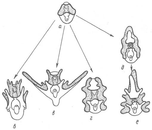
Fig. 170. Diagram of the formation of typical larval forms in echinoderms:
a — dipleurula; b — echinopluteus; c — ophiopluteus; d — auricularia; e, f — bipinnaria and brachiolaria
The integuments around the larval mouth become slightly depressed to form a circumoral cavity bordered by a ciliated band, which provides Nutrition for the larva and performs a locomotory function. At this stage, the larva is called a dipleurula, emphasizing its bilaterally symmetrical body plan.
Next, the contours of the circumoral cavity become more complex: lobes or long outgrowths (arms) bordered by a ciliated band appear along its margin. Through this complexification of the dipleurula structure, the characteristic larvae of various echinoderm classes arise (Fig. 170). The detailed structure of these larvae and their metamorphosis into adult animals will be examined in the descriptions of the respective classes.
Alongside sexual reproduction, cases of asexual reproduction are known, in which the body of sea stars, ophiuroids, and certain holothurians divides in half or into several parts. Each part then regenerates the missing body regions. The phenomenon of regeneration is highly characteristic of echinoderms. When escaping from predators, some echinoderms cast off body parts, viscera, or sometimes even break apart into pieces, after which the missing parts regenerate.
The phylum Echinodermata is divided into five classes grouped into three subphyla.
Last update: 13/08/2026
Editorial and Educational Adaptation: This material has been compiled based on the primary/original source text. The project team performed an editorial review, corrected technical inaccuracies, structured sections, and adapted the content for an educational format.
What was processed:
- elimination of formatting defects (OCR errors, structural breaks, corrupted characters);
- editorial organization of content;
- standardization of terminology in accordance with academic sources;
- verification of factual statements against the original source text.
All mentions of the author, publication year, and origin of the primary text have been preserved in accordance with the source.