PLANT MORPHOLOGY - T. A. Sautkina - 2012
CHAPTER 7. THE FLOWER AS A SPECIAL REPRODUCTIVE ORGAN OF ANGIOSPERMS
7.4. General Plan of Flower Structure
The flowers of angiosperms vary immensely in Structure, shape, and size, yet they share distinct common features (Fig. 150). A typical flower consists of a receptacle, a perianth (which is sometimes absent), stamens, and pistils. Flowers that bear both stamens and pistils are termed bisexual, whereas those containing only one of these elements are called unisexual (Fig. 151). Very rarely, both stamens and pistils are entirely absent (such as the funnel-shaped sterile flowers of cornflower — Centaurea cyanus).
Class="center">Fig. 150. Generalized diagram of a bisexual flower structure: 1—receptacle; 2—sepals of the calyx; 3—petal of the corolla; 4—stamen; 5—anther; 6—Ovary of the pistil; 7—style of the pistil; 8—stigma of the pistil; 9—ovules within the ovary

Fig. 151. Unisexual flowers: A—papaya (Carica papaya); B—starwort (Callitriche palustris); C—wax begonia (Begonia semperflorens): 1— male flower; 2—female flower; 3—stamens; 4—pistil; 5—calyx; 6—corolla; 7—upper leaves; 8—inferior ovary of the flower

The receptacle (Lat. receptaculum) is the axial part of the flower that bears all its other floral Organs. The receptacle may be elongated and conical—which confirms its axial origin—or shortened and flat. A conical receptacle is found in members of primitive families such as Magnoliaceae and Ranunculaceae. In most angiosperms, however, the receptacle is flat, while in members of the Rosaceae family it is saucer-shaped and expanded (wild strawberry — Fragaria vesca) or cup-shaped and concave (common plum — Prunus domestica), in which case it is called a hypanthium.
The floral organs are arranged on the receptacle in a definite, orderly sequence.
All floral organs can be divided into sterile (those not directly involved in reproductive processes) and fertile (those where reproduction takes place). The sterile organs include the perianth, while the fertile ones comprise the stamens and pistils.
The perianth, or floral envelope (Lat. perianthium), may be either double or simple, depending on its structure.
A double perianth consists of a calyx (Lat. calyx) and a corolla (Lat. corolla).
The calyx is formed by sepals, which are foliar in nature, as confirmed by anatomical data. Sepals typically possess three leaf traces—the exact same number that is characteristic of vegetative leaves. Sepals may remain free from one another, forming a polysepalous calyx (meadow buttercup — Ranunculus acris, marsh stitchwort — Stellaria graminea). Alternatively, when sepals are fused to a greater or lesser extent, they form a gamosepalous calyx (corncockle — Agrostemma githago, germander speedwell — Veronica chamaedrys). Sepals are most frequently green, but they can also exhibit other colors: yellow (meadow buttercup), blue (tall larkspur — Delphinium grandiflorum), orange (Asian globeflower — Trollius asiaticus), or white, pink, and violet (garden hydrangea — Hydrangea hortensis). During the bud stage, sepals perform a protective function; after the bud opens, green sepals participate in Photosynthesis, whereas brightly colored ones help attract insect pollinators.
Situated behind the calyx is the corolla, which is composed of petals. Like the calyx, the corolla can be polypetalous (marsh stitchwort — Stellaria graminea, biting stonecrop — Sedum acre) or sympetalous (canterbury bells — Campanula media, large-flowered foxglove — Digitalis grandiflora). The morphological nature of petals has been interpreted differently by various authors. According to some (J. W. Goethe and his followers), petals originated, like sepals, from foliage leaves, whereas other researchers (A. P. de Candolle et al.) considered them to be derivatives of stamens. E. Arber, J. Parkins, B. M. Kozo-Polyansky, and A. Ems suggested that petals may have a dual origin. This is supported by vascular anatomy data. For instance, in certain ranunculids, petals—like stamens—have only a single leaf trace, whereas in members of the Magnoliaceae and Schisandraceae families, petals, like sepals, possess three leaf traces. Petals perform a protective function and also serve to attract insect pollinators, a task facilitated not only by their bright coloration but also by The formation of nectaries of various structures either directly on the petals (Water crowfoot — Batrachium) or derived from them (monkshood — Aconitum, columbine — Aquilegia).
If the perianth is uniformly colored, it is termed simple. Depending on its coloration, a simple perianth can be sepaloid (greenish, as in garden beet — Beta vulgaris, or lamb's quarters — Chenopodium album) or petaloid (brightly colored, as in poet's narcissus — Narcissus poeticus, or autumn crocus — Colchicum autumnale). Occasionally, however, a simple perianth formed by sepals can be petaloid in appearance (wolfsbane — Aconitum napellus, larkspur — Consolida regalis).
The coloration of perianth elements is determined by various pigments. The primary pigments in flowers are Flavonoids (anthocyanins and flavones); they are water-soluble and located in The Cell sap of vacuoles. Depending on the pH of the cell sap, flavonoids change color: for example, the anthocyanin cyanidin appears red in an acidic environment, violet in a neutral environment, and blue in an alkaline one. A yellow hue is imparted to floral organs by flavones (such as lutein) and carotenoids. Carotenoids are lipid-soluble and reside within Plastids known as chromoplasts. The brown pigment—anthophaein—forms dark or brown spots on the petals of certain legumes (such as the broad bean) and many tropical orchids. The mixing of pigments across various pH levels produces the entire spectrum of floral colors. In some plants (such as lungworts and forget-me-nots), the corolla changes color during flowering due to shifts in cell sap pH. White flowers contain no pigments; their color is due to the presence of large, air-filled intercellular spaces within the Tissues of the petals and sometimes the sepals (such as Clerodendrum thomsoniae), which efficiently reflect sunlight.
The most vital reproductive Organs of the flower are the stamens and pistils.
7.4.1. Androecium and Its Types. Evolution and STRUCTURE OF THE Stamen
The aggregate of stamens constitutes the androecium of the flower. Depending on their arrangement, the androecium can be monadelphous, diadelphous, or polyadelphous. In a monadelphous androecium, all stamens are united into a single group by their filaments (cut-leaf mallow — Malva excisa, large-leaved lupine — Lupinus polyphyllus) or by their anthers (Asteraceae). When stamens are fused into two groups, the androecium is termed diadelphous (tufted vetch — Vicia cracca, field pumpkin — Cucurbita pepo). If several groups of fused stamens are formed (St. John's wort — Hypericum perforatum) or if all stamens remain free (eastern pasque flower — Pulsatilla patens), the androecium is considered polyadelphous. The androecium may also be didynamous—where a flower has four stamens, two of which are long and two short (Lamiaceae family)—or tetradynamous, featuring four long stamens and two short ones (Brassicaceae family) (Fig. 152).
Fig. 152. Types of androecium in representatives of various angiosperm families: 1—polyadelphous didynamous (Lamiaceae family); 2—polyadelphous tetradynamous (Brassicaceae family); 3—monadelphous (Malvaceae family); 4—monadelphous (Asteraceae family); 5—diadelphous (Fabaceae family)

A stamen consists of a filament, an anther formed by two halves (two thecae), and a connective—a parenchymatous tissue connecting the anther halves. The stamen represents a reduced microsporophyll, of which the filament and connective are remnants, bearing microsporangia located within the anther.
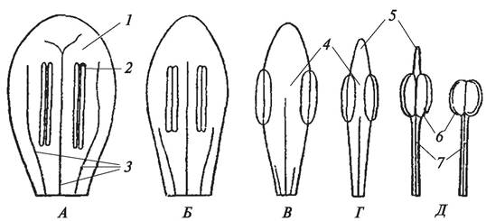
Comparative studies of stamens across various angiosperm taxa have provided a clear picture of the evolution of this reproductive organ (Fig. 153). In modern primitive angiosperms (especially in the genus Degeneria and certain magnolias), stamens have a very archaic structure: they are flat, broad, and undifferentiated into a filament, anther, and connective, bearing linear microsporangia arranged laminally—that is, directly On the surface of the laminar microsporophyll. The anthers are positioned either on the adaxial (inner) side, in which case they dehisce introrsely, or on the abaxial (outer) side, where they dehisce extrorsely. Primitive microsporophylls contain three well-developed vascular bundles (three Veins). Gradually, over the course of evolution, the microsporophyll underwent reduction: the number of vascular bundles in the mesophyll decreased, much of the lamina disappeared, and ultimately a specialized stamen differentiated into a filament, anther, and connective was formed. The filament contains a single vascular bundle that extends up to the connective, supplying nutrients to its living parenchymatous Cells. As the stamen differentiates, THE POSITION OF the anther changes. Gradually, the anther halves shift to a lateral position and then, As a result of further reduction of the connective, fuse into an anther composed of two halves (two thecae) that occupies a terminal position. Thus, in the majority of angiosperms, the anther contains four microsporangia and Functions as a synangium (an aggregate of sporangia).
Fig. 153. Evolutionary Stages of the microsporophyll from the primitive laminar type of Degeneria to the highly specialized stamen: A–D — reduction in microsporophyll size, vein reduction, and changes in microsporangia position; E — structure of the stamen: 1—microsporophyll; 2—microsporangia; 3—microsporophyll veins; 4—developing connective; 5—developing apical extension of the connective (superconnective); 6—microsporangia (anthers); 7—filaments
In some plants, as an adaptation to pollination, a portion of the stamens lose their anthers and transform into nectaries called staminodes (e.g., bogbean or bog star-flower — Parnassia palustris, globeflower — Trollius europaeus).
The number of stamens in bisexual flowers varies widely, ranging from indefinitely large (families Ranunculaceae, Magnoliaceae) down to two (germander speedwell — Veronica chamaedrys) or even a single stamen (lesser butterfly-orchid — Platanthera bifolia). In unisexual staminate flowers, the stamen count also varies (see Fig. 151, B, C; 159, B).
Stamens within a flower are arranged either in a spiral (families Magnoliaceae, most representatives of Ranunculaceae) or in whorls (families Rosaceae, Caryophyllaceae).
7.4.2. Gynoecium and Its Types. Evolution of the Carpels, FORMATION AND STRUCTURE of the Pistil
The central part of the flower is occupied by one or more pistils. The aggregate of pistils or fused carpels constitutes the gynoecium of the flower.
The pistil is a closed, modified, and highly specialized macrosporophyll (megasporophyll, carpel) that encloses the ovules within. It is formed by the fusion of the carpel margins and is differentiated into a hollow ovary, stylodia (stylodium — the narrowed portion of the carpel) or a style (stylos — a structure resulting from the fusion of several stylodia), and a stigma.
The foliar nature of carpels was scientifically established by A. P. de Candolle as early as 1827, although he mistakenly derived them from vegetative rather than sporogenous leaves. Indeed, the more primitive the flower, the more leaf-like the individual carpels appear, and their Vascular System resembles that of a leaf. According to A. L. Takhtajan's concept, the angiosperm carpel differs from an open, unfolded macrosporophyll in being folded along its midrib, i.e., conduplicate (Fig. 154).
Fig. 154. Stages of carpel (macrosporophyll) evolution: A — primitive conduplicate carpel of the Degeneria type with a stigmatic crest; B, C — stages of carpel margin fusion and reduction of the stigmatic crest; G, D — stages of pistil formation; E — formed pistil: 1 — conduplicate carpel;
2 — stigmatic crest; 3 — ovary of the pistil; 4 — stylodium; 5 — capitate stigma

The ovary of the pistil contains ovules. Due to the conduplicate Nature of the carpel, the ovules—whose nucellus functions as a macrosporangium—come to lie on the adaxial (inner) side, which enhances their protection against various adverse environmental factors (air dryness, high and low temperatures, abrupt Temperature fluctuations, insect damage, etc.).
Gradually, the closely appressed margins of the conduplicate carpel began to fuse, ultimately resulting in the formation of a typical closed pistil. Among extant angiosperms, the most archaic pistils are found in Degeneria, where each pistil consists of a carpel fused only at the base. The upper margins of the carpel are not only unfused but actually do not even Touch one another (Takhtajan, 1964). Incomplete fusion of carpel margins is also observed in some more advanced angiosperms (peony — Paeonia, plane tree — Platanus). In Resedaceae (wild mignonette — Reseda lutea), the apex of the ovary remains open.
Since the ovules in a conduplicate carpel became enclosed and inaccessible to pollen, a specialized surface capable of receiving pollen had to evolve. Thus, special glandular tissue gradually began to form along the margins of the conduplicate carpels in angiosperms, giving rise to the stigmatic surface, or stigmatic crest. The presence of a stigmatic crest is one of the most Characteristic Features of macrosporophylls in primitive angiosperms. Initially, the stigmatic crest extended along both margins of the folded, broad carpel, but as the margins fused, it shortened, changed shape, and ultimately transformed into the pistil stigma, which assumed a terminal position. During evolution, the carpel itself differentiated, gradually transforming into a typical pistil. Its upper part elongated and turned into a stylodium—a sterile structure. In primitive angiosperms, the stylodium is still conduplicate, bearing two stigmatic crests, but its margins gradually fuse, and a typical stigma forms at the apex.
A pistil may be formed by a single carpel or several fused carpels. A gynoecium in which each pistil is formed by a single unfused carpel is termed apocarpous. An apocarpous gynoecium may consist of a single pistil (field bean — Faba vulgaris, sour cherry — Cerasus vulgaris) or of several to many pistils (families Magnoliaceae, most Ranunculaceae). Thus, an apocarpous gynoecium can be polycarpellary or unicarpellary. A polycarpellary apocarpous gynoecium is characteristic of primitive families, whereas a unicarpellary one occurs in evolutionarily more advanced groups.
In the course of evolution, The structure of the gynoecium grew more complex. Through the fusion of multiple carpels, a syncarpous (coenocarpous) gynoecium developed. The number of fused carpels in a coenocarpous gynoecium ranges from two (families Cruciferae, Gramineae) to an indefinitely large number (mallow — Malva).
Depending on the pattern of carpel fusion, the coenocarpous gynoecium is subdivided into syncarpous, paracarpous, and lysicarpous types.
In a syncarpous gynoecium, the carpels fuse completely by their lateral (side) walls, forming a multilocular ovary whose number of locules corresponds to the number of fused carpels (two in black henbane — Hyoscyamus niger, five in cultivated flax — Linum usitatissimum).
In a paracarpous gynoecium, the carpels fuse in such a way that the ovary remains unilocular (families Papaveraceae, Cruciferae, Cucurbitaceae). In a lysicarpous gynoecium, the ovary is also unilocular, but it originates differently than in the paracarpous type. During the formation of a lysicarpous gynoecium, the partitions (walls of the fused carpels) break down, while a central Column—derived from the central part of the fused carpels—remains in the center of the ovary (family Caryophyllaceae).
The evolutionary relationships among the various types of gynoecium can be represented as follows (Fig. 155).
Fig. 155. Diagram of the evolution of the main gynoecium types (after Takhtajan, 1964, with additions): A — polycarpellary apocarpous gynoecium: 1 — ovary wall; 2 — ovary locule; 3 — central vascular bundle of the carpel; 4 — ovules; B — unicarpellary apocarpous gynoecium; C — syncarpous gynoecium; D — variants of paracarpous gynoecium; E — variants of lysicarpous gynoecium

During The Development of various coenocarpous gynoecium types, the stylodia either remain completely free (caraway — Carum carvi, common chickweed — Stellaria media) or partially fuse (apple — Malus domestica). In many species, complete fusion of the stylodia results in the formation of styles (wild radish — Raphanus raphanistrum, speedwell — Veronica officinalis).
7.4.3. METABOLISM/2.html">THE CONCEPT OF the Ovule. Structure and Classification of Ovules
The ovule is a specialized structure in gymnosperms and angiosperms consisting of a macrosporangium (megasporangium) protected by sterile protective envelopes, the integuments (Fig. 156).
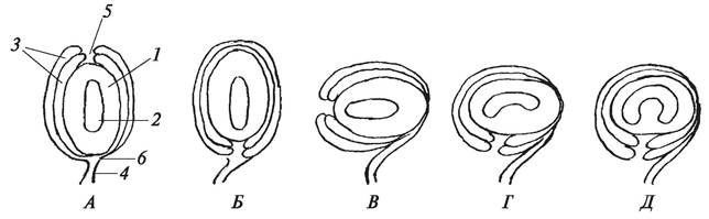
Fig. 156. Diagram of the STRUCTURE AND TYPES of ovules: A—atropous (or orthotropous) — straight: 1 — nucellus (macrosporangium); 2—embryo sac; 3—integuments; 4— funiculus; 5—micropyle; 6—chalaza; B—anatropous — inverted; C—hemitropous — half-turned; D—campylotropous — curved to one side; E—amphitropous — bilaterally curved
The term "ovule" (German: *Samenknospe*), introduced by J. W. Goethe in 1790 in his famous work *Metamorphosis of Plants* (*Versuch die Metamorphose der Pflanzen zu erklären*), has long been traditional. However, since the mid-19th century, plant morphologists and embryologists have used the concept of "ovule" (*semyazachatok* in Russian) in parallel with this term, thereby emphasizing that ultimately a seed develops from the ovule.
The macrosporangium is the central part of the ovule, known as the nucellus. Unlike the macrosporangia of HIGHER SPORE PLANTS, the nucellus is a massive, multicellular structure reliably protected by integuments and the walls of the pistil ovary. The structure of the nucellus in most studied species has been investigated predominantly at the light-optical level, which has prevented the identification of A number of essential features.
Electron microscopic studies have shown that the growing nucellus consists of meristematic cells interconnected by plasmodesmata. Based on the degree of Development of the nucellus, P. van Tieghem in 1898 divided ovules into two types: crassinucellate and tenuinucellate. Crassinucellate ovules are characterized by a massive, multi-layered nucellus. As electron microscopic studies have shown, the massive nucellus is histologically differentiated into an epidermis externally covered with a cuticle layer, and a parenchymatous tissue. In most flowering plants, the epidermis is single-layered because the meristematic cells forming the epidermis divide only anticlinally. At the same time, in representatives of certain families (e.g., Asparagaceae), meristematic cells can divide periclinally, forming a multi-layered nucellar cap in the micropylar region of the nucellus.
In tenuinucellate ovules, the nucellus is poorly developed. In the epidermal cells of this ovule type, plasmodesmata have been found only on the radial walls.
The Base of the ovule adjacent to the ovary wall is called the chalaza. In most angiosperms, a vascular bundle extending from the ovary into the ovule terminates in the chalaza, suggesting that the chalaza performs a trophic function by transferring metabolic products from the ovary to the ovule, as well as a protective function.
Extending from the chalaza is the stalk of the ovule, or funiculus, by means of which the ovule is attached to the ovary wall, along with the integuments. The funiculus features a well-developed conducting system through which solutions of organic and Mineral Substances are supplied from the pistil ovary to the tissues of the ovule.
The integuments protect the nucellus of the ovule and the structures developing within it from adverse conditions. Integuments vary in number and degree of development among different plants. According to the number of integuments, angiosperm ovules are classified as bitegmic (double-integumented), unitegmic (single-integumented), and ategmic (naked). Bitegmic ovules are characteristic of monocots and many dicots, unitegmic ones of sympetalous dicots, and ategmic ones are found in certain parasitic plants.
The integuments grow around the nucellus but do not fuse at the pole opposite the chalaza, resulting in the formation of a canal known as the micropyle.
Thus, the ovule is a polarized structure with two distinct poles: the micropylar and the chalazal.
The structure of ovules varies among different plants, which is reflected in their morphological classification. Depending on the position of the micropyle relative to the chalaza, ovules are distinguished as atropous or orthotropous (straight), anatropous (inverted), hemitropous (semi-inverted), campylotropous (curved), and amphitropous (transversely curved) (see Fig. 156).
In atropous ovules, the micropyle and chalaza lie on the same vertical straight line; in anatropous ovules, the micropyle and chalaza lie on the same horizontal straight line (on the same side); in hemitropous ovules, the micropyle is rotated by 90° relative to the chalaza. In atropous, anatropous, and hemitropous ovules, the nucellus is not curved. In campylotropous and amphitropous ovules, the nucellus is curved to varying degrees. Today, embryologists use a more detailed classification of ovules.
The shape of the ovule, the presence or absence of coverings, and The Nature of the nucellus are of major systematic and phylogenetic importance.
7.4.4. Types of Placentation
Placentation is the pattern of arrangement of placentas—the sites of attachment of ovules to the walls of the pistil ovary. Depending on the structure of the gynoecium, two MAIN TYPES OF placentation are distinguished: laminal (surface) and sutural.
In laminal placentation, the ovules are attached along the inner surface of the ovary. The most primitive type of laminal placentation is diffuse (laminal-diffuse), in which ovules are scattered across the entire adaxial surface of the ovary (flowering rush — *Butomus umbellatus*) (Fig. 157, B, 6).
In sutural placentation, ovules are attached either to the dorsal suture (the midrib of a fused carpel) or to the ventral suture (the fused margins of a carpel). Sutural placentation can be subdivided into median (laminal-median), lateral (laminal-lateral), axile, parietal, and free-central (columnar) (Fig. 157).
Fig. 157. Diagrams of Different types of ovule placentation: A—in various types of coenocarpous gynoecium; B—in apocarpous gynoecium: 1—axile (syncarpous gynoecium); 2—parietal (paracarpous gynoecium); 3—free-central (lysicarpous gynoecium); 4—parietal-dorsal (median); 5—parietal-marginal (ventral); 6—diffuse-laminal; 7—dorsal suture; 8—ventral suture

In an apocarpous gynoecium, placentation can be median or lateral. In median placentation, ovules are located along the midrib of the carpel (lotus genus — *Nelumbo*); in lateral placentation, they are located along the ventral suture (garden pea — *Pisum sativum*).
Axile placentation is characteristic of a syncarpous gynoecium. In this type of placentation, ovules are attached at the angles of fused carpels (tiger lily
— *Lilium tigrinum*, common pear — *Pyrus communis*). In a paracarpous gynoecium, placentation is parietal, but ovules may be attached in different ways. If they are located along the margins of fused carpels, parietal placentation is termed lateral (marginal-parietal); if along the midrib, it is termed median (dorsal-parietal) (wild pansy — *Viola tricolor*, cucumber — *Cucumis sativus*).
In a lysicarpous gynoecium, ovules are attached to a central column, in which case placentation is free-central (pink — *Dianthus*, soapwort — *Saponaria*).
During the development of the gynoecium, the Placenta can proliferate extensively and form false septa within the ovary (Brassicaceae family).
The number of ovules in both apocarpous and coenocarpous gynaecia varies from many to just one. The reduction in the number of ovules within a carpel ovary, alongside a decrease in the number of carpels and a more complex carpel structure, is a result of evolutionary specialization across various plant families.
7.4.5. Position of the Ovary in the Flower
The varying positions of the ovary within a flower, determined relative to the perianth (Fig. 158), are also a product of evolution. If the perianth is attached below the ovary, the ovary is termed superior. Such an ovary is attached to the receptacle solely by its base (genera such as strawberry — Fragaria, buttercup — Ranunculus, rye — Secale). If the perianth is situated above the ovary, the ovary is inferior (families Campanulaceae, Umbelliferae, Iridaceae). The ovary can also be semi-inferior, in which case the perianth is attached around its middle (Tartarian honeysuckle — Lonicera tatarica). In sour cherry (Cerasus vulgaris) and sweet cherry (Cerasus avium), the free ovary is deeply sunken into the receptacle, or hypanthium. Such an ovary is considered half-superior (or intermediate). Sometimes the terms "semi-inferior" and "half-superior" are used synonymously. The position of the ovary in the flower is an important taxonomic feature. The superior ovary is considered the most primitive type.
Fig. 158. Position of the ovary in the flower: A—superior ovary (hypogynous flower); B—semi-inferior ovary (perigynous flower with half-inferior ovary); C—half-superior ovary (perigynous flower); D—inferior ovary (epigynous flower): 1—receptacle; 2—calyx sepal; 3—corolla petal; 4—hypanthium (receptacle of rosaceous plants); 5—stamen

7.4.6. Flower Classification
Structural diversity allows flowers to be classified using the following criteria: presence and structure of the perianth; Symmetry type; Structural Features of the perianth elements; floral sexuality; position of the ovary; and arrangement of floral elements on the receptacle.
Based on the presence of a perianth, flowers are distinguished as achlamydeous (Fig. 159), achlamydeate (apochlamydeous), haplochlamydeous, and diplochlamydeous.
Fig. 159. Flowers without a perianth (achlamydeous): A, B—bisexual flowers of marsh calla (Calla palustris) and common ash (Fraxinus excelsior), respectively; C, D—unisexual flowers of goat willow (Salix caprea): 1—pistils; 2—stamens; 3—bracts; 4—nectaries

Achlamydeous, or naked, flowers lack a perianth and are primarily devoid of floral envelopes. Plants with such flowers are found among both insect-pollinated (genus willow — Salix) and wind-pollinated taxa (genera sedge — Carex, ash — Fraxinus).
In apochlamydeous flowers (such as the female flowers of birch), the perianth is also absent; however, since male flowers possess one, its absence in female flowers is viewed as the result of reduction and, consequently, as a secondary feature.
In haplochlamydeous flowers, the perianth elements are arranged in a single whorl. Such a perianth is simple and may be sepaloid (green) (stinging nettle — Urtica dioica) or petaloid, exhibiting various colors (marsh marigold — Caltha palustris, eastern pasqueflower — Pulsatilla patens).
In diplochlamydeous flowers, the perianth elements are arranged in two whorls. Depending on the coloration of the outer and inner whorl elements, the perianth is categorized as homochlamydeous or heterochlamydeous.
In a homochlamydeous perianth, the segments are uniformly colored, meaning the perianth is simple (wood tulip — Tulipa sylvestris, garden onion — Allium cepa, hairy wood-rush — Luzula pilosa). Depending on its color, a homochlamydeous perianth can be sepaloid (Norway maple — Acer platanoides) or petaloid (wood tulip — Tulipa sylvestris). In a heterochlamydeous perianth, the calyx and corolla are clearly distinguishable, making the perianth double (water forget-me-not — Myosotis palustris, bird cherry — Padus racemosa).
Based on symmetry type, flowers are classified as actinomorphic (regular), zygomorphic (irregular), and asymmetrical.
Symmetry refers to proportion, specifically The ability to divide a flower via longitudinal axes (planes of symmetry) into mirror-image halves. A flower is termed actinomorphic (Fig. 160) if multiple planes of symmetry can pass through it (eastern pasqueflower — Pulsatilla patens, silvery cinquefoil — Potentilla argentea). Only a single plane of symmetry can pass through a zygomorphic flower, dividing it into just two equal parts (snapdragon — Antirrhinum majus, monkshood — Aconitum napellus) (Fig. 161). An asymmetrical flower lacks any plane of symmetry (Indian shot — Canna indica) (Fig. 162).
Fig. 160. Actinomorphic flowers: A—diagram of structure (1—planes of symmetry); B—wild radish (Raphanus raphanistrum); C—meadow buttercup (Ranunculus acris); D—tiger lily (Lilium tigrinum)
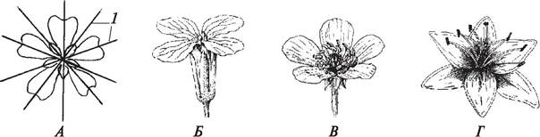
Fig. 161. Zygomorphic flowers: A—diagram of structure (1—plane of symmetry); B—rocket larkspur (Consolida regalis); C—snapdragon (Antirrhinum majus);
D—monkshood (Aconitum napellus)
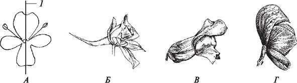
Fig. 162. Asymmetrical flowers: A—Indian shot (Canna indica); B—arrowroot (Maranta arundinacea): 1—inferior ovary; 2—sepals; 3—petals; 4—stamen; 5—staminodes; 6—pistil stigma
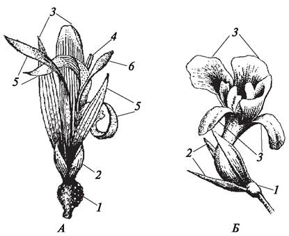
The structure of the perianth is also determined by how its elements are interconnected. The calyx may be chorisepalous (polysepalous) or gamosepalous (synsepalous), while the corolla may be choripetalous (polypetalous) or gamopetalous (sympetalous). Fusion can occur in both simple (lily of the valley — Convallaria majalis) and double perianths. In a double perianth, the calyx may be synsepalous while the corolla is polypetalous (corn cockle — Agrostemma githago, bladder campion — Silene vulgaris), or the elements of both whorls may be fused (snapdragon — Antirrhinum majus, potato — Solanum tuberosum).
The sex of a flower is determined by its reproductive organs—the stamens and pistils. Depending on the presence of these organs, flowers are classified as bisexual (hermaphroditic) or unisexual (diclinous). Bisexual flowers contain both stamens and pistils (e.g., meadow buttercup — Ranunculus acris, garden pea — Pisum sativum). Unisexual flowers possess only one type of organ—either stamens or pistils. Because the stamen determines the male sex (as reproductive processes culminate in the formation of male Gametes, or sperm cells), flowers containing only stamens are called male, or staminate. The pistil determines the female sex (since reproductive processes culminate in the Formation of the female gamete, or egg cell); therefore, flowers containing only pistils are called female, or pistillate (see Fig. 151, 159). In most plants, the sexual type of a flower is constant. However, in representatives of certain families, variations in floral structure and their arrangement on the plant are observed. For instance, in the Apiaceae family, both male and bisexual flowers may occur on the same plant (individual). This phenomenon is known as andromonoecy. Many Asteraceae exhibit gynomonoecy—the formation of female and bisexual flowers within a single flower HEAD. In some species (e.g., false helleborine — Veratrum lobelianum, bistort — Bistorta major), male and bisexual flowers develop on different individuals, manifesting androdioecy. Representatives of the Lamiaceae and Caryophyllaceae families typically feature bisexual flowers, yet gynodioecy is also common, where female and bisexual flowers develop on separate plants. In gynodioecy, Sexual Dimorphism often affects flower size: bisexual flowers are generally larger than unisexual ones.
Because certain species possess unisexual flowers that may develop on the same or separate individuals, The concepts of "monoecious" and "dioecious" plants arose. In monoecious plants, male and female flowers are located on the same individual (cucumber — Cucumis sativus, castor bean — Ricinus communis, corn — Zea mays). Dioecious plants are those in which male and female flowers develop on different individuals (sorrel — Rumex acetosa, goat willow — Salix caprea, hemp — Cannabis sativus). Polygamous plants, which bear male, female, and bisexual flowers, are very rare (e.g., Chinese magnolia vine — Schizandra sinensis), bearing both unisexual and bisexual flowers on a single individual.
An important characteristic considered in flower classification is the position of the ovary. Depending on the ovary position, flowers are categorized as hypogynous with a superior ovary (e.g., Fabaceae, Solanaceae), epigynous with an inferior ovary (e.g., Apiaceae, Asteraceae), and perigynous with a half-inferior or intermediate ovary (e.g., Caprifoliaceae) (see Fig. 158).
The elements of a flower are arranged on the receptacle in various ways, leading to three distinct floral types: acyclic (spiral), cyclic (whorled), and hemicyclic (spirocyclic). In acyclic flowers, all elements are arranged in a spiral (southern magnolia — Magnolia grandiflora, globeflower — Trollius europaeus). In cyclic flowers, all elements are arranged in whorls (maiden pink — Dianthus deltoides, large yellow foxglove — Digitalis grandiflora). In hemicyclic flowers, the perianth elements are arranged in whorls, whereas the stamens and pistils are arranged in a spiral (maunsegirdle — Myosurus minimus, creeping buttercup — Ranunculus repens).
The combination of floral traits allows one to assess whether a flower is primitive or advanced. For each trait, the presumed evolutionary direction can be outlined. For example, a simple perianth is considered more primitive than a double one. Free tepals and free petals evolved earlier than fused perianth segments (connation and sympetaly). A large number of stamens and pistils is a more primitive trait than their reduced, fixed number, and so on. However, it is evident that the development of floral traits in various angiosperm families occurred in parallel and independently of one another. Consequently, highly organized families often combine advanced organizational features with primitive traits in their flowers. This phenomenon is known as heterobathmy. For instance, in Rosaceae, progressive structural features include the cyclic arrangement of floral elements, the presence of an inferior ovary, and a syncarpous gynoecium in certain representatives. At the same time, many Rosaceae retain a superior ovary, an apocarpous polycarpellary gynoecium, and an aggregate follicle fruit. Very frequently, simplification of floral structure is not a sign of primitiveness, but rather the result of adaptation to specific environmental conditions.
7.4.7. Floral Formulas and Diagrams
Floral structure is the most crucial diagnostic feature of angiosperms. A flower can be characterized not only descriptively, but also through formulas and diagrams.
A floral formula is a concise representation of a flower's structure using symbols and numerical indices. The symbols are derived from the initial letters of the Latin names of all floral parts:
P — Perigonium — simple perianth;
K — Lat. Calyx, Ger. Kelch — calyx;
С — Corolla — corolla;
A — Androeceum — androecium;
G — Gynoeceum — gynoecium.
The type of floral symmetry is indicated by the following symbols:
«*» — actinomorphic (regular, polysymmetric) flower;
«↑» — zygomorphic (irregular, monosymmetric) flower.
Numbers beside the symbols indicate the count of each floral element (sepals, petals, stamens, pistils, or fused carpels).
The symbol «∞» denotes an indefinitely large number of elements (more than 12). The tilde symbol «~» is used when the number of elements varies across different flowers of the same species (variable or unstable count); «+» indicates that homologous floral elements (sepals, petals, stamens) are arranged in separate whorls. Parentheses ( ) enclose fused elements. Commas are used when necessary to separate elements within the same whorl if they exhibit specific morphological differences.
A line placed below, above, or beside the number characterizing the gynoecium indicates the position of the ovary: 2_ — superior ovary, (5&mac5;) — inferior, 1¬ — intermediate or half-inferior.
Special symbols can indicate the sex of the flower: «♂» — male; «♀» — female.
All floral elements are entered into the formula in a specific sequence, from the periphery to the center, matching their actual arrangement on the receptacle.
Examples of floral formulas:

While formulas provide a comprehensive characterization of a flower, they do not reflect the spatial arrangement of its elements, the specific type of simple perianth, or the exact subtype of coenocarpous gynoecium. These details are provided by a floral diagram.
A floral diagram is a graphic representation of a flower constructed as a projection of a bud's transverse cross-section onto a two-dimensional plane; consequently, the overall outline of an actinomorphic versus a zygomorphic flower will differ (Fig. 163). Standard conventions are adopted for each floral part. Sepals are typically designated by crescent-shaped brackets with a keel, and petals by plain brackets. Stamens and pistils, if few in number, can be drawn to reflect the shape of their cross-section. When stamens and pistils are numerous, they are depicted as small circles. When constructing a diagram, the relative spatial arrangement of all floral elements must be strictly maintained. Petals generally alternate with sepals.
Fig. 163. Floral diagrams: A, B—actinomorphic flowers; C, D—zygomorphic flowers: A—grass lily (*Ornithogalum umbellatum*); B—primrose (*Primula vulgaris*); C—rocket-larkspur (*Consolida orientalis*); D—broad bean (*Vicia faba*)

In cyclic flowers, stamens are arranged in a strictly regular pattern. In most angiosperms, the stamens of the outer whorl alternate with the petals of the corolla, an arrangement known as diplostemony. Less common is obdiplostemony, where the stamens of the outer whorl are positioned opposite the petals (Caryophyllaceae, Primulaceae, and sugar beet (*Beta vulgaris*)).
Having both a FLORAL FORMULA AND a diagram allows for a comprehensive Description of the flower.
Last update: 07/08/2026
Editorial and Educational Adaptation: This material has been compiled based on the primary/original source text. The project team performed an editorial review, corrected technical inaccuracies, structured sections, and adapted the content for an educational format.
What was processed:
- elimination of formatting defects (OCR errors, structural breaks, corrupted characters);
- editorial organization of content;
- standardization of terminology in accordance with academic sources;
- verification of factual statements against the original source text.
All mentions of the author, publication year, and origin of the primary text have been preserved in accordance with the source.