MODERN BOTANY - P. RAVEN - 1990
SECTION I. THE PLANT CELL
CHAPTER 2. THE EUKARYOTIC CELL
Endoplasmic Reticulum
The Endoplasmic reticulum is a complex, three-dimensional membrane system of indefinite extent. In cross-section, the endoplasmic reticulum appears as two unit membranes enclosing a narrow, translucent space (Figs. 2-5, C and 2-20). The shape and extent of the endoplasmic reticulum depend on The Cell type, its metabolic activity, and its stage of differentiation. For example, in Cells that secrete or store Proteins, the endoplasmic reticulum takes the form of flattened sacs, or cisternae, with numerous Ribosomes attached to its outer surface. The ribosome-bearing endoplasmic reticulum is called rough endoplasmic reticulum (Figs. 2-5, C, 2-19, and 2-20). Polysomes and rough endoplasmic reticulum are the primary sites of Protein Synthesis. In contrast, cells that secrete Lipids possess an extensive system of tubules lacking ribosomes. The endoplasmic reticulum devoid of ribosomes is called smooth endoplasmic reticulum. Smooth endoplasmic reticulum typically has a tubular shape. Both rough and smooth endoplasmic reticulum may be present within the same cell, and numerous connections typically exist between them.
Class="center">Fig. 2-20. Parallel strands of rough endoplasmic reticulum (endoplasmic reticulum studded with ribosomes) visible in a section of a frond cell of the fern Vittaria guineensis. The relatively light areas are vacuoles

The endoplasmic reticulum apparently Functions as the cell's communication system. In some electron micrographs, it can be seen connected to the outer nuclear envelope; in fact, these two structures form a single continuous membrane system. When the nuclear envelope breaks down during Cell Division, its fragments resemble pieces of the endoplasmic reticulum. It is easy to view the endoplasmic reticulum as a transport system for substances such as proteins and lipids to various PARTS OF THE cell. In addition, the endoplasmic reticulum of adjacent cells is interconnected through cytoplasmic strands called plasmodesmata, which traverse the cell walls (see p. 39).
The endoplasmic reticulum is the primary site of cellular membrane synthesis. In some plant cells, it appears to give rise to vacuolar and microbody membranes, as well as dictyosome cisternae.
Golgi Apparatus
The term Golgi apparatus is used to designate all the dictyosomes, or Golgi bodies, within a cell. Dictyosomes are groups of flattened, disk-shaped vesicles, or cisternae, whose edges branch into a complex tubular system (Fig. 2-21). In higher plant cells, dictyosomes typically consist of four to eight cisternae stacked together.
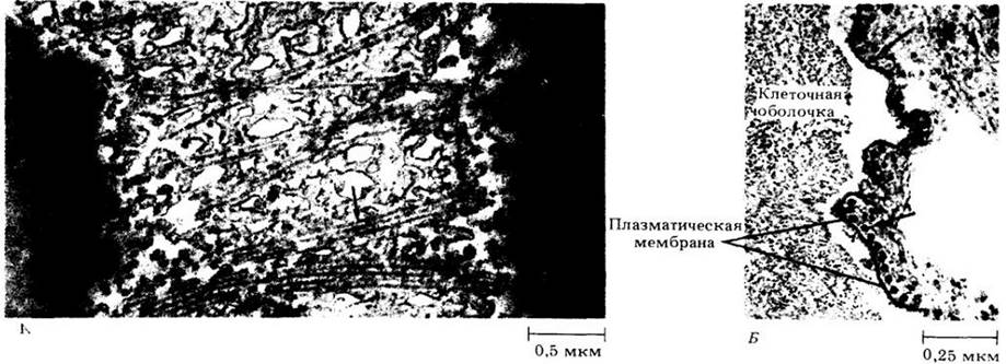
Fig. 2-21. A dictyosome consists of a group of flat membrane-bound sacs associated with vesicles that apparently bud off from the sacs. The dictyosome serves as the "packaging center" of The Introduction/5.html">Eukaryotic Cell and plays a key role in secretory processes. A. Cisternae of a dictyosome from a parenchymatous stem cell of the horsetail Equisetum hyemale, seen in cross-section. B. A single cisterna, surface view. The arrows point to numerous secretory vesicles along the margins of the cisternae

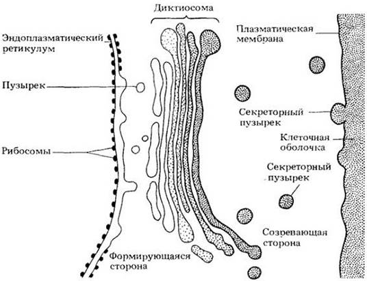
A forming face and a maturing face can usually be distinguished in a stack of cisternae. The membranes of the forming cisternae structurally resemble those of the endoplasmic reticulum, whereas the membranes of the maturing cisternae resemble The Plasma Membrane (Fig. 2-22).
Dictyosomes are involved in secretion and, in most higher plants, in Cell wall formation. Cell wall Polysaccharides synthesized by dictyosomes accumulate in vesicles that subsequently pinch off from the maturing cisternae. These secretory vesicles migrate and fuse with the plasma membrane (Fig. 2-22), thereby incorporating their contained polysaccharides into The cell wall. The products that accumulate in dictyosomes are not always synthesized by them. Some substances are formed in other structures, such as the endoplasmic reticulum, and then transported to the dictyosomes, where they are modified prior to secretion. A good example of such substances is Glycoproteins (carbohydrate-Structure/178.html">Protein Complexes), an important structural material of the cell wall. The protein portion is synthesized by the polysomes of the rough endoplasmic reticulum, while the carbohydrate portion is synthesized in the dictyosomes, where both parts combine to form glycoproteins.
The Endomembrane Concept
Membranes are dynamic, fluid structures that constantly change their shape and surface area. METABOLISM/2.html">THE CONCEPT OF the endomembrane system is based on this mobility of cellular membranes. According to this concept, the internal membranes of the Cytoplasm (except for those of Mitochondria and Plastids) form a single integrated whole and originate from the endoplasmic reticulum. New dictyosome cisternae are formed from the endoplasmic reticulum via an intermediate vesicle stage, and the secretory vesicles pinching off from the dictyosomes ultimately contribute to The formation of the plasma membrane (Fig. 2-22). Thus, the Endoplasmic reticulum and dictyosomes form a functional unity in which dictyosomes act as intermediate structures in The process of converting endoplasmic reticulum-like membranes into plasma membrane-like membranes.
It is important to note that even in Tissues whose cells grow and divide slowly, membrane components undergo continuous renewal.
Fig. 2-22. Schematic illustration of the endomembrane concept. New membranes are synthesized on the rough endoplasmic reticulum. Small vesicles bud off from the smooth surface of the endoplasmic reticulum and transport their contained substances and membranes to the forming face of the dictyosome. Secretory vesicles budding off from the cisternae of the maturing face of the dictyosome then migrate to the plasma membrane and fuse with it, adding a fresh Complement of membranes and building Materials
Microtubules
Found in virtually all Eukaryotic cells, microtubules are slender cylindrical structures with a diameter of about 24 nm and variable lengths. Each microtubule consists of subunits of a protein called tubulin. The subunits form 13 longitudinal protofilaments surrounding a central lumen. Microtubules are dynamic structures that regularly disassemble and reassemble at specific Stages of the Cell Cycle (see Fig. 2-15). Their assembly occurs at specialized sites called microtubule-organizing centers, which in plant cells have a poorly defined amorphous structure.
Microtubules have many functions. In elongating and differentiating cells, microtubules located near the inner surface of the plasma membrane apparently participate in cell wall formation by controlling the deposition pattern of Cellulose microfibrils laid down by the cytoplasm onto the growing cell wall (Fig. 2-23). The direction of cell elongation is, in turn, determined by the orientation of the cellulose microfibrils within the cell wall. Microtubules guide dictyosome vesicles to the forming wall—much like spindle fibers formed in a dividing cell—and apparently play a role in the Formation of the cell plate (the initial boundary between daughter cells). In addition, microtubules are an important component of flagella and cilia, in the movement of which they appear to play a significant role.
Fig. 2-23. Microtubules (indicated by arrows) visible in longitudinal (A) and cross (B) sections of leaf Cells of the fern Botrychium virginianum. A. The section passed more or less parallel to the cell wall near the inner surface; the plasma membrane is visible. B. Microtubules can be seen separated from the cell wall by the plasma membrane
Microfilaments
Microfilaments, much like microtubules, are found in virtually all eukaryotic cells. They appear as long filaments, 5 — 7 nm in diameter, composed of the contractile protein Actin. Bundles of microfilaments occur in many higher plant cells (Fig. 2-24) and appear to play a decisive role in cytoplasmic streaming. Together with microtubules, microfilaments form a flexible network known as the Cytoskeleton.
Fig. 2-24. A bundle of microfilaments in a leaf cell of the staghorn fern (Platycerium bifurcatum)

Ground Substance
Until recently, the ground substance of the cell was considered to be a homogeneous, protein-rich solution containing few structural elements or even to be completely structureless. However, recent studies of animal cells demonstrate that the ground substance possesses a complex Organization. Under a high-voltage Electron microscope, the ground substance is revealed as a three-dimensional lattice constructed of thin strands (3 — 6 nm in diameter) that pervade the entire cell. Other cytoplasmic components, including microtubules and microfilaments, are suspended within this microtrabecular lattice.
The microtrabecular lattice divides the cell into two phases: a protein-rich phase (the lattice strands) and a Water-rich phase that fills the spaces between the strands. Together with water, the lattice has the consistency of a gel—it is a truly living gel.
It is believed that Organelles are anchored to the microtrabecular lattice, which serves to connect different parts of the cell and direct Intracellular Transport. There is also evidence that plant cells possess a microtrabecular lattice.
Lipid Droplets
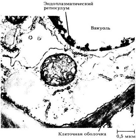
Lipid droplets are spherical structures that give The plant cell cytoplasm a granular appearance under the Light Microscope. In electron micrographs, lipid droplets appear amorphous (Fig. 2-25). Similar, though generally smaller, droplets occur within plastids (Fig. 2-7, A).
Fig. 2-25. Cytoplasmic components in a parenchyma cell from a thickened stem, or corm, of the spikemoss (Isoetes muricata). Flanking and below the mitochondrion, located near the center of the electron micrograph, two lipid droplets are visible. Above the mitochondrion and slightly to the side lies a cisterna of the endoplasmic reticulum, which appears swollen. Some vacuoles may have originated in this manner from the endoplasmic reticulum. A portion of a vacuole is visible at the top of the micrograph. The dense material within the vacuole is tannin
Lipid droplets were initially mistaken for organelles and termed spherosomes. It was thought that they were bounded by a single or double membrane. However, recent findings indicate that lipid droplets lack a membrane, though they may be coated with a layer of protein.
Ergastic Substances
Ergastic substances are "passive" products of the protoplast: reserve materials or waste products. They may appear and disappear at various stages of the cell cycle. We have already discussed several ergastic substances, such as starch grains, crystals, anthocyanin pigments, and lipid droplets. Other Examples include resins, Gums, Tannins, and protein bodies. Ergastic substances are incorporated into the cell wall, the cytoplasmic ground substance, and various organelles, including vacuoles.
Flagella and Cilia
Flagella and cilia are Hair-like structures that project from The surface of many eukaryotic cells. They are relatively slender, maintaining a constant diameter of about 0.2 µm, while their length ranges from 2 to 150 µm. Conventionally, the longer and less numerous ones are called flagella, whereas the shorter and more numerous ones are called cilia. Nevertheless, no sharp structural distinction exists between these two types, and we will use the term flagellum to refer to both.
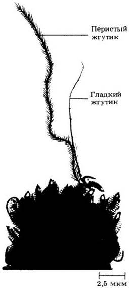
In certain Algae and Fungi, flagella serve as locomotory Organs that propel the organisms through water. In plants (such as mosses, liverworts, ferns, and some gymnosperms), only reproductive cells (Gametes) bear flagella. Some flagella (tinsel flagella) bear one or two rows of small lateral projections, whereas others (whiplash flagella) lack such appendages (Fig. 2-26).
Fig. 2-26. Two Types of flagella—tinsel and whiplash—found on a single cell of the colonial chrysophyte alga Synura petersenii. The tinsel flagellum (left) is longer than the whiplash flagellum (right)
The internal architecture of flagella was elucidated only through the advent of the electron microscope. Each flagellum exhibits a characteristic organization (Fig. 2-27). An outer ring of nine pairs of microtubules surrounds two central microtubules running down the axis of the flagellum. Enzyme-containing "arms" project from one microtubule of each outer pair. This fundamental 9+2 organizational pattern is conserved across all eukaryotic flagella.
The movement of flagella and cilia is autonomous; they are capable of beating even after being detached from the cells. Some researchers propose that flagellar movement relies on a sliding microtubule mechanism, whereby the outer pairs of microtubules slide past one another without contracting. This relative sliding of microtubule pairs induces localized bending of the flagellum. This sliding motion is apparently driven by the interaction between the "arms" of one microtubule pair and the adjacent pair.
Flagella emerge from cylindrical cytoplasmic structures known as basal bodies, which form the proximal Base of the flagellum (Fig. 2-27). Basal bodies share an internal organization reminiscent of flagella, except that their outer tubules are arranged in triplets rather than doublets, and the two central microtubules are absent.
Fig. 2-27. A. Longitudinal section through the flagellum of a green alga gamete, Ulvaria. Note that the membrane enclosing the flagellum proper is continuous with the plasma membrane. B. Cross section through a flagellum of Ulvaria, showing the typical 9+2 structure. C. Cross section through the basal body of a Ulvaria flagellum. Note that the basal body possesses a ring of 9 microtubule triplets and lacks central microtubules

Last update: 07/08/2026
Editorial and Educational Adaptation: This material has been compiled based on the primary/original source text. The project team performed an editorial review, corrected technical inaccuracies, structured sections, and adapted the content for an educational format.
What was processed:
- elimination of formatting defects (OCR errors, structural breaks, corrupted characters);
- editorial organization of content;
- standardization of terminology in accordance with academic sources;
- verification of factual statements against the original source text.
All mentions of the author, publication year, and origin of the primary text have been preserved in accordance with the source.