MODERN BOTANY - P. RAVEN - 1990
SECTION I. THE PLANT CELL
CHAPTER 2. THE EUKARYOTIC CELL
Mitochondria
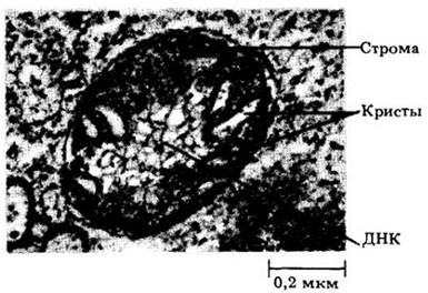
Like METABOLISM/14.html">Chloroplasts, Cell/35.html">Mitochondria are bounded by two elementary membranes (Figs. 2-14 and 2-15). The inner membrane forms numerous folds and projections called cristae, which significantly increase the internal surface area of the mitochondrion. Mitochondria are generally smaller than Plastids, being about half a micrometer in diameter, and exhibit A wide variety of lengths and shapes. As a rule, mitochondria are barely visible under a Light Microscope, but can be clearly observed using an Electron microscope.
Class="center">Fig. 2-14. Section of a mitochondrion from a spinach (Spinacia oleracea) leaf cell. DNA strands are visible. The mitochondrial envelope consists of two separate membranes. The inner membrane forms cristae projecting into the stroma. Small granules in the stroma are Ribosomes
Mitochondria are the sites of cellular Respiration, a process in which organic molecules are broken down to release energy, transferring it to ATP (adenosine triphosphate) molecules—the primary energy reserve of all Eukaryotic Cells. (Respiration is discussed further below in Chapter 6.) Most plant cells contain hundreds or thousands of mitochondria, their number in a given cell being determined by The Cell's demand for ATP.
Time-lapse cinematography reveals that mitochondria are in constant motion. They twist, bend, move from one part of the cell to another, and frequently fuse and divide. Mitochondria typically congregate wherever energy is in high demand.
If The Plasma Membrane is actively transporting substances into or out of the cell, mitochondria tend to align along the membrane's surface. In motile unicellular Algae, mitochondria generally cluster near the bases of flagella, presumably supplying the energy required for their movement.
Like plastids, mitochondria are semiatonomous Organelles containing the components necessary for the synthesis of their own Proteins. The inner membrane encloses a fluid matrix containing proteins, RNA, DNA strands, bacterial-like ribosomes, and various dissolved substances. DNA exists as circular molecules located within one or more nucleoids (Fig. 2-14).
Origin of Mitochondria and chloroplasts
Based on the similarities between Bacteria and the mitochondria and chloroplasts of eukaryotic cells, it is hypothesized that mitochondria and chloroplasts originated from bacteria that found "refuge" within larger heterotrophic cells—the precursors of eukaryotes. Bacteria capable of utilizing molecular oxygen to oxidize nutrients and harnessing light energy imparted these advantageous traits to larger cells, which, aided by such "helpers," gained a distinct advantage over their contemporaries and undoubtedly soon multiplied at their expense. With very few exceptions, all living eukaryotes contain mitochondria, and all autotrophic eukaryotes also contain chloroplasts. These organelles appear to have been acquired through independent symbiotic events (Symbiosis being a close association of two or more organisms that may, but does not necessarily, benefit each). Larger and more complex eukaryotic cells presumably protected their symbiotic organelles from adverse environmental influences. Consequently, eukaryotes were able to colonize land and acidic waters where prokaryotic cyanobacteria (or "blue-green algae") are absent, but eukaryotic green algae thrive.
Microbodies
Unlike plastids and mitochondria, which are bounded by two membranes, Microbodies are spherical organelles surrounded by a single membrane. Their diameter ranges from 0.5 to 1.5 µm. Microbodies have a granular matrix, and crystalline protein inclusions are occasionally found within them (Fig. 2-15). Microbodies are typically associated with one or two regions of The Endoplasmic reticulum.
Fig. 2-15. Organelles in a tobacco (Nicotiana tabacum) leaf cell. A microbody with a large crystalline inclusion is surrounded by a single membrane, distinguishing it from two mitochondria and a plastid, each bounded by two membranes. The double membrane of the plastid is visible only at the bottom of the micrograph. Part of a vacuole (light area at upper right) is separated from the rest of the Cytoplasm by a single membrane, the tonoplast
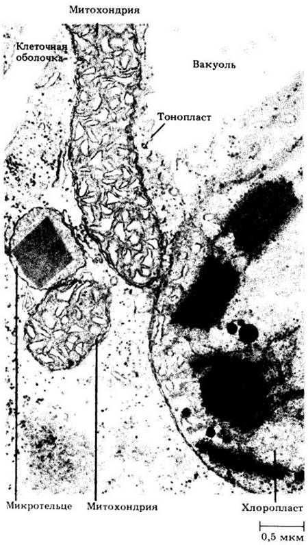
Certain microbodies, known as Peroxisomes, play a crucial role in glycolate metabolism, which is closely linked to Photorespiration (see p. 105). In green leaves, they are typically associated with mitochondria and chloroplasts (Fig. 2-15). Other microbodies, called glyoxysomes, contain Enzymes necessary for The conversion of fats into CARBOHYDRATES, a process that occurs in many seeds during germination (see p. 92).
Vacuoles
Vacuoles are membrane-bound cellular compartments filled with a fluid called cell sap. They are enclosed by the tonoplast, or vacuolar membrane (Figs. 2-6 and 2-15).
A young plant cell typically contains numerous small vacuoles that increase in size and fuse into a single large vacuole as the cell matures. In a mature cell, up to 90% of the volume may be occupied by the vacuole, while the cytoplasm is pressed against The Cell wall as a thin peripheral layer (Fig. 2-6). By filling most of the cell with "inexpensive" vacuolar contents, plants economize on nitrogen-demanding, "expensive" cytoplasm while maintaining a large surface area. Cell enlargement is driven primarily by the growth of the vacuole. This generates turgor pressure and maintains tissue rigidity, which is one of the principal Functions of the vacuole and tonoplast (see Chapter 4).
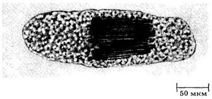
The main component of cell sap is Water, while other constituents vary depending on the plant type and its physiological state. Vacuoles typically contain salts and sugars, and occasionally soluble proteins. The tonoplast plays a vital role in The Active Transport and accumulation of certain ions within the vacuole. Consequently, the concentration of ions in the cell sap can be higher than in the surrounding cytoplasm. When certain substances reach high concentrations, crystals may form within the vacuoles. Calcium oxalate crystals of various shapes are particularly common (Figs. 2-16 and 2-17). The contents of vacuoles are generally mildly acidic, and occasionally very acidic, as in lemon juice.
Fig. 2-16. Druses of calcium oxalate crystals in epidermal cells of a redbud (Cercis canadensis) cotyledon. Scanning electron micrograph
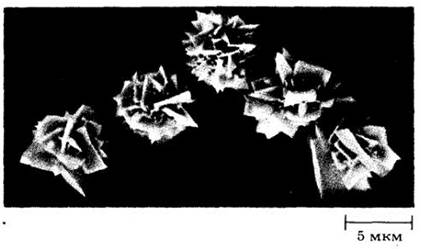
Fig. 2-17. Raphides—needle-like crystals of calcium oxalate—in the vacuole of a Sansevieria leaf cell. The tonoplast is not visible in this micrograph. The granular substance surrounding the crystals is the cytoplasm
Vacuoles serve as storage sites for various metabolites (metabolic products), such as Reserve Proteins in seeds and malic acid in CAM plants (see p. 106). They also sequester toxic secondary metabolites from the cytoplasm, such as the alkaloid nicotine (see ch. 29).
Vacuoles frequently store pigments. Blue, violet, purple, dark red, and crimson hues are imparted to plant cells by pigments belonging to the anthocyanin group (see ch. 29). Unlike most other plant pigments, anthocyanins are readily water-soluble and reside within the cell sap. They determine the red and blue coloration of numerous vegetables (radishes, turnips, cabbage), fruits (grapes, plums, cherries), and a multitude of flowers (cornflowers, geraniums, delphiniums, roses, and peonies). Sometimes these pigments mask chlorophyll in leaves, as seen, for example, in ornamental red maples. Anthocyanins color autumn leaves a bright red; they develop during cold, sunny weather when chlorophyll synthesis in the leaves ceases. As the existing chlorophyll breaks down, the anthocyanins become visible. In leaves where these pigments are not formed, the yellow-orange carotenoids of the chloroplasts emerge after the chlorophyll degrades. Leaves exhibit their most vibrant coloration during a clear, cold autumn.
Vacuoles also participate in the degradation of macromolecules and the recycling of their components within the cell. Individual organelles, such as ribosomes, mitochondria, and plastids, can be taken up by vacuoles and broken down there. Due to this digestive activity, vacuoles are comparable to Lysosomes—organelles found in animal cells.
It has long been hypothesized that vacuoles originate from the endoplasmic reticulum. Once it became clear that vacuoles are analogous to lysosomes, attempts were made to determine whether vacuoles, at least in some plants, form in the same manner as lysosomes. In animal cells, lysosome formation is associated with a specialized region of the cytoplasm designated as GERL (Golgi-endoplasmic reticulum-lysosome complex). GERLs have been identified in the ROOT tip cells of radishes and spurge, as well as in the cotyledons of mung bean embryos (Phaseolus aureus). Fig. 2-18 illustrates the stages of vacuole formation associated with GERL. The Golgi apparatus is discussed below.
Fig. 2-18. Stages of vacuole formation according to F. Marty. Tubular provacuoles originating from the GERL complex initially appear as pear-shaped vesicles, which then elongate and branch throughout the cell (I). The provacuoles enclose Regions of the cytoplasm like the bars of a cage (II). The tubular "bars" of each "cage"
fuse and completely isolate the entrapped cytoplasm (III). The inner membrane of the "cage" and the enclosed cytoplasm are digested by lysosomal enzymes (IV). Up to this point, the digestive enzymes were confined within the interconnected tubules. The outer membrane of the "cage" remains intact, and digestive activity is sustained within the developing vacuole. The resulting numerous small vacuoles fuse (IV), ultimately giving rise to one or more larger vacuoles characteristic of the mature plant cell (V)

Ribosomes
Ribosomes are small particles, only about 17–23 nm in diameter, composed of roughly equal amounts of Protein and RNA. In ribosomes, Amino Acids are linked together to form proteins; they are most abundant in the cytoplasm of metabolically active cells. Ribosomes occur freely in the Cell Cytoplasm or attached to the endoplasmic reticulum. They are also found in The Nucleus. As noted above, plastids and mitochondria contain ribosomes similar to those of prokaryotes.
Ribosomes actively engaged in Protein Synthesis form complexes known as polyribosomes, or Polysomes (Fig. 2-19). Cells synthesizing large quantities of proteins possess an extensive polysome system. Polysomes are frequently attached to the outer surface of the nuclear envelope (Fig. 2-5, B).
Fig. 2-19. Clusters of ribosomes (polysomes) On the surface of the endoplasmic reticulum. The endoplasmic reticulum is a network of membranes permeating the cytoplasm of eukaryotic cells and partitioning it into distinct compartments. Various Chemical Reactions can take place on The surface of the endoplasmic reticulum. Ribosomes are the structures in which amino acids are assembled into proteins. This transmission electron micrograph shows a region of a leaf parenchyma cell of the fern Regnellidium diphyllum

Last update: 07/08/2026
Editorial and Educational Adaptation: This material has been compiled based on the primary/original source text. The project team performed an editorial review, corrected technical inaccuracies, structured sections, and adapted the content for an educational format.
What was processed:
- elimination of formatting defects (OCR errors, structural breaks, corrupted characters);
- editorial organization of content;
- standardization of terminology in accordance with academic sources;
- verification of factual statements against the original source text.
All mentions of the author, publication year, and origin of the primary text have been preserved in accordance with the source.