MODERN BOTANY - P. RAVEN - 1990
SECTION I. THE PLANT CELL
CHAPTER 2. THE EUKARYOTIC CELL
The Plant Cell
A plant Cell typically consists of a more or less rigid Cell wall1 and a protoplast. The term protoplast is derived from protoplasm, a word long used to describe the living contents of a cell. The protoplast is the protoplasm of an individual cell; in a plant cell, it is the protoplasm bounded by The Cell wall.
1Along with the term "cell wall" (клеточная оболочка), the equivalent term "cell wall" (клеточная стенка) is also encountered. However, in Russian literature on plant Cytology and anatomy, the term "cell wall" (клеточная оболочка) is adopted as the priority term. — Trans. Note.
The protoplast consists of the Cytoplasm and The Nucleus (see Table 2-1). The cytoplasm contains Organelles (such as Ribosomes, microtubules, Plastids, and Mitochondria) and membrane systems (the Endoplasmic reticulum and dictyosomes). These can be observed in detail only with an Electron microscope. In addition, the cytoplasm includes the cytoplasmic matrix, or ground substance, in which the organelles and membrane systems are embedded. The cytoplasm is separated from the cell wall by The Plasma Membrane, which is a unit membrane. Unlike most animal Cells, plant cells contain one or more vacuoles in their cytoplasm. Vacuoles are fluid-filled vesicles surrounded by a unit membrane called the tonoplast.
In a living plant cell, the ground substance is in constant motion (Fig. 2-4). Organelles and other inclusions can be seen being swept along in this motion, known as cytoplasmic streaming, or cyclosis. Cyclosis ceases in dead cells. It undoubtedly facilitates the movement of substances within the cell and the exchange of Materials between the cell and its environment. However, The primary function of cyclosis remains unknown.
Class="center">Table 2-1. Components of the Plant Cell

Fig. 2-4. An Elodea cell. A. Cell surface. B. Cell center. The numerous disk-like structures are METABOLISM/14.html">Chloroplasts located along the cell wall. When viewed from above (A), the chloroplasts appear circular in outline. B. The chloroplasts present their broad faces to the wall and appear elongated. Note the absence of chloroplasts in the center of the cell (B), i.e., in the vacuole

The Plasma Membrane
Among the numerous cellular membranes, the plasma membrane exhibits the trizonal Structure (dark — light — dark layers) most clearly. Invaginations of the plasma membrane are common in plant cells.
The plasma membrane performs the following Functions: (1) it participates in the Metabolic exchange between the cell and its environment (see Chapter 4); (2) it coordinates the synthesis and assembly of Cellulose microfibrils of the cell wall; (3) it transmits hormonal and external signals that control cell growth and differentiation.
The Nucleus
The nucleus is often the most prominent structure in the cytoplasm of a Introduction/5.html">Eukaryotic Cell. The nucleus performs two vital functions: (1) it controls cellular activity by determining which Proteins are synthesized and when (see Chapter 8); (2) it stores Genetic information and transmits it to daughter cells during Cell Division.
The nucleus of a eukaryotic cell is surrounded by two unit membranes that form the nuclear envelope. It is pierced by numerous pores, ranging from 30 to 100 nm in diameter, which are visible with an electron microscope (see Fig. 2-5). These pores are not simply holes in the envelope; they have a complex structure. In some places, the outer membrane of the nuclear envelope merges with The endoplasmic reticulum. The nuclear envelope can be viewed as a specialized, locally differentiated region of the endoplasmic reticulum.
Fig. 2-5. Nuclear pores (A) visible On the surface of the nuclear envelope in an onion ROOT tip cell (Allium cepa). The preparation was obtained using the freeze-etching technique. B, C. ELECTRON MICROGRAPHS OF nuclei from the seedless vascular plant Selaginella kraussiana. B. Pores viewed from the surface; C — in cross-section (indicated by arrows). Note the Polysomes (spirals of granules) on The surface of the nuclear envelope (B) and the rough endoplasmic reticulum arranged parallel to the nuclear envelope (C)

In a nucleus stained with specific Dyes, fine threads and clumps of Chromatin, as well as the nucleoplasm (the ground substance of the nucleus), can be distinguished. Chromatin consists of DNA bound to A large number of specialized proteins called Histones. During cell division, chromatin becomes increasingly condensed and ultimately organizes into Chromosomes. It has recently been suggested that in non-dividing (interphase) nuclei, chromosomes (chromatin) are attached to the nuclear envelope at one or more points. Genetic information in both PROKARYOTES AND EUKARYOTES is encoded in DNA molecules. The DNA content of a single eukaryotic cell is significantly higher than that of a bacterial cell. In Bacteria, DNA molecules lie freely in the cytoplasm, whereas in eukaryotes, they are organized into chromosomes contained within the nucleus.
Organisms vary in the number of chromosomes found in their somatic (non-sex) cells. The desert annual Haplopappus gracilis has 4 chromosomes; cabbage has 20; sunflower, 34; wheat, 42; humans, 46; and a species of fern, Ophioglossum, has about 1250. Sex cells, or Gametes, contain only half the number of chromosomes characteristic of the Organism's somatic cells. The chromosome number in gametes is termed haploid (single), and in somatic cells, diploid (double). Cells containing more than two sets of chromosomes are called polyploid.
Spherical structures known as nucleoli can be distinguished within the nucleus under a Light Microscope. Each nucleus contains one or more nucleoli, which are prominent in non-dividing nuclei (Fig. 2-1). Ribosomal RNAs are synthesized within the nucleoli. Typically, the nuclei of diploid organisms contain two nucleoli, one for each haploid set of chromosomes. Composed mainly of protein, nucleoli contain about 5% RNA.
Plastids
Along with vacuoles and the cell wall, plastids are characteristic components of plant cells. Each plastid is surrounded by its own envelope, consisting of two unit membranes. Within the plastid, one can distinguish a membrane system and a more or less homogeneous substance called the stroma. Mature plastids are generally classified based on the pigments they contain.
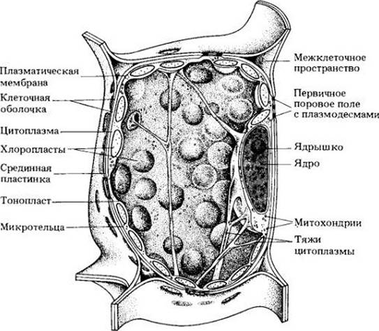
Chloroplasts, which are the sites of Photosynthesis (see Chapter 7), contain chlorophylls and carotenoids. Plant chloroplasts are typically disk-shaped, ranging from 4 to 5 µm in diameter. A single mesophyll cell (the "middle of the leaf") may contain 40 to 50 chloroplasts, amounting to about 500,000 per square millimeter of leaf surface. Within the cytoplasm, chloroplasts are generally arranged parallel to the cell wall, as illustrated in Fig. 2-6.
Fig. 2-6. Three-dimensional diagram of a plant cell containing chloroplasts. Numerous chloroplasts lie in the cytoplasm along the cell wall, facing it with their broad surfaces. The main volume of the cell is occupied by a vacuole traversed by cytoplasmic strands. The nucleus is located along the wall, though in other cases it may be suspended on cytoplasmic strands in the center of the vacuole and surrounded by a thin layer of cytoplasm.
The Internal Structure of a chloroplast is quite complex (Fig. 2-7). The stroma is pervaded by an elaborate membrane system consisting of flattened vesicles called thylakoids. Each thylakoid, much like the chloroplast envelope, is composed of two membranes. It is believed that thylakoids form a continuous, interconnected system. Typically, thylakoids are stacked into structures known as grana, which resemble piles of coins. The thylakoids of individual grana are interconnected by stroma thylakoids, or intergranal thylakoids. Chlorophylls and carotenoids are embedded within the thylakoid membranes. Chloroplasts of green Algae and plants frequently contain starch grains and small lipid (fat) droplets. Starch grains serve as temporary storage depots for the products of photosynthesis (Fig. 2-1). They can disappear from the chloroplasts of a plant kept in total darkness for just 24 hours, and reappear within 3 to 4 hours after the plant is returned to the light.
Fig. 2-7. A. Chloroplast of a maize leaf (Zea mays). B. Grana consisting of stacks of disk-like thylakoids. The grana thylakoids are connected by other thylakoids, commonly referred to as stroma thylakoids.

Chloroplasts are semi-autonomous organelles and in some respects resemble bacteria. For instance, the ribosomes of both bacteria and chloroplasts are one-third smaller than eukaryotic ribosomes. Protein Synthesis on bacterial and chloroplast ribosomes is inhibited by the antibiotic chloramphenicol, which has no such effect in Eukaryotic cells. Furthermore, both bacteria and chloroplasts possess one or more nucleoids—light-appearing, grana-free regions containing strands of DNA. Plastid and bacterial DNA are organized in a similar fashion: they are not enclosed by a membrane, lack associated histones, and typically exist in a circular form.
The Genetic Code of plastid DNA is currently being investigated in several laboratories. Isolated chloroplasts carry out RNA Synthesis, a process that, as will be shown in Chapter 8, is normally controlled exclusively by chromosomal DNA. The formation of chloroplasts and the synthesis of their resident pigments are largely regulated by chromosomal DNA, which interacts in a yet poorly understood manner with chloroplast DNA. Nevertheless, in the absence of their own DNA, chloroplasts fail to develop.
Chloroplasts can be considered premier cellular organelles because they initiate The conversion of solar energy that ultimately provides our food and fuel. However, chloroplasts do more than just carry out photosynthesis; they also participate in the synthesis of Amino Acids and Fatty acids and serve as temporary storage sites for starch.
Chromoplasts (from the Greek chroma, meaning color) are pigmented plastids (Fig. 2-8). Diverse in shape, chromoplasts lack chlorophyll but synthesize and accumulate carotenoids, which impart yellow, orange, and red colors to flowers, senescent leaves (see Chapter 7), fruits, and roots. Chromoplasts may develop from green chloroplasts; in the process, the latter lose their chlorophyll and internal membrane structures while accumulating carotenoids, as occurs during the ripening of many fruits. The exact function of chromoplasts remains unknown, although in some cases they serve to attract insects and other animals with which they have coevolved (see Chapter 29).
Fig. 2-8. Chromoplasts from a marigold petal (Tagetes). Each chromoplast contains numerous lipid droplets that store the pigments responsible for the petal color.

Leucoplasts (Fig. 2-9) are non-pigmented plastids. Some synthesize starch (amyloplasts; Figs. 2-10 and 2-11), while others appear capable of producing various substances, including Lipids and proteins. When exposed to light, leucoplasts can transform into chloroplasts.
Fig. 2-9. Leucoplasts clustered around the nucleus in epidermal cells of a Zebrina leaf.

Fig. 2-10. Scanning electron micrograph showing spherical and ovoid starch grains in a potato cell (Solanum tuberosum). Starch grains are formed within amyloplasts, with one grain per plastid.

Fig. 2-11. Amyloplast from an embryo sac of soybean (Glycine max). The rounded, light-colored bodies are starch grains, and the smaller dark bodies are lipid droplets.

Proplastids are small, colorless or pale green, undifferentiated plastids found in the meristematic (dividing) cells of roots and shoots. They serve as precursors to other, more differentiated plastids—chloroplasts, chromoplasts, and amyloplasts (Fig. 2-12). If The Development of proplastids into more differentiated structures is delayed due to a lack of light, one or more prolamellar bodies may appear within them; these are semi-crystalline aggregates of tubular membranes (Fig. 2-13). Plastids containing prolamellar bodies are termed etioplasts. Etioplasts develop into chloroplasts upon exposure to light, as the membranes of the prolamellar bodies organize into thylakoids. Etioplasts form in the leaves of plants kept in the dark. Seed embryo proplastids first differentiate into etioplasts, which then develop into chloroplasts in the light. Plastids are characterized by relatively easy transitions from one type to another.
Fig. 2-12. Plastid developmental cycle according to J. M. Whatley. Successive stages of plastid development. Stages 1–3: the eoplast (eo-, "early"), amyloplast, and amoeboid plastid can be considered non-green proplastid stages. Stage 4: the pre-granal plastid may be green or non-green. In the absence of light, the pre-granal stage may be represented by an etioplast (Stage 4'). Chromoplasts (V) can originate from several types of plastids.

Fig. 2-13. Etioplast with a semi-crystalline prolamellar body constructed of tubular membranes in a leaf cell of a dark-grown plant. In the light, the membranes of the prolamellar body transform into thylakoids.

Plastids multiply by binary fission, in this respect also resembling bacteria. In meristematic cells, the time of proplastid division roughly coincides with the timing of cell division. However, in mature cells, the majority of plastids are formed through the division of pre-existing mature plastids.
Last update: 07/08/2026
Editorial and Educational Adaptation: This material has been compiled based on the primary/original source text. The project team performed an editorial review, corrected technical inaccuracies, structured sections, and adapted the content for an educational format.
What was processed:
- elimination of formatting defects (OCR errors, structural breaks, corrupted characters);
- editorial organization of content;
- standardization of terminology in accordance with academic sources;
- verification of factual statements against the original source text.
All mentions of the author, publication year, and origin of the primary text have been preserved in accordance with the source.