BIOCHEMISTRY - L. Stryer - 1984
VOLUME 1
PART I. CONFORMATION AND DYNAMICS
ANSWERS TO QUESTIONS AND PROBLEMS
Chapter 2
1. a) Phenyl isothiocyanate.
b) Dansyl chloride.
c) Urea; β-mercaptoethanol for disulfide reduction.
d) Chymotrypsin.
e) CNBr.
f) Trypsin.
2. a) 3; b) 12; c) 4.28; d) 9.8; e) 2.4.
3. 0.01; 0.1; 1; 10; 100.
4. 477 Å (318 residues per chain; 1.5 Å per residue).
5. Each amino acid residue, except for the C-terminal one, yields a hydrazide upon reaction with hydrazine. The C-terminal residue can be identified by the fact that it yields a free amino acid.
6. a) Approximately +1.
b) Two Peptides.
7. The side chain of S-aminoethylcysteine resembles the side chain of Lysine; the only difference is that the S-aminoethylcysteine molecule has a sulfur atom instead of a methylene group.
8. The native conformation of Insulin is not the thermodynamically most stable form. For a Structure/133.html">Discussion of this, see Sec. 35.9.
Chapter 3
1. a) 2.96 • 10-11 g.
b) 2.71 • 108 molecules.
c) No. If Hemoglobin were in the form of cubic crystals, a single erythrocyte would accommodate 3.22-108 molecules. Therefore, the actual packing density of hemoglobin in the erythrocyte is 84% of the maximum possible.
2. 2.65 g (or 4.75 • 10- 2 mol) Fe.
3. a) In humans, 1.44 • 10 - 2 g (4.49 • 10- 4 mol) O2 per 1 kg of Muscle. In the sperm whale, 0.144 g (4.49 • 10-3 mol) O2 per 1 kg of muscle.
b) 128.
4. a) Approximately +36 at pH 2, +4 at pH 7, and +2 at pH 9.
b) Approximately 10. However, interaction between titratable groups occurs, so the actual isoelectric point is 8.2.
5. a) Three peptides consisting of residues 1 to 55, 56 to 131, and 132 to 153. b) Short α-helices are marginally stable in aqueous solution. In Myoglobin, they are stabilized by tertiary interactions.
Class="center">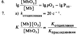
b) The average lifetime is 0.05 s (the reciprocal of koff).
8. A 1 mg/ml myoglobin solution (myoglobin mass = 17.8 kDa) corresponds to a concentration of 5.62 • 10-5 M. The absorbance of this solution at a path length of 1 cm is 0.84, which corresponds to an I0/I ratio of 6.96. Therefore, 14.4% of the incident light is transmitted through the solution.
Chapter 4
1. a) Increase.
b) Decrease.
c) Decrease.
d) Decrease oxygen affinity.
2. a and b) Less H+ will bind.
3. Inositol hexaphosphate.
4. a) ∆Y = 0.977 - 0.323 = 0.654, b) AY = 0.793 - 0.434 = 0.359.
5. a) pK will decrease.
b and c) pK will increase.
6. The additional methyl group prevents the movement of the iron atom into the plane of the porphyrin ring upon oxygenation. This model compound mimics the T-state of hemoglobin.
7. a) Yes. KAB=KBA(KB/KA) = 2.10 • 5M.
b) The presence of A enhances the binding of B, and similarly, the presence of B enhances the binding of A.
8. Carbon monoxide, by binding to one heme, alters the oxygen affinity of the other Hemes in the same hemoglobin molecule. In particular, CO increases the affinity of hemoglobin for oxygen, thereby reducing The amount of O2 released in the Tissues. Carbon monoxide has a stabilizing effect on The quaternary structure of oxyhemoglobin. In other words, CO mimics O2 as an allosteric effector.
9. a) Transport is maximal at K = 10-5 M. In general, transport reaches a maximum at 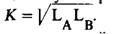
b) Maximum Oxygen transport occurs at P50 = 44.7 torr, which is significantly higher than the physiological value of 26 torr. It should be emphasized that this calculation does not account for cooperative binding and the Bohr effect.
Chapter 5
1. a) Lysine or Arginine at position 6.
p) b) At pH 8, this hemoglobin will migrate toward the anode more slowly than the other two Hemoglobins because it has the lowest net negative charge.2. a) HbC; b) HbD; c) HbJ; d) HbN.
3. Mutations in the α Gene affect all three hemoglobins because these hemoglobins have the following subunit structures, respectively: α2β2, α2δ2, and α2γ2. Mutations in the β, δ, and γ genes will affect only one of the hemoglobin types.
4. The reaction
2(α2ββS) ⇄ α2β2 + α2βS2
is rapid compared to the duration of Electrophoresis. The Separation of α2α2 and α2αS2 in an electric field shifts the reaction equilibrium to the right.
5. Deoxy-HbA contains a complementary site and therefore can bind to the deoxy-HbS fiber. Further elongation of the fiber will then be impossible, as the terminal deoxy-HbA molecule lacks a sticky patch.
6. The Hydrogen bond between aspartate and Tyrosine at the α1α2 contact site, characteristic of deoxyhemoglobin A, is absent in hemoglobin Kempsey (p. 100). Consequently, the deoxy form of this mutant hemoglobin should dissociate into dimers more readily than normal hemoglobin. On the other hand, the oxygenated forms of both hemoglobins dissociate to approximately the same extent, because the hydrogen bond in question does not participate in stabilizing either hemoglobin.
Chapter 6
1. a) 31.1 • 10-6 mol.
b) 5 • 10- 8 mol.
c) 622 s-1.
2. a) Yes. KM = 5.2 • 10-6 M.
b) Vmax = 6.84 • 10-10 mol.
c) 337 s-1.
3. a) In the absence of inhibitor, Vmax = 47.6 μmol/min and KM = 1.1 • 10- 5 M. In the presence of inhibitor, Vmax does not change, while the apparent KM = 3.1 • 10-5 M.
b) Competitive inhibition.
c) 1.1 • 10-3 M.
d) fES = 0.243 and fEI = 0.488.
e) In the absence of inhibitor, fES = 0.73, and in the presence of 2 • 10- 3 M inhibitor, fES = 0.49. The ratio of these values, which is 1.49, is equivalent to the ratio of the reaction rates under the same conditions.
4. a) Vmax = 9.5 μmol/min. KM = 1.1 • 10-5 M, the same as in the absence of inhibitor.
b) Noncompetitive.
c) 2.5 • 10-5 M.
d) 0.73 both in the presence and absence of inhibitor.
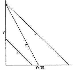
5. a) V = Vmax - (V/S) KM.
b) Slope = — KM; y-intercept equals Vmax; x-intercept corresponds to Vmax/KM.
c) 1 - without inhibitor,
2 - competitive inhibitor,
3 - non-competitive inhibitor.
6. Binding of a competitive inhibitor to one Active Site of an allosteric enzyme can lead to an increased affinity for the substrate at another site within the same enzyme molecule. According to the concerted model, such a competitive inhibitor promotes the T → R transition.
7. Potentially, in hydrogen bond formation at pH 7, the side chains of the following amino acid residues can serve as Donors: arginine, asparagine, glutamine, Histidine, lysine, Serine, Threonine, Tryptophan, and tyrosine.
8. The rates of consumption of A and B are given by the equations
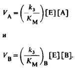
Hence, the ratio of the rates is
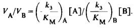
Consequently, the enzyme selects between competing substrates depending on the value of k3!!!!KM, and not just KM.
Chapter 7
1. (b) is hydrolyzed the fastest, (a) the slowest,
2. a) B—C; b) A—B and E F; c) A—B—C (one sugar residue does not interact with the enzyme, As a result of which site D, being energetically unfavorable, remains unoccupied.
3. 18O will end up in the hydroxyl group at C-4 in di-NAG (residues E—F).
4. Lacking a bulky substituent at C-5, this analog apparently can bind to site D without undergoing deformation. Consequently, the binding of residue D of this analog, unlike that of residue D in tetra-NAG, does not require an input of Free energy.
5. a) In oxyhemoglobin, Fe is coordinated to five nitrogen atoms and one oxygen atom. In Carboxypeptidase A, Zn is coordinated to two nitrogen atoms and two oxygen atoms.
b) In oxymyoglobin, one of the nitrogen atoms bound to Fe belongs to the proximal histidine residue, and the other four belong to the heme. The oxygen atom bound to Fe originates from O2. In carboxypeptidase A, two nitrogen atoms coordinated to Zn belong to histidine residues. One of the oxygen atoms bound to Zn belongs to the glutamate side chain, and the other to a Water molecule.
c) Aspartate, Cysteine, and Methionine.
6. Water labeled with 18O is formed when carboxypeptidase A catalyzes the synthesis of a peptide bond between N-benzoylglycine and L-phenylalanine. In the presence of L-β-phenyllactic acid, 18O is not incorporated into H2O, because this compound lacks an amino group, and thus peptide bond synthesis does not occur.
Chapter 8
1. a) Carboxypeptidase A.
b) Lysozyme and carboxypeptidase A.
c) Chymotrypsin.
d) Carboxypeptidase A and chymotrypsin.
2. a) Yes.
b) In chymotrypsin - histidine-57, in lysozyme - glutamate-85, in carboxypeptidase A - tyrosine-248.
3. In chymotrypsin - serine-195, in carboxypeptidase A - glutamate-270 (or hydroxyl ion).
4. Precise positioning of the catalytic residues and substrates, geometric strain (substrate distortion), electron transfer, and substrate desolvation.
5. a) Tosyl-L-lysyl chloromethyl ketone (TLCK).
b) First, determine whether substrates can prevent the inactivation of trypsin by TLCK. Then, test whether the D-isomer of TLCK is capable of inactivating trypsin.
c) Thrombin.
6. a) Serine.
b) Hemiacetal between the aldehyde group of the inhibitor and the hydroxyl group of the active-site serine.
7. The boron atom binds to the oxygen atom of the active-site serine. The geometry of the resulting tetrahedral intermediate resembles the Transition State.
8. Activated factor X remains bound to platelet membranes, which accelerates its activation of prothrombin.
9. Antithrombin III appears to be a transition-state analog, so its interaction with thrombin requires a fully formed active site.
10. Amino acid residues a and d are located inside the α-helical coiled coil, near its axis. Hydrophobic interactions between the side chains of these residues stabilize the coiled coil.
Chapter 9
1. a) Every third residue in each of the Collagen chains must be Glycine, as there is simply no room for a larger amino acid residue.
b) The melting Temperature of poly(Gly-Pro-Gly) is lower than that of poly(Gly-Pro-Pro).
c) No. Glycine does not occupy every third position.
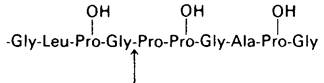
2. a) and b)
The arrow indicates the scissile peptide bond.
3. a) Disulfides.
b) No.
c) Peptide bonds between specific glutamine and lysine side chains.
d) Aldol cross-link, histidine-aldol cross-link, and lysinonorleucine.
e) Aldol cross-link, lysinonorleucine, and desmosine.
4. Decarboxylation of α-oxoglutarate represents half of the physiological reaction. Apparently, α-oxoglutarate is first attacked by oxygen to form a peroxy acid, which then reacts with the Proline-containing substrate.
Chapter 10
1. 2.86 • 106 molecules.
2. The cyclopropane ring prevents the orderly packing of hydrocarbon chains, thereby increasing membrane fluidity.
3. 2 • 10-7 cm; 6.32 • 10-6 cm and 2 • 10-4 cm.
4. The radius of this molecule is 3.08 • 10-7 cm and the diffusion coefficient is 7.37 • 10-9 cm2/s. The mean distance in 1 μs is 1.72 • 10-7 cm; in 1 ms, 5.42 • 10-6 cm; and in 1 s, 1.72 • 10-4 cm.
5. The initial decrease in the amplitude of the paramagnetic Resonance spectrum is due to the reduction of spin-labeled phosphatidylcholines in the outer leaflet of the bilayer. Under the experimental conditions, ascorbate does not cross the membrane and therefore does not reduce the Phospholipids of the inner leaflet. The slow decay of the residual spectrum is attributed to the reduction of phospholipids that have transversely diffused into the outer leaflet.
Last update: 06/08/2026
Editorial and Educational Adaptation: This material has been compiled based on the primary/original source text. The project team performed an editorial review, corrected technical inaccuracies, structured sections, and adapted the content for an educational format.
What was processed:
- elimination of formatting defects (OCR errors, structural breaks, corrupted characters);
- editorial organization of content;
- standardization of terminology in accordance with academic sources;
- verification of factual statements against the original source text.
All mentions of the author, publication year, and origin of the primary text have been preserved in accordance with the source.