Peptide Self-Regulation of Living Systems (Facts and Hypotheses) - Shataeva, L. K. 2003
Interaction of Peptides with Cell Nucleus Chromatin
Model and Natural Nucleoprotein Complexes
Besides the fact that the DNA molecule serves as a repository of Genetic information, it is, as shown in Sections 3.1 and 3.2, a macromolecule with distinct physicochemical properties and unique regulatory Functions. Specifically, DNA is known to cross cellular and nuclear membranes in Cells and Tissues (Simon, 1992). Following this, the macromolecule can be cleaved by specific Nucleases, and its fragments can participate in regulating certain Cell Cycle processes. For example, the biological activity of DNA fragments was utilized in developing Derinat, an immunomodulator consisting of a 1.5% aqueous solution of highly purified native DNA sodium salt (sodium deoxyribonucleotide) derived from sturgeon milt, depolymerized by ultrasound, and dissolved in a 0.1% aqueous sodium chloride solution (Ursova, 2000).
Enhancing the ability of bioactive macromolecules to cross cell membranes represents one of the major challenges in modern Biotechnology and Medicine. Difficulties associated with overcoming the cellular membrane barrier limit the Prospects of targeted drug delivery, genetic cell transformation, and The regulation of intracellular processes by exogenous protein factors (Maryanovich, Polyakov, 1991). One approach to overcoming these obstacles is the complexation of BIOLOGICALLY ACTIVE SUBSTANCES with polymers, which prevents the degradation of protein factors by hydrolytic Enzymes or imparts the missing properties required for cellular penetration. Nucleic Acids satisfy all the requirements for carrier polymers, most notably biocompatibility (Panarin, 1989). When interpolyelectrolyte complexes (IPECs) are formed between Nucleic Acids and linear synthetic polycations, a Cooperative binding of electrostatically complementary chains takes place, accompanied by the shielding of the charge of the nucleic acid phosphate groups, which leads to The formation of a hydrophobic region within the macromolecule. The number and extent of such regions are determined by the degree of polymerization of the blocking polycation and the COMPOSITION OF THE polycomplex. The expression of the biological activity of a nucleic acid requires its specific recognition by regulatory Proteins and Enzymes. The formation of IPECs does not always result in the loss of such recognition capability. For instance, studies have shown that poly-N-ethyl-4-vinylpyridinium bromide bound to DNA does not hinder its specific recognition. The polycation shifts along the DNA chain from one site to another, exposing restriction sites. Transformation efficiency experiments indicate the promising potential of applying such complexes in Introduction/32.html">Genetic Engineering (Kabanov et al., 1989).
At the same time, foreign DNA can integrate into The Genome of other cells and alter THE SPECTRUM OF endogenous Protein Synthesis products. Several studies (Kabanov, Kabanov, 1994; Debabov, 1997) have outlined the prospects of using this property for DNA vaccination and Gene Therapy, as well as for delivering genetic material into The Cell. However, the surface-active properties of DNA have not yet been fully investigated. Such studies could elucidate the mechanisms by which macromolecules of this size penetrate the cytoplasmic membrane and substantiate, from a colloid chemistry perspective, the feasibility of introducing exogenous information into the cellular genome.
For the targeted delivery of foreign genetic material into a cell without membrane damage, The Use of Insulin-poly-L-Lysine-DNA IPECs has been proposed (Rosenkranz et al., 1990). Insulin acts as a Ligand that specifically recognizes cell-surface receptors, while poly-L-lysine provides its non-covalent linkage to DNA. Such a conjugate ensures the selective and targeted delivery of foreign genetic material into mammalian and plant cells via receptor-mediated internalization (endocytosis). The efficacy of this transfection method has also been proven, and the feasibility of its in vivo application has been demonstrated.
Investigating the selectivity of DNA binding to model nonspecific proteins—which differ in acid-base properties and are not typical Components of the Cell Nucleus—is of interest for understanding The properties of DNA AS A carrier polymer that forms nucleoprotein complexes with diverse physiological characteristics. We studied the quantitative regularities and mechanisms of formation of such complexes in model systems (Ryadnova et al., 2000a); we investigated the binding patterns of DNA extracted from bovine Spleen with Globular proteins varying in acid-base properties: Pepsin (pK = 2.0), insulin (pK = 5.4), cortexin (pK = 9.5), cytochrome c (pK = 10.6), and protamine (pK = 11.5). Under physiological conditions, the net charge of these proteins ranged from 36- for pepsin to (30–32)+ for protamine. Despite the net negative surface charge of DNA, stable nucleoprotein complexes form between DNA and all the studied proteins across an Ionic strength range of 0.1 to 0.5 M. Furthermore, complexation does not cause "unraveling" (Denaturation) of the double-stranded nucleic acid molecule, which would otherwise be indicated by a hyperchromic effect—a sharp increase in UV absorbance at a wavelength of 260 nm. Fig. 13 illustrates the Chromatography and rechromatography of the DNA–insulin complex on Sephadex G-150 at ionic strengths of 0.3 and 0.5 M. Two peaks are registered on the chromatogram. The first peak elutes with the exclusion volume of the Column, while the second peak elutes with a volume significantly exceeding the exclusion volume of free, unbound protein. The rechromatogram (Fig. 13, B) of the high-molecular-weight fraction reveals only the peak corresponding to the nucleoprotein complex, demonstrating its stability even at an elevated ionic strength (Shataeva et al., 1999).
In all the systems studied, the complexation process followed a cooperative mechanism, but in each case, the stoichiometry and binding strength of DNA to proteins depended on the concentration ratio of the components within the complex and The Nature of the protein (Ryadnova et al., 2000a).
Class="center">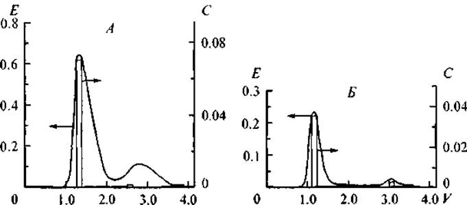
Fig. 13. Gel filtration chromatography of the DNA–insulin complex (Sephadex G-150).
A — gel filtration of the initial solution, ionic strength 0.3 M; B — rechromatography of the first peak from the DNA–insulin complex chromatogram, ionic strength 0.5 M. E — optical density at a wavelength of 260 nm; V — relative elution volume; C — protein concentration (mg/mL).
As noted earlier, nucleic acids hold great promise as polymeric carriers for biologically active substances to protect the latter from Enzymatic Hydrolysis and facilitate their delivery to target Organs or cells. We investigated the resistance of DNA complexes with insulin, pepsin, and cortexin to hydrolysis by DNase and Trypsin. It was found that these enzymes do not degrade the complexes nor hydrolyze the complex-bound protein. This undoubtedly highlights the potential of using DNA–protein and DNA–peptide complexes for developing oral drug formulations with regulatory functions.
It is well known that DNA conformation is a crucial factor both for executing the cellular genetic program and for the penetration of exogenous DNA into The Nucleus. Several studies have shown that the interaction of DNA with basic histone proteins results in macromolecular compaction, the extent of which depends on the DNA/histone weight ratio (Martynkina et al., 1991, 1998). We have likewise demonstrated that the DNA complexation process depends on the initial quantitative ratio of components in solution and determines the degree (density) of DNA occupancy by the protein. In turn, the occupancy degree governs the Morphology of the resulting nucleoprotein complexes (NPCs). Fig. 14 shows the Changes in the intrinsic viscosity of DNA solutions as a function of the packing density of the rigid-chain DNA macromolecule with globular protein molecules. Evidently, as the protein packing density on DNA increases, the intrinsic viscosity of the NPC solutions decreases. The limiting values of NPC intrinsic viscosity at maximum DNA saturation with globular Proteins are very similar across all investigated systems. However, the DNA compaction processes during its gradual saturation with proteins of differing acid-base properties proceed differently. Specifically, the interaction of DNA with insulin leads to a progressive compaction of the complex Structure, followed by the formation of a "beads-on-a-string" configuration, similar to that observed in DNA–histone interactions (Kiseleva et al., 1998). Apparently, it is precisely this compaction of the DNA–insulin complex structure that ensures its stability under increased ionic strength and the action of hydrolytic enzymes. Given that obtaining new insulin formulations is a pressing problem in endocrinology (Vlasov et al., 1988; Valuev et al., 1998), it seems advisable to continue investigating DNA–insulin complexes and to study the biological activity of insulin incorporated into NPCs.
Upon DNA binding to cytochrome c, the compaction process proceeds much more intensively. Nonlinear "beads-on-a-string" structures form even at low protein concentrations, as the basic Nature of the side groups of cytochrome c facilitates its local multipoint interactions with the phosphate groups of DNA. Consequently, supramolecular NPC structures do not arise from the interaction of DNA with cytochrome c.
In the case of the DNA–pepsin complex (the most acidic among all studied proteins), a decrease in the intrinsic viscosity of NPCs is observed even at a low protein content in the system. Obviously, the mechanism underlying the formation of complexes between DNA and acidic proteins requires further research and an expansion of methodological approaches, since it is precisely the acidic regulatory Chromatin proteins that are involved in unwinding the DNA double helix (Ryadnova et al., 2000a).
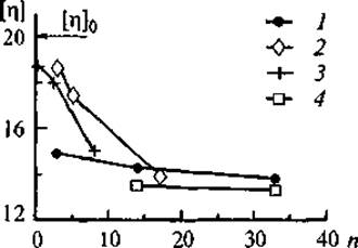
Fig. 14. Dependence of the intrinsic viscosity of DNA [η] (dL/g) and its complexes on The amount of bound protein.
[η]₀ — free DNA; nucleoprotein complexes; 1 — DNA–cytochrome c, 2 — DNA–insulin, 3 — DNA–pepsin, 4 — DNA–cortexin. n — average number of protein molecules bound per DNA molecule.
Investigating the concentration regularities of model DNA–protein complex formation and evaluating their stability under physiological conditions allow these patterns to be extrapolated to natural NPCs isolated from various tissues. The medical application of model or natural NPCs for correcting the functions of damaged or Aging tissues is facilitated by the high resistance of these preparations to gastrointestinal hydrolytic enzymes.
The promise of research conducted in this direction is confirmed by the efficacy of natural NPCs isolated from various organs and tissues—cytamines, which are used in medical practice to correct physiological disorders of individual body functions, particularly in cases of weakened Immunity and memory resulting from stress and physical exertion (Morozov et al., 2000a; Khavinson et al., 2001g).
The technological approach underlying The production of natural nucleoprotein complexes consists of preserving the Structural elements of chromatin under mild alkaline cell hydrolysis conditions, wherein endogenous regulatory Peptides are naturally assembled and linked to specific corresponding DNA sites. Simultaneously, during the extraction stage, partial hydrolysis of the DNA linker regions is carried out by concomitant endogenous DNases. The products of such hydrolysis (oligomers) are removed during the NPC precipitation step.
The presence of membrane glycolipid and phospholipid components in preparations obtained at the initial isolation stage hinders complex solubility. Delipidation of the preparations using chloroform—a universal solvent for all lipid classes (lecithins, cephalins, and sphingomyelins)—increases NPC solubility and enhances the quality of chromatographic analysis of their composition (Preparative BIOCHEMISTRY OF Lipids, 1981).
Depending on the type of tissue from which NPCs are isolated, preparation compositions vary in their protein-to-lipid ratios. The nucleic acid content is virtually identical across all preparations, since the cells of all differentiated body tissues contain an identical set of chromosomal nucleic acids.
To evaluate the resistance of the obtained natural NPC preparations to enzymatic hydrolysis in the gastrointestinal tract, enzymatic Treatment of these complexes was performed. As in the case of model NPCs, following the enzymatic treatment of natural NPCs, Polypeptides tightly bound to DNA are preserved in their high-molecular-weight fraction. This serves as indirect confirmation that the selected conditions for cell and nuclear disruption during NPC preparation allow stable chromatin structural elements to be preserved. Chromatographic analysis of Brain NPC preparations revealed proteins with molecular masses in the 20–24 kDa range, which can presumably be identified as acidic Proteins of the BASP group localized in the synaptic region of Neurons (Mosevitsky et al., 1994). Furthermore, all natural NPC preparations contain low-molecular-weight peptides that are analogs of cytomedins (Kuznik et al., 1995).
These physicochemical studies served as the basis for utilizing natural NPCs as oral drug formulations and dietary supplements (Ryadnova et al., 2000b).
Last update: 06/08/2026
Editorial and Educational Adaptation: This material has been compiled based on the primary/original source text. The project team performed an editorial review, corrected technical inaccuracies, structured sections, and adapted the content for an educational format.
What was processed:
- elimination of formatting defects (OCR errors, structural breaks, corrupted characters);
- editorial organization of content;
- standardization of terminology in accordance with academic sources;
- verification of factual statements against the original source text.
All mentions of the author, publication year, and origin of the primary text have been preserved in accordance with the source.