Biochemistry of Amino Acids - A. Majster 1961
Natural Amino Acids
Issues in the Stereochemistry of Amino Acids
Asparagine was the first amino acid to be isolated from natural sources [14] and one of the earliest in which optical activity was identified. Over a century ago, Pasteur [446, 447] discovered that natural asparagine rotates the plane of polarization to the left, whereas aspartic acid obtained by the Hydrolysis of natural asparagine is a dextrorotatory compound, while chemically synthesized aspartic acid is optically inactive. These investigations by Pasteur were apparently prompted by Dessaignes' assertion that aspartic acid prepared chemically from fumaric acid and ammonia is identical to natural aspartic acid [448]. Pasteur strongly insisted that natural and synthetic aspartic acids are non-identical, but he failed to realize at the time that the synthetic product is a racemic mixture of two isomers, much like the racemic form of tartaric acid he had previously studied.
It was not until 34 years later that Schulze and Bosshard [449, 450] demonstrated that optically inactive Amino Acids are mixtures of equal quantities of dextrorotatory and levorotatory forms. These authors found that 1) alkaline Hydrolysis of Proteins yields optically inactive amino acids; 2) such Amino acids differ in solubility from the Optically active amino acids obtained via acid Protein Hydrolysis; and 3) treating optically active amino acids with baryta at high temperatures causes them to lose their optical activity. If such an optically inactive amino acid is introduced into a nutrient medium for mold cultivation, an optically active amino acid can be isolated from the medium once Microbial growth is complete.
The racemates of leucine and glutamic acid were exposed to a growing culture of Penicillium glaucum, which, as Pasteur had previously established [451, 452], primarily utilizes the dextrorotatory isomer of tartaric acid. The optical Rotation of the leucine and glutamic acid isolated from the medium following microbial growth was opposite in sign and approximately equal in magnitude to that of the leucine and glutamic acid preparations obtained from products of acid protein hydrolysis.
It is now well established that the amino acids commonly found in protein hydrolysates (with the exception of Glycine) are optically active, and all of them share the same configuration at the a-carbon atom—specifically, the L-configuration.
This Conclusion is supported by evidence derived from three distinct approaches. First, it is possible to interconvert Certain amino acids or transform them into identical products using reactions that do not involve the a-carbon atom. A classic example is The conversion of L-Serine into L-Alanine and L-Cysteine, accomplished by Fischer and Raske [453]:
Class="center">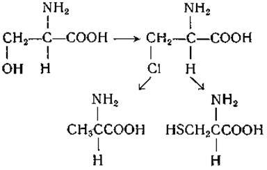
A second line of evidence is based on studies examining how the optical rotation of amino acids varies with their degree of ionization and the wavelength of polarized light [454]. For instance, a-amino acids (and a-hydroxy acids) of the L-configuration exhibit a higher positive optical rotation in acidic solution than in Water (Table 5). Finally, the premise that amino acids isolated from proteins possess a uniform configuration is reinforced by data obtained using Enzymes with strict optical Specificity.
In older literature, the direction of optical rotation in amino acids was denoted by the lowercase letters d (dextro, or right) and l (levo, or left), indicating the direction of rotation in aqueous solution. Later, as it became clear that amino acids isolated from proteins share an identical configuration, the prefix l was introduced to designate amino acids of the "natural" steric series, while the direction of rotation was indicated by the signs (+) and (—) enclosed in parentheses, such as l(—)-alanine or l(+)-glutamic acid. Using two concurrent notation systems frequently led to confusion, which was further compounded by the fact that some Natural Amino Acids are levorotatory while others are dextrorotatory. Furthermore, for certain amino acids, the signs of optical rotation in aqueous solution and in the presence of acid are opposite. Additional difficulties arose regarding amino acids containing more than one asymmetric carbon atom in their molecules; a case in point is Threonine isolated from protein, which was initially designated as d(—)-threonine due to its structural relationship with D-threose.
To standardize amino acid nomenclature, a new system was proposed wherein capital letters L and D are used to denote the configuration of the a-carbon atom [455, 456]. This notation system has almost entirely superseded the older one; however, ambiguities regarding amino acid designations still occasionally appear in the literature. When the configuration of an amino acid is unknown, it is customary to assign it the designation d or l based on the sign of its optical rotation in aqueous solution1, with the solvent used for the measurement explicitly stated. Tables 5, 6, and 7 list the specific rotations in water and Hydrochloric acid for Amino Acids Commonly Found in Proteins, as well as for various Other Amino Acids.
Table 5 Specific optical rotation (at 25° ±1°) of amino acids commonly found in protein hydrolysates
|
L-Amino Acid |
[a]D (in H2O) |
[a]D (in 5 N HCl) |
|
Alanine * |
+ 1.8 |
+14.6 |
|
+12.5 |
+27.6 |
|
|
Asparagine |
+33.2 ** |
|
|
Aspartic acid * |
+ 5.0 |
+25.4 |
|
Valine * |
+ 5.6 |
+28.3 |
|
—38.5 |
+ 11.8 |
|
|
Glutamine |
+ 6.3 |
+31.8*** |
|
Glutamic acid * |
+12.0 |
+31.8 |
|
Isoleucine * |
+12.4 |
+39.5 |
|
Leucine * |
—11.0 |
+ 16.0 |
|
Lysine * |
+ 13.5 |
+26.0 |
|
—10.0 |
+23.2 |
|
|
4-Hydroxyproline |
—76.0 |
—50.5 |
|
Proline * |
—86.2 |
—60.4 |
|
Serine * |
— 7.5 |
+15.1 |
|
Tyrosine * |
—10.0 |
|
|
Threonine * |
—28.5 |
—15.0 |
|
—33.7 |
+ 2.8 *** |
|
|
Phenylalanine * |
—34.5 |
— 4.5 |
|
Cysteine * |
—16.5 |
+ 6.5 |
|
Cystine * |
—232 |
* Determinations performed using isomers obtained by enzymatic resolution of racemates [585].
** In 3 N HCl.
*** In 1 N HCl.
1 In such cases, it is preferable to use the signs (+) and (—) in parentheses. — Ed. note.
Table 6 Specific optical rotation of Other Naturally Occurring Amino Acids
|
Amino Acid |
[a]D (in HCl) |
[a]D (in 5 N HCl) |
|
(+)-S-Allyl-L-cysteine sulfoxide |
+62.8 |
|
|
L-a-Aminoadipic acid * |
+ 3.2 |
+25.0 |
|
L-a-Aminobutyric acid * |
+ 9.3 |
+20.6 |
|
L-Homoserine * |
— 8.8 |
+ 18.3 |
|
L-Djenkolic acid |
—44.5 ** |
|
|
L-a,y-Diaminobutyric acid * |
+ 7.2 |
+31.7 |
|
LL-a,e-Diaminopimelic acid *..................... |
+ 8.1 |
+45.1 |
|
L-a,ß-Diaminopropionic acid * |
0 |
+34.0 |
|
3,5-Diiodo-L-tyrosine |
+ 2.9*** |
|
|
L-Canavanine |
+ 7.9 |
|
|
O-Carbamyl-D-serine |
+ 2.0 |
—19.6 *** |
|
L-Kynurenine |
—30.5 |
|
|
a-Methyl-D-serine * |
+ 4.5 |
+ 2.0 |
|
(+)-S-Methyl-L-cysteine sulfoxide |
+125 |
+ 168*** |
|
5-Hydroxy-L-lysine * |
+ 9.2 |
+17.8 |
|
allo-Hydroxy-L-proline |
—59.5 |
—18.8 |
|
L-Ornithine * |
+ 12.1 |
+28.4 |
|
D-Penicillamine |
—56.0 |
|
|
L-Pipecolic acid ...................... |
—24.6 |
|
|
L-Thyroxine |
—4.4****** |
|
|
L-Theanine |
+ 6.3 |
|
|
D-Cycloserine |
+116 |
|
|
L-Cystathionine |
23.7 *** |
|
|
L-Citrulline * |
+ 4.0 |
+24.2 |
* Determinations performed using isomers obtained by enzymatic resolution of racemates [585].
** In 1% HCl.
*** In 1.1 N HCl.
**** In 1 N HCl.
***** In a 1% solution (based on the free amino acid) of The amino acid hydrochloride in 1 N NaOH.
****** In a 0.13 N NaOH solution in 70% alcohol.
Table 7 Specific optical rotation of certain amino acids
|
L-Amino Acid |
[a]D (in H2O) |
[a]D (in 5 N HCl) |
|
Allylglycine * |
—37.1 |
—5.7 |
|
a-Aminoheptanoic acid * |
+6.8 |
+23.3 |
|
a-Amino-d-hydroxy-n-valeric acid * |
+6.0 |
+28.8 |
|
a-Amino-e-hydroxy-n-caproic acid * |
+4.0 |
+23.7 |
|
a-Aminocaprylic acid * |
+9.1 |
+23.0 |
|
a-Aminotricarballylic acid (A) * |
+7.5 |
+36.4 |
|
a-Aminotricarballylic acid (B) * |
—32.8 |
—48.0 |
|
a-Aminophenylacetic acid * |
+ 114 |
+168 |
|
a-Aminocyclohexylacetic acid * |
+6.7 |
+35.5 |
|
a-Aminocyclohexylpropionic acid * |
—9.0 |
+15.0 |
|
Homoglutamine |
+2.6 |
+21.0 ** |
|
Homolanthionine |
+37.3 |
|
|
Homocystine * |
+78.0 |
|
|
Isoleucine * |
+11.2 |
+6.7 |
|
allo-Isoleucine * |
+15.9 |
+39.6 |
|
Lanthionine |
+43.8 *** |
|
|
tert-Leucine * |
—9.7 |
+7.4 |
|
Norvaline * |
+7.0 |
+24.1 |
|
Norleucine * |
+4.7 |
+24.5 |
|
allo-5-Hydroxylysine * |
+ 10.9 |
+31.4 |
|
ß-2-Thienylalanine |
—31.7 |
|
|
allo-Threonine * |
+10.0 |
+31.7 |
|
ß-Phenylserine * |
—33.1 |
—50.3 |
|
allo-ß-Phenylserine * |
+8.2 |
+81.3 |
|
allo-Cystathionine |
—25.0 ** |
|
|
Ethionine * |
—9.2 |
+23.7 |
* Determinations were performed on isomers obtained by the Enzymatic Cleavage of racemates [585].
** In 1 N HCl.
*** In a 2% aqueous solution containing 1 equivalent of NaOH.
It should be noted that even The Use of the capital letters D and L is not always unambiguous. For instance, mannosaminic acid derived from D-mannose should, strictly speaking, be considered D-mannosaminic acid; however, this compound is simultaneously an L-amino acid. To avoid ambiguity, the lowercase subscripts "s" (for serine) and "g" (for glyceraldehyde) are appended to the right of the letters D and L. Thus, Dg-mannosaminic acid = Ls-mannosaminic acid.
The D-glyceraldehyde molecule, conventionally adopted as the reference Structure (standard of comparison) to which the configuration of sugars is correlated, can be chemically converted without configurational change into (+)-malic acid, (—)-lactic acid, and (+)-tartaric acid. The L-serine molecule—the levorotatory serine commonly present in proteins—was arbitrarily chosen as the standard of comparison for determining the configuration of amino acids.
Both standards are mutually consistent: the identity of the configurations of D-glyceraldehyde and D-serine has been unambiguously proven by carrying out the following series of Chemical Reactions [457–460]:

The absolute optical configuration of the amino acids discussed above was established based on data obtained by X-Ray Diffraction and other Methods [461, 462]. These data confirmed that the conventional designations proposed by Fischer coincide with the true absolute configuration. Thus, the molecules of Dg-glyceraldehyde and Ls-serine have the following configuration:
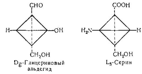
The molecules of most natural amino acids possess only a single asymmetric carbon atom having the L-configuration, but certain natural amino acids contain more than one asymmetric carbon atom and therefore exist in more than two stereoisomeric forms. The formulas of the isoleucine isomers serve as an example of this.
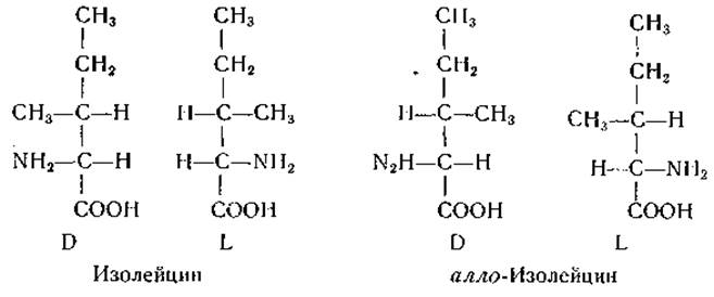
The ß-carbon atom of L-isoleucine has the L-configuration [463–466], whereas the ß-carbon atom of L-threonine has the D-configuration [467]. In hydroxy-L-proline and its mirror image isomer, the hydroxyl group is in the *trans* position relative to the carboxyl group, whereas in the corresponding allo-forms, it is in the *cis* position [468–470].
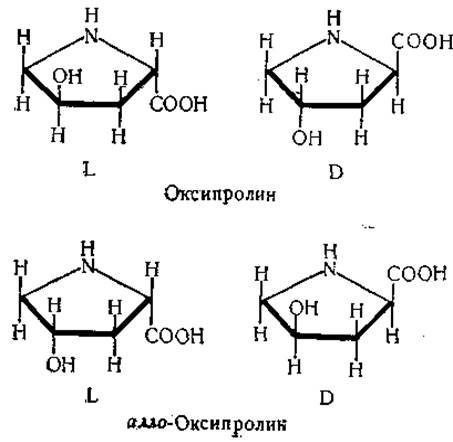
The y-carbon atom of hydroxy-L-proline (and allo-hydroxy-D-proline) possesses the L-configuration; this conclusion follows from considerations based on The Study of the relationships among the stereoisomers of hydroxyornithine and hydroxyproline [586, 587]:
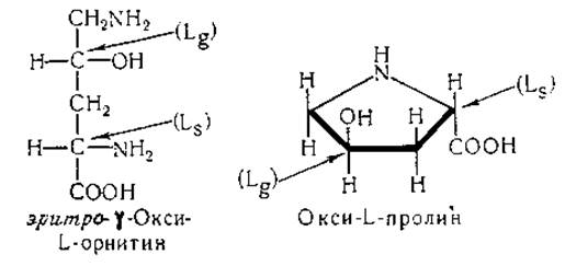
The configuration of the second asymmetric center of δ-hydroxy-L-lysine has not yet been definitively elucidated; however, based on available data, it is suggested that this asymmetric center has the L-configuration. Both asymmetric carbon atoms of natural δ-hydroxypipecolic acid have this same configuration [586, 588].
Natural octopine is known to contain residues of D-alanine and L-arginine in its molecule (p. 57). The methyl derivative of lanthionine obtained from subtilin apparently contains D-a-aminobutyric acid and L-alanine, while the configuration of the third asymmetric carbon atom remains unknown (p. 53). Natural argininosuccinic acid, canavanosuccinic acid, and cystathionine presumably possess the L-configuration.
The molecules of certain amino acids occur in an internally compensated or meso-form. For example, the racemization of cystine in acidic solution yields (along with a small amount of DL-cysteine) a meso-form that cannot be resolved into optically active forms by the methods used to separate DL-cystine [471, 472]. Meso-forms of lanthionine [242–245], homolanthionine [473], and cystathionine [474] have also been obtained. Meso-a,ε-diaminopimelic acid has been isolated from Bacteria (p. 51), but it also occurs in nature in the LL-form [475]; the meso-, DD-, and LL-forms of this acid have been synthesized [476]. Chemical degradation of streptomycin yields a,γ-diamino-ß-hydroxyglutaric acid, which can exist in two meso- and one racemic form [477]. The configuration of natural a,ε-diamino-ß-hydroxypimelic acid is unknown. Two meso-forms are possible for this amino acid, as well as for the methyl derivative of lanthionine isolated from subtilin (p. 53).
A number of amino acids of interest to biochemists do not occur in nature. Some of these have been resolved into optical isomers and utilized in biochemical research. The Chemical synthesis of ß-hydroxyglutamic acid—which was at one time considered a constituent of proteins (p. 11)—has been accomplished. Racemates of two diastereomers of this compound have been obtained [478, 479]. One of them, allo-ß-hydroxyglutamic acid, apparently undergoes enzymatic decarboxylation more readily than the other (p. 205). Data indicate that the Decarboxylases of allo-ß-hydroxyglutamic and glutamic acids are not identical [480].
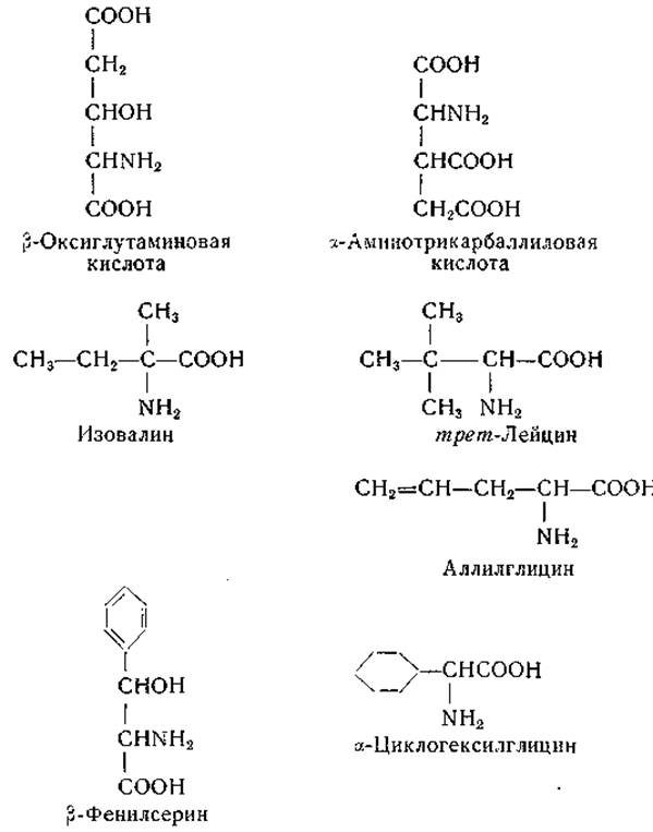
The existence of a distinct allo-ß-hydroxyglutamic acid decarboxylase seemingly points to the occurrence of this amino acid in nature. However, since reinvestigation of the natural product presumed to contain ß-hydroxyglutamic acid failed to detect the latter [320], further evidence is required to confirm the presence of this amino acid in biological systems [320].
α-Aminotricarballylic acid—the amino analog of isocitric and oxalosuccinic acids—has been prepared synthetically, and its four isomers have been obtained [481]. Treatment of the latter with nitrous acid converted them into isomeric isocitric acids. The action of nitrous acid on one of the levorotatory isomers of α-aminotricarballylic acid possessing the L-configuration [481] yields natural d-isocitric acid. The latter is subject to the action of isocitrate dehydrogenase [482]; its carbon atom evidently has the L-configuration. As for the ß-carbon atom of isocitric acid, it apparently possesses the D-configuration [483]. Four isomers of ß-phenylserine have also been obtained [484–487]. This amino acid does not occur in nature, but its D-threo-isomer is stereochemically related to the antibiotic chloramphenicol (levomycetin) (p. 76).
A number of other non-natural DL-Amino acids have also been resolved into optical isomers, including norvaline (a-aminovaleric acid), norleucine (a-aminocaproic acid), tert-leucine (ß,ß'-dimethyl-a-aminobutyric acid), isovaline (a-methyl-a-aminobutyric acid), ethionine (a-amino-y-ethylthiobutyric acid), ß-2-thienylalanine, a-phenylglycine, a-cyclohexylglycine, ß-cyclohexylalanine, allylglycine, etc. It is quite possible that one or another of these listed amino acids will eventually be discovered in natural products; after all, many amino acids, such as alanine [2], a,ß-diaminopropionic acid [488], γ-aminobutyric acid [489], a,ε-diaminopimelic acid [490], and aspartic acid [448], were first obtained synthetically and only later identified among natural products.
Special considerations arise when choosing designations for amino acids whose molecules contain an a-methyl group [491]. For example, in (+)-isovaline, the methyl group occupies the same spatial position as the a-hydrogen atom in the L-a-aminobutyric acid molecule. At the same time, (+)-isovaline can be viewed as D-alanine in which the hydrogen atom has been replaced by an ethyl group:
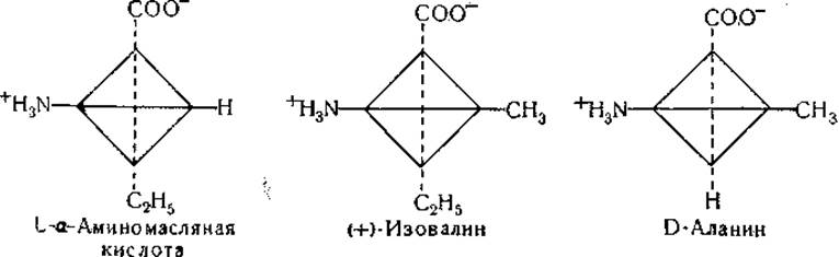
Therefore, dextrorotatory isovaline can be designated either as a-methyl-L-a-aminobutyric acid or as a-ethyl-D-alanine. The specific optical rotation of (+)-isovaline is +13.1° in water and +7.8° in hydrochloric acid; this direction of change in specific rotation is typical of D-amino acids. However, the molar rotation of (+)-isovaline in glacial acetic acid is greater than in water, which is characteristic of L-amino acids.
Chloroacetyl-(+)-isovaline is hydrolyzed by renal acylase, whereas its enantiomorphic derivative is resistant to this enzyme. Based on such studies, (+)-isovaline should be designated as L-isovaline. Applying this biological criterion to determine the optical configuration appears to be fully justified, yet the foregoing Discussion illustrates the difficult challenges that arise in establishing the configuration of a-alkyl-a-amino acids.
The question of isomerism in S-stereoisomers of methionine sulfoxide [492] deserves attention. In the methionine sulfoxide molecule, the sulfur atom represents an additional center of Asymmetry, and therefore 4 isomers of this compound are possible. The following have been obtained: L-methionine-l-sulfoxide, L-methionine-d-sulfoxide, and the corresponding d-sulfoxide. There is evidence that the d- and l-isomers of the sulfoxide differ in biological activity (p. 149). An example of a natural amino acid containing an asymmetric sulfur atom in its molecule is alliin (p. 54).
Other types of stereoisomerism are also known. For example, introducing an isotope atom into a molecule can lead to The formation of an asymmetric carbon atom. This type of isomerism is possible for a serine molecule containing two deuterium atoms [589]:
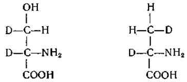
Of interest in this regard are studies concerning the enzymatic Decarboxylation of amino Acids in a D2O medium (p. 257). Finally, it should be noted that identical groups in fully symmetrical molecules, such as citric acid, behave differently in enzymatic and non-enzymatic reactions with optically active compounds. This type of asymmetry and the reactions of such substances were discussed in detail by Schwartz and Carter [493].
Last update: 06/08/2026
Editorial and Educational Adaptation: This material has been compiled based on the primary/original source text. The project team performed an editorial review, corrected technical inaccuracies, structured sections, and adapted the content for an educational format.
What was processed:
- elimination of formatting defects (OCR errors, structural breaks, corrupted characters);
- editorial organization of content;
- standardization of terminology in accordance with academic sources;
- verification of factual statements against the original source text.
All mentions of the author, publication year, and origin of the primary text have been preserved in accordance with the source.