Harper's Illustrated Biochemistry, Volume 1 - Murray R. 1993
Biomolecules and Biochemical Methods
The Cell
Schleiden and Schwann, along with other 19th-century pioneers of science such as Virchow, considered The Cell to be the fundamental unit of biological activity. However, immediately following the end of World War II, three developments marked the beginning of a period during which biochemistry and cell biology pursued separate paths: 1) the widespread adoption of electron microscopes; 2) The Development of Cell Disruption techniques under relatively mild conditions that preserve the functions of cellular components; and 3) the widespread availability of high-speed refrigerated ultracentrifuges, which made it possible to generate centrifugal forces sufficient to separate the components of disrupted Cells while preventing overheating. Electron Microscopy revealed numerous previously unknown or poorly resolved cellular components, whereas cell disruption and ultracentrifugation enabled their Separation and in vitro investigation.
Class="center">Table 2.3. Normal Chemical composition of a 65 kg human body1)
|
Components |
Mass, kg |
Content, % |
|
11 |
17.0 |
|
|
9 |
13.8 |
|
|
1 |
1.5 |
|
|
Water 2) |
40 |
61.6 |
|
Minerals |
4 |
6.1 |
1) From Davidson S. D., Passmore R., Brock J. F.: Human Nutrition and Dietetics, 5th ed. Churchill Livingstone, 1973. Reprinted by permission.
2) Water content varies significantly across different Tissues: in Bone tissue lacking Bone Marrow, it accounts for 22.5%. Water content also decreases with fat accumulation.
Rat Liver cell
Figure 2.1 shows a schematic diagram of a rat liver cell (hepatocyte). The biochemical properties of these cells have been studied in the greatest detail—partly because liver cells can be obtained in relatively large quantities, and partly because they are well-suited for fractionation and perform a wide range of functions. The hepatocyte contains all The major types of Organelles present in Eukaryotic cells (Table 2.4): The Nucleus, Mitochondria, Endoplasmic reticulum, free Ribosomes, Golgi apparatus, Lysosomes, Peroxisomes, Plasma Membrane, and cytoskeletal elements.

Fig. 2.1. Schematic diagram of a rat liver cell showing major cellular organelles.
Subcellular Fractionation
To thoroughly investigate the function of any organelle, one must first obtain these organelles in a relatively pure state, such that the preparation is minimally contaminated with other organelles. The Procedure typically used to achieve this is called subcellular fractionation, which consists of three main steps: extraction, homogenization, and centrifugation. Much of the pioneering work in this field was performed using rat liver.
A. Extraction. The first step in isolating specific organelles (or molecules) is their extraction from the cells in which they reside. Most organelles and many Biomolecules (particularly proteins) are highly labile and readily lose their biological activity.
Consequently, they must be extracted under mild conditions (In aqueous solutions, avoiding extreme pH levels, osmotic pressures, and high temperatures). Most operations involving organelle isolation are carried out between 0–4° C (in a cold room or on ice). At room Temperature, a significant loss of activity may occur, partly due to the action of various hydrolytic Enzymes (proteases, Nucleases, etc.) released upon cell disruption. Typically, a 0.25 M sucrose solution (an isoosmotic solution) containing K+ and Mg2+ ions at near-physiological concentrations is used for organelle extraction; the pH of the solution is adjusted to 7.4 with 0.05 M Tris-HCl buffer (tris[hydroxymethyl]aminomethane hydrochloride). This solution is commonly referred to as STKM. Not all Solvents provide as gentle extraction conditions as STKM; for instance, organic solvents are used for the extraction of lipids and carbohydrates.
B. Homogenization. To isolate organelles (or biomolecules) from cells, it is first necessary to disrupt the cells under mild conditions. Homogenization is a convenient method for breaking down Organs (such as the liver, Kidney, or Brain) and their constituent cells. In this procedure, minced fragments of the respective organ are placed in a suitably sized Glass tube filled with a homogenization medium (e.g., STKM), and a pestle is rotated within the mixture (either manually or using a motor). Rotating the pestle at a controlled speed generates shear forces that disrupt the cells and release their contents into the sucrose solution. The resulting suspension, which contains many intact organelles, is called a homogenate.
C. Centrifugation. Subfractionation of the homogenate by differential centrifugation is one of the most important techniques in biochemistry. Classically, it involves three successive centrifugation steps at increasing speeds (Fig. 2.2). Each step yields a pellet and a supernatant. The supernatant obtained at each stage is subjected to centrifugation in the next step. This procedure produces Three types of pellets, designated as the nuclear, mitochondrial, and microsomal fractions. None of these fractions represents absolutely pure organelles. However, using electron microscopy as well as identifying characteristic marker enzymes and chemical components (such as DNA or RNA), it has been established that the major components of these three fractions are nuclei, mitochondria, and microsomes, respectively. A marker enzyme or chemical compound is a component that is present almost exclusively within a specific type of organelle; for example, acid phosphatase is localized in lysosomes, and DNA is found in the nucleus (Table 2.4). Thus, a marker serves as an indicator of the presence or absence of the organelle fraction in which it resides. The microsomal fraction (microsomes) consists primarily of a mixture of smooth endoplasmic reticulum fragments, rough endoplasmic reticulum (i.e., endoplasmic reticulum with attached ribosomes), and free ribosomes. The Contents of the final supernatant correspond roughly to the COMPOSITION OF THE cell sap (Cytosol). Modifications of this basic approach—utilizing different homogenization media, conditions, or centrifugation Methods (such as continuous or discontinuous sucrose gradients)—have made it possible to isolate, in a more or less pure state, all the organelles shown in Fig. 2.1 and listed in Table 2.4. Although the scheme described above is applicable to most organs and cells, standardizing the subcellular fractionation procedure in each case requires a series of assays for marker enzymes or other chemical components, alongside electron microscopic observations.
Table 2.4. Major cellular organelles and their functions. Only the primary functions of each organelle type are listed. In certain cases, these organelles may also support other processes, reactions, and metabolic pathways.
|
Organelle or fraction1 |
Marker |
Main functions |
|
Nucleus |
DNA |
Site of chromosome localization Site of DNA-dependent RNA Synthesis (METABOLISM/31.html">Transcription) |
|
Mitochondrion |
Citric Acid Cycle, ATP synthesis |
|
|
Ribosome1) |
High RNA content |
|
|
Endoplasmic reticulum |
Glucose-6-phosphatase |
Membrane-bound ribosomes serve as the primary site of protein synthesis Synthesis of various lipids Oxidation of many xenobiotics (by cytochrome P-450) |
|
Lysosome |
Acid phosphatase |
Site of Action for numerous Hydrolases (enzymes catalyzing hydrolytic Cleavage) |
|
Plasma membrane |
Na/K+-ATPase, 5'-nucleotidase |
Transport of Molecules into and out of the cell, cell-Cell Adhesion and communication |
|
Golgi apparatus |
Galactosyltransferase |
Intracellular protein sorting Sulfation reactions |
|
Peroxisome |
Catalase Urate oxidase |
Degradation of certain Fatty acids and Amino Acids Generation and breakdown of hydrogen peroxide |
|
Specific marker enzymes 2) absent |
Structural support (microfilaments, microtubules, Intermediate filaments) |
|
|
Cytosol 1) |
Glycolysis, fatty acid synthesis |
1) An organelle can be defined as a membrane-bounded subcellular Structure that can be isolated by high-speed centrifugation. According to this definition, ribosomes, the cytoskeleton, and the cytosol are not strictly organelles. However, they are listed alongside organelles in this table because they are likewise routinely isolated by centrifugation and can be regarded as subcellular entities or fractions. An organelle preparation obtained through a single round of differential centrifugation is rarely pure; obtaining a clean fraction typically requires repeated centrifugation steps.
2) The cytoskeletal fraction is identified via electron microscopy or electrophoretic analysis for fraction-specific proteins.
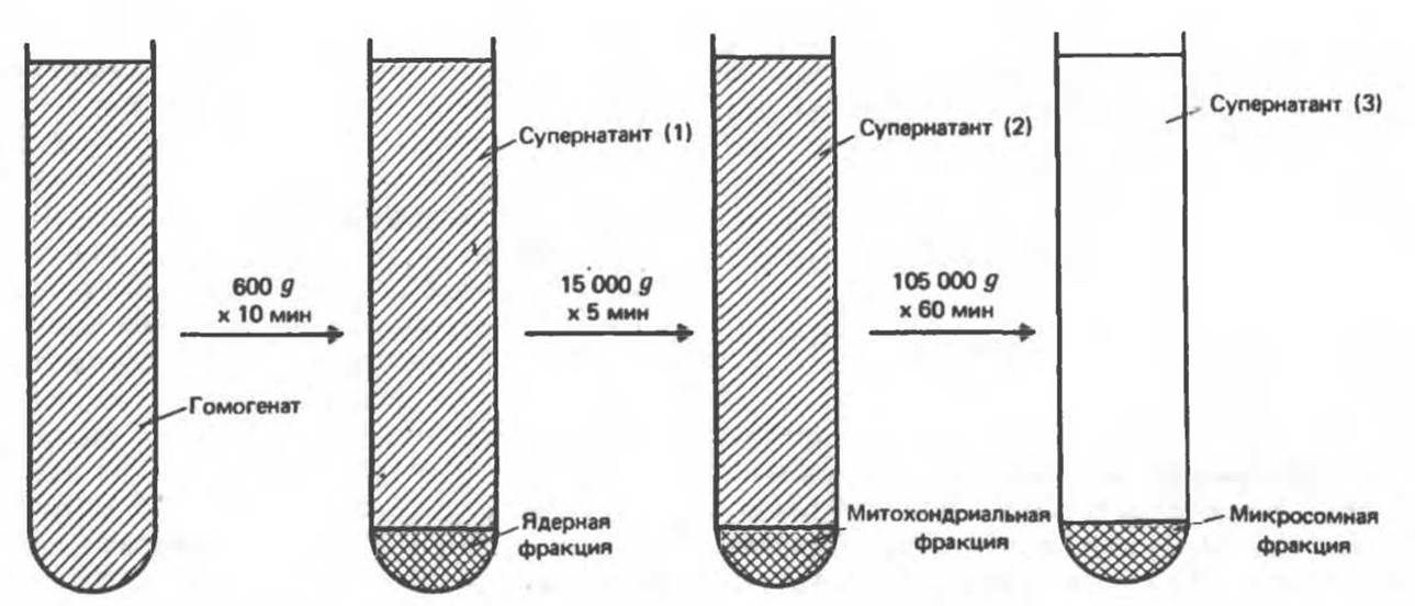
Fig. 2.2. Scheme for the separation of subcellular fractions by differential centrifugation. Homogenized tissue (e.g., liver) is first centrifuged at a low speed, which sediments the nuclear fraction (containing nuclei and unbroken cells) and leaves a supernatant (1). The supernatant is carefully decanted and centrifuged at a higher speed to separate the mitochondrial fraction (containing mitochondria, lysosomes, and peroxisomes) and supernatant (2). This supernatant is decanted and centrifuged at high speed to pellet the microsomal fraction (containing a mixture of free ribosomes and fragments of smooth and rough endoplasmic reticulum), leaving a clear, transparent solution—the final supernatant (3)—which represents the cytosol, or cell sap. By employing various modifications of the approach outlined here, each cellular organelle can generally be isolated in a relatively pure form.
The Importance of subcellular fractionation to the development of biochemistry and cell biology cannot be overstated. It constitutes a key component of the general experimental strategy (see below) that enabled the elucidation of the organelle functions listed in Table 2.4. Acquiring this information represents one of the major triumphs of biochemical research (see below).
Last update: 06/08/2026
Editorial and Educational Adaptation: This material has been compiled based on the primary/original source text. The project team performed an editorial review, corrected technical inaccuracies, structured sections, and adapted the content for an educational format.
What was processed:
- elimination of formatting defects (OCR errors, structural breaks, corrupted characters);
- editorial organization of content;
- standardization of terminology in accordance with academic sources;
- verification of factual statements against the original source text.
All mentions of the author, publication year, and origin of the primary text have been preserved in accordance with the source.