BIOCHEMISTRY - L. Stryer - 1984
VOLUME 1
PART I. CONFORMATION AND DYNAMICS
CHAPTER 8. ZYMOGEN ACTIVATION: DIGESTIVE ENZYMES AND BLOOD CLOTTING FACTORS
8.16. The major classes of proteolytic enzymes are serine proteases and carboxyl proteases
Thus far, we have considered two classes of Proteolytic Enzymes, namely zinc-containing proteases (exemplified by Carboxypeptidase A—see Section 7.8) and Serine proteases. Another widespread group is the thiol proteases. For example, Papain, obtained from papaya, has a Cysteine residue in its Active Site whose role is analogous to that of serine-195 in Chymotrypsin. Papain catalysis proceeds via a thioester intermediate, with a nearby Histidine side chain participating in the catalysis. Another large group of proteolytic enzymes comprises carboxyl proteases, also known as acid proteases because most of them are active only in an acidic environment. The best-known representative of this group is Pepsin, the principal proteolytic enzyme of gastric juice. Pepsin, with a Molecular Weight of 34.6 kDa, is formed from pepsinogen (a proenzyme) by the Cleavage of a 44-amino-acid N-terminal peptide. This activation either occurs spontaneously (at pH 2 and below) or is catalyzed by pepsin. Consequently, pepsinogen secreted into the gastric lumen is converted to pepsin within seconds. The Active Site of pepsin contains two aspartate residues. For enzymatic activity, one aspartate residue must be ionized and the other non-ionized; this determines the pH optimum of pepsin, which is between 2 and 3. Structurally and enzymatically similar carboxyl proteases have been isolated from Lysosomes, which carry out intracellular degradation (Section 20.8), as well as from various Molds (Fig. 8.25). All carboxyl proteases are inhibited by pepstatin (an analog of a hexapeptide transition-state intermediate) added at very low concentrations (on the order of 10-10 M).
Class="center">Fig. 8.25. Structure of an acid protease from the mold Rhizopus. The α-carbon atoms are shown. The active site is located in a cleft between the two lobes of the molecule, which accommodates 8 amino acid residues of the substrate. Pepsin has a very similar structure

8.17. Blood clotting as a cascade of zymogen activations
The Activation of a precursor protein by the cleavage of a peptide bond is a major regulatory mechanism in various biological systems. We will now examine The Role of zymogen activation in Blood clot formation, which is one of the three mechanisms of hemostasis. The other two regulatory mechanisms are the rapid constriction of the damaged blood vessel and the aggregation of platelets at its wall to protect the injured vascular surface.
Blood clot formation occurs through a cascade of sequential transformations involving more than 10 different Proteins. A striking feature of this process is that it involves a series of zymogen activation steps. In this cascade of enzymatic reactions, the active form of one clotting factor catalyzes the activation of the next. Because of the catalytic nature of this process, the factors acting in the Cytology/cytology/16.html">Early stages of the pathway are required in very small amounts. Their effect is amplified many times over through the numerous subsequent steps, ultimately ensuring a rapid response to injury.
8.18. Blood clotting requires the interaction of two pathways of enzymatic cascades
In 1863, Joseph Lister discovered that blood remains fluid in an excised bovine jugular vein, whereas it clots immediately in a Glass vessel. Contact with a foreign surface led to the activation of components originally present in the blood. Accordingly, this clotting mechanism (pathway) was called the intrinsic pathway. However, substances not normally present in the blood can also trigger clotting. For instance, adding extracts of many Tissues, especially the Brain, to plasma results in rapid clot formation. This clotting mechanism (pathway) is called the extrinsic pathway.
During blood clotting, the extrinsic and intrinsic pathways act in concert (Fig. 8.26). Both pathways are required for normal Blood Coagulation, as evidenced by various bleeding disorders associated with a deficiency in any of the proteins involved in only one of the pathways. The extrinsic and intrinsic pathways differ only in their initial steps, after which they merge into a common pathway that leads to The formation of a fibrin clot.
Fig. 8.26. Fibrin clot formation involves the interaction of three pathways

8.19. Fibrinogen is converted into a fibrin clot by thrombin
The most thoroughly studied step in the clotting process is The conversion of fibrinogen to fibrin by the proteolytic enzyme Thrombin. Fibrinogen differs from the proteins discussed earlier (such as Lysozyme and chymotrypsin) by its much larger mass and elongated shape. Electron Cell/15.html">Microscopy reveals that fibrinogen consists of three nodules connected by two strands (Fig. 8.27). The molecule is 460 A long and has a molecular weight of 340 kDa, which is about 10 times the mass of chymotrypsin. Fibrinogen consists of six polypeptide chains of three distinct types, paired as Aα, Bβ, and γ.
Fig. 8.27. Schematic representation of a fibrinogen molecule based on electron microscopy data

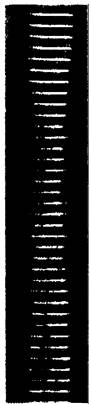
Fig. 8.28. Electron micrograph of modified fibrinogen with an ordered structure. The repeat distance along the fiber axis is 225 A, the same as in fibrin
Fibrinogen, a highly soluble plasma protein, is converted into an insoluble fibrin monomer by the proteolytic action of thrombin. Thrombin cleaves four peptide bonds between Arginine and Glycine in fibrinogen. This cleavage releases four Peptides: an A-peptide of 18
residues from each of the two α-chains and a B-peptide of 20 residues from each of the two β-chains. These A- and B-peptides are called fibrinopeptides. The fibrinogen molecule stripped of its fibrinopeptides is a fibrin monomer. Its subunit structure is _ (α, β, γ)2; it contains about 97% of The amino acid residues of fibrinogen.
Last update: 06/08/2026
Editorial and Educational Adaptation: This material has been compiled based on the primary/original source text. The project team performed an editorial review, corrected technical inaccuracies, structured sections, and adapted the content for an educational format.
What was processed:
- elimination of formatting defects (OCR errors, structural breaks, corrupted characters);
- editorial organization of content;
- standardization of terminology in accordance with academic sources;
- verification of factual statements against the original source text.
All mentions of the author, publication year, and origin of the primary text have been preserved in accordance with the source.