BIOCHEMISTRY - L. Stryer - 1984
VOLUME 1
PART I. CONFORMATION AND DYNAMICS
CHAPTER 8. ZYMOGEN ACTIVATION: DIGESTIVE ENZYMES AND COAGULATION FACTORS
8.20. Fibrin monomers spontaneously assemble into fibrils
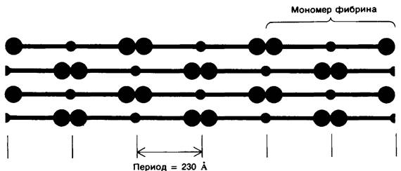
The solubility of fibrin monomers is much lower than that of fibrinogen. They spontaneously aggregate to form fibrin in the shape of long, insoluble strands (fibrils). Electron Cell/15.html">Microscopy and low-angle X-Ray Diffraction have shown that fibrin has a periodic Structure with a repeat length of 230 A (Fig. 8.29). Since the length of fibrinogen is about 460 A, which is twice as long, it seems likely that upon aggregation, fibrin monomers form parallel rows shifted relative to one another by half a molecule (Fig. 8.30).
Class="center">Fig. 8.29. Electron micrograph of fibrin. The axial repeat period of the fiber is 230 A, which is half the length of the fibrinogen molecule
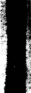
Fig. 8.30. Proposed arrangement of fibrin monomers in a fibrin clot. In this packing of fibrin monomers, where parallel rows are staggered by half a molecule, the repeat period is 230 A, consistent with the observed pattern
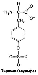
Why is it that fibrin monomers can aggregate, while the fibrinogen from which they are derived cannot? Ongoing detailed structural studies will provide the definitive answer. All fibrinopeptides studied to date from all vertebrate species carry a high net negative charge, being rich in glutamic and aspartic acid residues. In addition, fibrinopeptide B contains an unusual negatively charged Tyrosine derivative, tyrosine-O-sulfate. Apparently, the presence of these and other negatively charged groups causes fibrinogen molecules to repel one another. The fibrin monomers released by Thrombin have a surface with entirely different electrical properties that enable aggregation. Recall that the substitution of just a single charged group—glutamate to valine—causes the aggregation of deoxyhemoglobin in Sickle-Cell Anemia.
8.21. The fibrin clot is stabilized by covalent cross-links
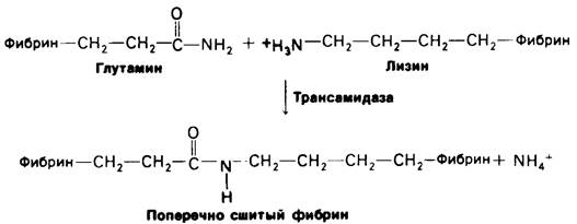
The clot formed by the spontaneous aggregation of fibrin monomers is initially very weak and loose. It is subsequently stabilized by covalent cross-links between the side chains of individual molecules in the fibrin strands. It has been established that during a transamidation reaction, peptide bonds are formed between the side chains of specific glutamine and Lysine residues (Fig. 8.31). The enzyme catalyzing this reaction is transamidase. This type of cross-linking is extremely rare in Proteins. Its significance is illustrated by the fact that patients with congenital transamidase deficiency exhibit increased bleeding tendencies.
Fig. 8.31. Cross-linking in fibrin occurs via transamidation
8.22. Thrombin is homologous to trypsin
The ability of thrombin to specifically cleave the bond between Arginine and Glycine residues suggests a similarity between thrombin and Trypsin. Indeed, such a similarity exists, as is evident from a comparison of the Amino acid sequences of these proteins. Thrombin has a mass of 33.7 kDa and consists of two chains. The 49-residue A chain shows no significant Homology to pancreatic Enzymes. In contrast, the B chain is highly homologous to trypsin, Chymotrypsin, and Elastase in its Amino Acid Sequence. The amino acid sequence around the active-site Serine in thrombin is Gly-Asp-Ser-Gly-Gly-Pro, which is identical to that in pancreatic Serine proteinases. Furthermore, thrombin also possesses a charge-Relay system. Its three-dimensional structure has not yet been fully solved, but one important feature of the Specificity pocket has been established: in thrombin, as in trypsin, an aspartate residue lies at the bottom of the substrate-binding pocket. This negatively charged group must electrostatically bind the positively charged arginine side chain of the substrate. The specificity of thrombin is much higher than that of trypsin: thrombin cleaves only specific arginine-glycine bonds, whereas trypsin hydrolyzes almost any peptide bond following arginine or lysine residues.
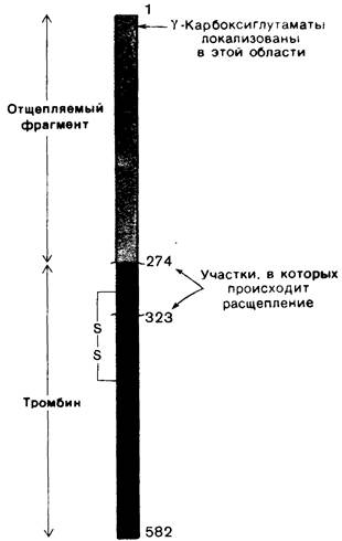
Like pancreatic serine proteinases, thrombin is synthesized as a zymogen, prothrombin, which has a molecular mass of 66 kDa. Proteolytic Cleavage of an arginine–Threonine bond releases a 32-kDa fragment from the amino terminus of prothrombin (Fig. 8.32). Subsequent cleavage of an arginine–leucine bond yields active thrombin. Thrombin also contains an ion pair analogous to the one in chymotrypsin between the positively charged amino group of isoleucine-16 and the negatively charged aspartate-194.
Fig. 8.32. Structure of prothrombin. Thrombin is formed by the cleavage of two peptide bonds (Arg-274—Thr-275 and Arg-323—Ile-324). The cleaved N-terminal fragment of prothrombin, which contains all the γ-carboxyglutamate residues, is shown in red. The A and B chains of thrombin are linked by a disulfide bond
The similarity in amino acid sequences indicates that thrombin is evolutionarily closely related to pancreatic serine proteinases. This is further supported by the similar activation mechanisms of these enzymes and the presence of a charge-relay system in both. Notably, prothrombin is synthesized in the Liver, which shares a common embryological origin with the Pancreas.
8.23. Vitamin K is required for prothrombin synthesis
For many years, it has been known that vitamin K is required for the synthesis of prothrombin and several other clotting factors. However, the MECHANISM OF ACTION of this vitamin was elucidated only recently through studies of abnormal prothrombin synthesized in the absence of vitamin K or in the presence of a vitamin K antagonist, such as dicoumarol. Dicoumarol is found in spoiled sweet clover, and cattle eating moldy hay die of hemorrhagic disease. A derivative of dicoumarol is used clinically as an anticoagulant to prevent thrombosis in patients with a tendency to form clots. Dicoumarol and warfarin (Fig. 8.33), a structural analog and antagonist of vitamin K, are also widely used as potent rodenticides. Under METABOLISM/18.html">The Influence of dicoumarol, a defective prothrombin appears in bovine Blood which, unlike normal prothrombin, cannot bind Ca2 +. For a long time, this was a mystery because the Amino Acid Composition and residue count of the defective prothrombin, determined after acid Hydrolysis, were identical to those of the normal protein. When normal prothrombin was cleaved into fragments, Ca2+ binding was shown to occur in the N-terminal region of the protein (Fig. 8.34). Furthermore, the N-terminal peptide of abnormal prothrombin was found to differ significantly from the corresponding peptide of normal prothrombin in electrophoretic mobility. Nuclear magnetic Resonance studies of these Peptides revealed that normal prothrombin contains a previously unknown amino acid, γ-carboxyglutamate, which is absent in the abnormal prothrombin. It turned out that the first ten glutamate residues in the N-terminal region of prothrombin are carboxylated to γ-carboxyglutamate, a carboxylation carried out by a vitamin K-dependent enzymatic system. This amino acid had escaped detection until recently because acid hydrolysis causes the loss of the γ-carboxyl group, converting γ-carboxyglutamate to Glu-.
Fig. 8.33. Structures of vitamin K2 and two of its antagonists, dicoumarol and warfarin
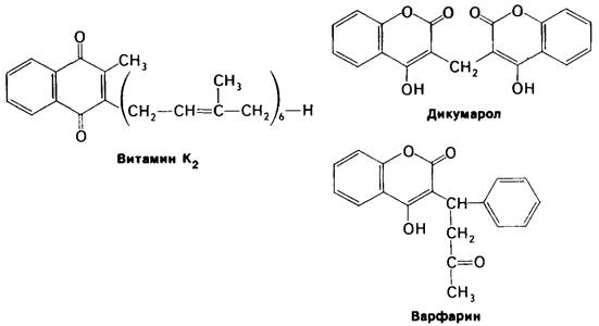
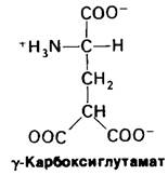
Fig. 8.34. Amino acid sequence of the N-terminal region of prothrombin [γ-carboxyglutamate (GL) residues are shown in red]

Last update: 06/08/2026
Editorial and Educational Adaptation: This material has been compiled based on the primary/original source text. The project team performed an editorial review, corrected technical inaccuracies, structured sections, and adapted the content for an educational format.
What was processed:
- elimination of formatting defects (OCR errors, structural breaks, corrupted characters);
- editorial organization of content;
- standardization of terminology in accordance with academic sources;
- verification of factual statements against the original source text.
All mentions of the author, publication year, and origin of the primary text have been preserved in accordance with the source.