BIOCHEMISTRY - L. Stryer - 1984
VOLUME 1
PART I. CONFORMATION AND DYNAMICS
CHAPTER 8. ZYMOGEN ACTIVATION: DIGESTIVE ENZYMES AND BLOOD CLOTTING FACTORS
8.13. Divergent and convergent evolution of serine proteases
Chymotrypsin, Elastase, and Trypsin share a generally similar Amino Acid Sequence because they arose from a common ancestor. The Gene for the ancestral enzyme apparently underwent several duplications. Subsequent Mutations in these genes eventually gave rise to the modern Enzymes. The Emergence of different specificities through mutations in genes derived from a common ancestor is a process of divergent evolution.
In comparing chymotrypsin with the bacterial enzyme subtilisin, we encounter a different type of evolutionary process.
Subtilisin is also a Serine protease. However, the Amino acid sequences of chymotrypsin and subtilisin are completely different, indicating their independent evolutionary origins. For example, the chymotrypsin molecule contains five disulfide bridges, whereas subtilisin has none. The amino acid sequence around the active-site serine in subtilisin and chymotrypsin is as follows:
-Gly-Thr- Ser *-Met-Ala-Ser in subtilisin
-Gly-Asp- Ser *-Gly-Gly-Pro in chymotrypsin
The three-dimensional structures of these enzymes are completely different. This makes it all the more surprising that subtilisin contains a charge-Relay system consisting, as in chymotrypsin, of aspartate, Histidine, and serine residues. Furthermore, subtilisin possesses an oxyanion hole that stabilizes the tetrahedral intermediate. The similarity of the charge-relay system, the oxyanion hole, and the catalytic mechanism in subtilisin and chymotrypsin is a striking example of independent, convergent evolution at the level of enzyme molecules. Why did independent, parallel Evolutionary Processes lead to the same solution in such diverse cases as vertebrate pancreatic serine proteases and bacterial subtilisin? As discussed below (Section 8.16), there are apparently very few ways to cleave peptide bonds. Studies of several hundred Proteolytic Enzymes have shown that there are perhaps only four basic types of proteolytic mechanisms, with serine proteases being the most widely distributed.
8.14. Coordinated activation of pancreatic zymogens
Protein Digestion in the duodenum requires the simultaneous action of several proteolytic enzymes, as each is specific for a limited number of side chains. Consequently, all zymogens must be converted into active enzymes at the same time. Coordinated control of the activation process is achieved because all pancreatic zymogens—trypsinogen, chymotrypsinogen, proelastase, and procarboxypeptidase—share a single common activator: trypsin. Where, then, does the large amount of trypsin required to activate these zymogens come from? The Cells lining the duodenum secrete the enzyme enteropeptidase1, which hydrolyzes a specific Lysine-isoleucine peptide bond in trypsinogen as it enters the duodenum. The small amount of trypsin formed in this way activates trypsinogen and the other zymogens. Thus, The formation of trypsin by enteropeptidase is the activation step of the activator:
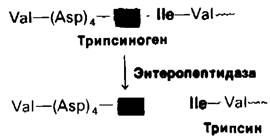
8.15. Premature activation of zymogens can be fatal, particularly in pancreatitis
There are many ways in which the Pancreas is protected from the proteolytic action of its own enzymes. First, all pancreatic enzymes that hydrolyze Proteins are synthesized as inactive zymogens. Second, within pancreatic cells, they are stored in granules surrounded by a lipoprotein membrane. Another protective measure is the presence of an inhibitor in pancreatic secretions (Section 8.12) that inactivates any small amounts of trypsin that might be formed there.
Class="center">Fig. 8.24. Schematic diagram of the pancreas and its connection to the Small Intestine

The pancreas also secretes lipases. One of these, phospholipase A2, is synthesized as prophospholipase A2, which is then activated by trypsin. Other lipid-cleaving enzymes are active in the presence of Bile salts, which enter the duodenum from the Liver. These Steroids act by solubilizing Lipids, thereby making them accessible to enzymatic Cleavage by lipases. Bile is synthesized in the liver, stored in the Gallbladder, and released into the duodenum, where it works together with pancreatic lipases to break down fats.
Acute pancreatitis is a severe, sometimes fatal, disease. It is characterized by the premature activation of pancreatic proteolytic and lipolytic enzymes. In pancreatitis, these enzymes become active while still within the pancreas itself. This results in the destruction of the pancreatic tissue and the walls of the Blood Vessels running through it. Acute pancreatitis can be caused by trauma to the acinar tissue.
Last update: 06/08/2026
Editorial and Educational Adaptation: This material has been compiled based on the primary/original source text. The project team performed an editorial review, corrected technical inaccuracies, structured sections, and adapted the content for an educational format.
What was processed:
- elimination of formatting defects (OCR errors, structural breaks, corrupted characters);
- editorial organization of content;
- standardization of terminology in accordance with academic sources;
- verification of factual statements against the original source text.
All mentions of the author, publication year, and origin of the primary text have been preserved in accordance with the source.